Lightness
We already know that low birth weight is a problem, but what it is is not yet fully understood. From the name itself it is clear that the main problem here is the child’s insufficient weight. When labor begins on time, and the baby’s weight is no more than 2500 grams, the fetus is a low-weight fetus. Hypotrophy is not necessarily accompanied by a simultaneous decrease in the size of the baby. Often, insufficient weight is a signal of fetal abnormality in the womb.
Organizational aspects
Often in an “interesting situation” the question arises: “At what stage of pregnancy is one registered?” Experts say: the sooner the better. There are three reasons supporting this belief:
- The main systems and organs begin to form precisely in the first 14 days after fertilization. The mother is strictly prohibited from drinking alcohol, smoking and taking medications. The need to take vitamins increases, so immediately after a positive test result, you need to visit a gynecologist as soon as possible so that he can prescribe a course of vitamins.
- Immunity during pregnancy is significantly weakened, so the risk of exacerbation of chronic diseases increases. During this period, poor health is dangerous for a developing baby.
- When registering in gynecology before the twelfth week, a woman in an official job will receive a lump sum benefit of 1/2 of her salary.
If the expectant mother is in excellent health and has no complaints, then you can register from the seventh to tenth week. At the slightest discomfort, and even more so pain in the lower abdomen, urgently visit an obstetrician-gynecologist. However, registration should be made no later than the 12th week, because from this day serious and necessary examinations begin. There is no maximum period for the start of observation, but if you come just before birth, the doctor has the right to refuse observation. Moreover, it is very irresponsible, both towards yourself and the baby.
Deviation or peculiarity of the body
In medicine, there is a term “constitutionally low-weight fetus.” In some families, over the course of entire generations, babies appear with a body weight below normal, but despite this, there are no significant deviations in further development. Low birth weight children often appear in women of short stature (up to 160 centimeters). In such a situation, even in the early stages of pregnancy, the girl is diagnosed with possible low weight. Fortunately, a baby born with insufficient body weight, in most cases is completely healthy, does not experience any difficulties on the way to growing up due to being born with low weight.
However, sometimes the diagnosis of a low birth weight fetus indicates a severe abnormality of the fetus during pregnancy - fetoplacental insufficiency. This pathology is a consequence of a delay in the proper development and formation of the child. In other words, fetal malnutrition. She needs to pay special attention. Hypotrophy is divided into two types: symmetrical and asymmetrical. In the first form of deviation, all organs are equally reduced in relation to each other. Asymmetrical malnutrition implies a delay in the growth of all organs, with the exception of the brain and skeleton, which correspond to the period. This pathology can be prevented if you do not drink alcohol, drugs, do not smoke and take care of your health so as not to pick up viral infections.
The fetus does not meet the gestational age - causes and consequences
At the next examination, when measurements are taken of the woman’s abdomen or on an ultrasound, you can hear that the fetus does not correspond to the gestational age. Is this diagnosis so scary? Is it possible to eliminate this condition?
Scientific approach In gynecology, the discrepancy between the size of the fetus and the gestational age is called malnutrition. In this condition, his physical development slows down. It is for this reason that the size of the baby differs from those that should be at a certain stage of pregnancy. Often malnutrition is also called intrauterine growth retardation. But regardless of the difference in name, the meaning of this pathology remains unchanged.
Types of fetal development delay Doctors distinguish two types of this pathology: symmetrical and asymmetrical. If the fertilized egg does not correspond to the gestational age already in the first weeks, then we can talk about a symmetrical form of malnutrition. It is characterized by a uniform decrease in all organs and systems of the baby.
Asymmetric malnutrition develops after 28 weeks of pregnancy. In this case, not all fetal sizes do not correspond to the gestational age, but only some organs. For example, the legs and abdominal circumference may lag behind other organs by 1 - 2 weeks, and the parameters of the head correspond to the gestational age set by obstetricians. In addition, developmental delay comes in three degrees of severity. With the first, the baby lags behind in development by 1-2 weeks, with the second by 2 - 4, and with the third for more than 4 weeks.
Causes of malnutrition The reasons for the discrepancy between the fetus and the gestational age can be very different: these are the characteristics of the woman’s body and the presence of chronic diseases, difficult living and working conditions, emotional and mental stress, as well as poor nutrition, lack of normal rest, the presence of concomitant pathologies of pregnancy, etc. d. In addition, the causes of symmetrical malnutrition are often: intrauterine infection, smoking, chromosomal pathologies and fetal malformations. The occurrence of asymmetric malnutrition can be provoked by age-related and social factors, gynecological diseases of the mother and complications of previous pregnancies.
Diagnosis and treatment If malnutrition is present, the baby does not receive sufficient oxygen and nutrients due to disruption of the uteroplacental blood flow. An accurate diagnosis is made by a doctor based on an examination and the results of an ultrasound scan performed by fetometry. The latter makes it possible to measure all the parameters of the child and assess the condition of the placenta. To prescribe treatment, it is necessary to duplicate the ultrasound to establish the dynamics of the delay in fetal development.
Often, Doppler ultrasound is used to diagnose this condition, aimed at studying not so much the fetus and its organs, but rather the blood flow in the uterine vessels, as well as in the arteries of the brain and umbilical cord.
If the pregnancy is more than 30 weeks, then CTG (cardiotocography) is additionally prescribed, which allows you to assess the baby’s well-being and his cardiovascular activity. Treatment of fetal malnutrition is always carried out in a hospital setting and is selected depending on the characteristics of pregnancy and the maternal body, the type of developmental delay and its severity.
Girls, good evening. I had an ultrasound today at the LCD, the obstetric period is 22 weeks, and according to today’s ultrasound they put it at 20 weeks in total. The fetus is developed symmetrically, no anomalies were detected. The ultrasound specialist said for a re-examination in 2 weeks. The doctor, having looked at the results, said not to worry, it could be because the cycle was too long and ovulation was not in the middle of the cycle, but at the end. nothing else. Maybe someone had a discrepancy in terms, share your story, I really need it now
Girls, good evening. I had an ultrasound today at the LCD, the obstetric period is 22 weeks, and according to today’s ultrasound they put it at 20 weeks in total. The fetus is developed symmetrically, no abnormalities.
I had the following situation. At 30 weeks they set a gap of one week. They offered to go to the hospital, but I refused. A month later I did it again at Flotskaya - the doctor said that everything was normal and corresponded to the deadline, but when she found out (and whoever was pulling my tongue) that I had previously been diagnosed with fetal development retardation + fetoplacental insufficiency - she changed her mind and wrote the same diagnosis. You see, she saw that the child’s tummy was small, at the very bottom of the norm. In general, go to another ultrasound, preferably for a fee, and don’t say anything, let them look. But not earlier than in a month. By the way, I was diagnosed with asymmetry. As a result, the girl was born just small - only 3 kg. But healthy and everything is fine. yahoo
I had the following situation. At 30 weeks they set a gap of one week. They offered to go to the hospital, but I refused. A month later I did it again to the Navy.
Many thanks to everyone who responded. You girls helped me a lot. My husband and I were in Harmony (the day after the LCD), we had an ultrasound there, everything corresponded to the due date (on average it turned out to be 21.5 weeks). The head is small, but the ultrasound specialist said that this is normal for breech presentation. In general, everything is very good, I looked at it in detail, we liked it very much. No, I won’t do ultrasound at the LCD anymore, that way you can go gray before your time.
Many thanks to everyone who responded. You girls helped me a lot. My husband and I were in Harmony (the day after the residential complex), we had an ultrasound there, everything was fine.
Paula:
The obstetric term is 22 weeks, but according to today’s ultrasound they put it at 20 weeks in total
The obstetric date is set on the first day of the last menstruation - well, you couldn’t get pregnant on the first day of your menstruation. Therefore, the ultrasound may not coincide by 1-3 weeks.
It’s the other way around for me - the obstetric period today is 27 weeks, but according to the ultrasound they said at least 29. dntknw That is. It turns out that during my last period I was already pregnant. Well, okay, we'll see when it's born! happy
Paula:
, The obstetric date is set according to the first day of the last menstruation - well, you couldn’t get pregnant on the first day of your menstruation. Therefore, the ultrasound may not coincide by 1-3.
I’m being seen at a paid center, where at 36-37 weeks they set it at 34-35.
Today I went to the doctor in the district, they also did an ultrasound and according to the terms of week 37-38, but the ultrasound shows 36-37 and that the baby is kind of thin, that there is not enough fat, in general, she came and sat there all evening. The doctor also said that it’s like... They give birth to 3 kg, but because of the small amount, will the baby be able to participate in childbirth... in general, I’m in a trance... before this I thought that everything was fine, but now I’m sitting in tears... I’ve also read about FPN.
I’m being seen at a paid center, where at 36-37 weeks they put it at 34-35.. Today I saw a doctor in the RD, they also did an ultrasound there and according to the timing it was 37-38 weeks, but the ultrasound shows 3.
Based on materials from zachatierody.ru
Makarov I.O.
Doctor of Medical Sciences, Professor. Medical center Art-Med.
It is generally accepted that during a normal pregnancy, its average duration in the population is about 280 days
.
if you count from the first day of the last menstruation
.
which is called the “obstetric gestational age”
.
This calculation is used to determine the timing of prenatal leave and the expected date of birth, as well as to estimate the size of the fetus according to ultrasound data during pregnancy more than 12 weeks. To determine the expected date of birth, according to the obstetric period, three months are counted back according to the calendar from the date of the first day of the last menstruation and 7 days are added
.
However, it should be borne in mind that the expected due date is not a fixed date. This is just a certain period of time, ± 10-12 days, when childbirth is most likely to occur
. The fact is that the gestation period for each woman in each of her pregnancies is strictly individual, varying within certain limits around an average duration of 280 days.
Sometimes they use the calculation of the so-called “embryonic period”
pregnancy from the moment of conception, which often almost
coincides with the date of ovulation
.
The fact is that a woman’s menstrual cycle (the period from the 1st day of one menstruation to the 1st day of the next menstruation) lasts on average 28-30 days. During the first half of the menstrual cycle, a follicle matures in one of the ovaries, and ovulation occurs on days 14-15. In this case, a mature egg is released from the follicle. Taking into account that a mature egg is capable of fertilization within 2 days after ovulation, and sperm have fertilizing activity within 4 days after ejaculation, the total period of the most probable possibility of conception is about 6 days. The embryonic period is shorter than the obstetric period by approximately 14 - 15 days
.
In clinical practice, doctors mainly use the calculation of the “obstetric period”, since most pregnant women know well when the first day of the last menstruation was, but not all of them have a clear idea of the date of conception.
It is also possible to determine the gestational age by the size of the uterus and the movement of the fetus. However, these criteria are not precise, as they have a pronounced individual character. The fact is that the size of the uterus at the same stages of pregnancy in different women varies within quite a wide range, which does not allow us to estimate the gestational age of each specific patient with an accuracy of up to a week.
The sensation of fetal movement is also very subjective and depends on the sensitivity threshold of each woman. So some women may, for example, begin to feel fetal movements from 18 weeks, while others only from 22 weeks. But in fact, the fetus begins to move from 7-8 weeks.
A fairly typical and widespread misconception is that the gestational age is determined using ultrasound (ultrasound).
. This doesn't actually happen. In the process of this study, another equally important question is solved - for what period of pregnancy are the fetal sizes obtained during the study typical, provided that the expected period of pregnancy is known, which, in turn, is calculated from the first day of the last menstruation or from the date of conception. Therefore, if in some observation, for example, it is known that the expected gestational age is 32 weeks, and according to ultrasound data, the size of the fetus is characteristic of a gestational age of 29 weeks, then no one will argue that the gestational age is 29 weeks and not 32. It will it was concluded that there is a delay in fetal size by 3 weeks, which will require a more in-depth examination to determine the reasons. In addition, in dynamics, ultrasound assesses the rate (speed of fetal growth) in accordance with the gestational age.
During the normal course of pregnancy, which occurs in most patients, the size of the fetus and the gestational age coincide, which creates a false assumption that ultrasound can determine the gestational age.
It is also important to take into account the following circumstances: up to 12 weeks of pregnancy, when the length of the embryo (its coccygeal-parietal size - CTE) and the average internal diameter of the fetal egg are assessed using ultrasound, the correspondence of the measurement results to the gestational age is assessed using tables calculated for the “embryonic period” ", which is 2 weeks less than obstetric.
At a later date, to compare the results of fetal measurements using ultrasound, tables calculated for the obstetric period
(from the first day of the last menstruation).
If the doctor who conducted the study up to 12 weeks did not make an adjustment by 2 weeks for the obstetric period, then discrepancies of 2 weeks may arise between the gestational ages that were suggested by ultrasound data before 12 weeks and after. In reality there is no difference. You should only add 2 weeks to the result obtained before 12 weeks, if this has not been done.
Thus, the gestational age in weeks is determined only on the basis of a set of data, including the date of the last menstruation, the date of conception, dynamic ultrasound results, and data on the rate of fetal growth.
Make an appointment with specialists by phone:
+7495 637-8167 (metro station Shchukinskaya) or 636-2946 (metro station Ulitsa 1905 Goda). You can also make an appointment with a doctor on our website, we will call you back!
In obstetrics, there are many indices that can be used to determine the gestational age and the presence or absence of abnormalities in fetal development. The biparietal size of the fetal head is one of such indices; it can tell more accurately about the gestational age than others. The biparietal size of the fetal head can be determined using ultrasound, and its information content is especially high in the period from 12 to 28 weeks. In our article we will look at how to measure the biparietal size of the head, what its indicators are at different stages of fetal development and its possible deviations from the norm.
The BDP of the fetal head is the distance between the outer and inner contours of both parietal bones; the line connecting the outer contours of the parietal bones should pass above the thalamus. Deviation from the measurement rules leads to distortion of the results obtained and, as a result, incorrect determination of the gestational age. Each stage of pregnancy corresponds to a certain value of the normal fetal BDP. As the gestational age increases, the biparietal size of the fetal head increases; by the end of pregnancy, its growth rate noticeably decreases. Thus, the fetal BDP at 12 weeks is, on average, 21 mm, the fetal BDP at 13 weeks is 24 mm, at 16 weeks - 34 mm, at 24 weeks - 61 mm, BDP at 32 weeks is 82 mm, at 38 weeks - 84 mm, and at 40 weeks – 96 mm.
The biparietal size of the fetal head is assessed together with the fronto-occipital size (FOR), measuring them in the same plane (at the level of the cerebral peduncles and visual thalamus). The change in the size of these two indicators is directly proportional to the duration of pregnancy.
After 38 weeks, the configuration of the fetal head may change, and the biparietal size of the fetal head will also depend on this. So, with a dolichocephalic configuration, the BPR of the fetal head will be less than normal.
Ultrasound during pregnancy BPD of the fetal head, normal and pathological
The biparietal size of the fetal head, together with other indicators, makes it possible to determine such deviations in fetal development as intrauterine growth retardation, hydrocephalus and a large fetus. If the BPR of the head is higher than normal, then you should not rush to conclusions; you need to measure other parts of the fetal body. A uniform increase in all body sizes (head, chest, abdomen) suggests a large fetus.
If only the biparietal and fronto-occipital dimensions are increased (the distance from the most protruding outer edge of the frontal bone to the outer edge of the occipital bone), then this confirms the diagnosis of hydrocephalus. The cause of hydrocephalus in the fetus is intrauterine infection.
In cases where the BPD of the fetus is less than normal and all its other dimensions do not correspond to the gestational age, then a diagnosis is made - intrauterine growth retardation (IUGR). The causes of IUGR are intrauterine infection of the fetus, chronic hypoxia due to fetoplacental insufficiency. If intrauterine growth retardation is diagnosed, then the woman must undergo treatment aimed at eliminating the cause: improving uteroplacental blood flow, increasing the delivery of oxygen and nutrients to the fetus (Curantil for pregnant women. Actovegin, Pentoxifyline).
A decrease in fetal BPD together with LZR without a decrease in other body sizes indicates microcephaly.
We examined the values of the biparietal size of the fetal head, its values in normal conditions and with pathological abnormalities.
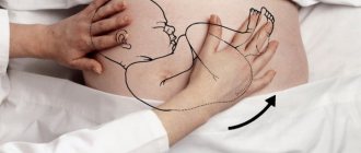
Usually, the expectant mother is informed about the low position of the fetus during pregnancy during an examination by a gynecologist or after the next ultrasound. All this can happen at different stages - from 20 to 36 weeks of pregnancy. It is considered normal for the fetus to descend into the birth canal at about 38 weeks, but in cases where this happens a little earlier, be more attentive to yourself.
You need to clearly understand that this is simply a feature of your body, and not a pathology. Therefore, having learned that the fetus is low during pregnancy, you should not panic and worry unnecessarily.
This feature of pregnancy requires increased attention from doctors and extreme caution from the expectant mother. We should also not forget that such an arrangement can lead to premature birth. One of the reasons for this danger is the pressure that the fetus puts on the cervix, thereby promoting its earlier opening.
There are several symptoms that accompany this feature. First of all, it is aching pain in the lower abdomen. There are situations when no symptoms appear at all. It is generally accepted that a low position of the fetus during pregnancy indicates that a girl should be expected. However, science does not confirm this fact. Everything depends, as a rule, on the individual characteristics of the mother’s body, lifestyle and the presence of any pathologies and abnormalities.
Usually, a low position of the fetus does not pose any particular danger. But, for safety reasons, doctors recommend: a
) expectant mothers should wear a special prenatal bandage,
b
) do not lift heavy objects,
c
) do not walk on stairs,
d
) rest more often. For more careful observation and examination, the expectant mother may be admitted to a hospital.
In any case, there is no need to panic. The main thing is to follow all the doctor’s instructions as accurately as possible. Moreover, according to statistics, the vast majority of births following such a pregnancy proceed and end quite successfully. And for both the mother and her baby.
Future mothers are very concerned about the question: what danger can a placenta that is too low pose during pregnancy? Under normal conditions, the placenta is the organ through which the blood of the fetus and mother is exchanged. It is attached to the back wall of the uterus or closer to it.
Displacement of the uterus is also associated with disruption of all functions of the pelvic organs. This also leads to problems with reproductive function, which is also scary. That is why the slightest feeling of discomfort in the uterine area should lead you to a gynecologist for an urgent diagnosis. IN.
Caesarean section is an abdominal operation or surgical delivery in which the baby is delivered by cutting the abdominal wall of the uterus. Initially, a caesarean section was the most extreme measure when the mother was completely unable to give birth on her own. Realizing the consequences of a cesarean section for the mother, the operation was already done.
Based on materials from vovremiaberemennosti.ru
Causes of low birth weight babies
Often, a pathology such as asymmetrical malnutrition appears in the third trimester due to any difficulties during gestation or hereditary diseases in the expectant mother. Various diseases that affect changes in blood circulation lead to a slowdown in intrauterine growth of the fetus and its correct and normal development. Hereditary/chronic diseases of a girl, during which the female body experiences intoxication and lack of oxygen, affect the baby, which increases the development of malnutrition.
The question of maternal nutrition during pregnancy and its influence on the development in the womb and the birth of a child with low body weight is still open. Statistically, girls who constantly adhere to a low-calorie diet most often reproduce underweight babies. But we should not forget the data that show that even during the Second World War, during the siege of Leningrad, children appeared (quite often) with completely normal indicators. The age of the expectant mother also occupies an important place here: girls under 18 and after 35 have a much greater chance of fetal malnutrition during the period of bearing a baby. This is difficult for the body of a fairly young girl, but an older mother’s body most likely already has chronic diseases.
Treatment of non-developing pregnancy
After diagnosing a non-developing pregnancy, the doctor will prescribe treatment in accordance with the results of the studies. Usually the presence of this pathology leads to miscarriage or surgical termination of pregnancy.
There are several treatment methods:
- Wait for a spontaneous miscarriage. This is a natural process during a non-developing pregnancy. Miscarriage occurs after a decrease in the level of placental hormones.
- The use of special hormonal drugs that provoke miscarriage. This method of treatment is possible up to 8 weeks of pregnancy.
- The procedure for curettage of the uterus. Surgery is a radical method that will help get rid of a frozen embryo. After the operation, mandatory therapy with antibacterial drugs will follow. After 2 weeks, an ultrasound is recommended to ensure that the uterus is restored after the operation.
Diagnosis
When diagnosing the fetus, the best option is ultrasound. During the process, the doctor looks at many parameters - the state of amniotic fluid, disruption of the placenta, and with Doppler monitoring, traces changes in blood circulation. To clarify the diagnosis of a low birth weight fetus, the doctor performs a Doppler examination of the baby’s umbilical cord and blood vessels, and also monitors the speed of blood flow.
The doctor also conducts a cardiotocographic study, which shows what the nature of the child’s heart contractions is, what the reaction is to its own movements or contractions of the uterus. If normal data is obtained as a result of these two studies (even if the baby is low birth weight), the child is healthy.
Hospital assistance
Provided that during a low-weight pregnancy the baby develops without deviations, then there is no need to resort to treatment. However, if there is a possible risk, complications or any developmental abnormalities, the expectant mother is prescribed therapy for her illnesses, and placental insufficiency is also restored. The girl is prescribed medications that dilate blood vessels to increase the child's blood supply. In addition, medications are prescribed that create a slight hypertonicity of the uterus so that the vessels do not become pinched and blood flow improves. This will be home treatment or hospital leave will be prescribed depending on the condition of the baby in the womb.
If treatment is successful, the onset of labor should not be rushed with medication. As a rule, when the due date approaches, the child manages to gain normal weight. Otherwise (lack of fetal weight), labor is induced artificially. If the baby is too weak to pass through the birth canal, the woman in labor is given a cesarean section.
The perfect time to take care of yourself
Therefore, the expectant mother, in order to take care of the child and not deprive herself during the period of pregnancy, needs to eat a variety of foods, spend a lot and often in the fresh air, and not limit her physical activity. However, we want to warn especially zealous mothers - there is no need to eat for two! This will have a bad effect on the pregnant woman’s figure, and if you overeat in the last month, it will also affect the baby’s proportions. In the period from the 36th to the 40th week, babies no longer develop - their body is ready for birth, they are just gaining strength and accumulating fat deposits. But good things should be in moderation, and if the mother enthusiastically eats flour, sweets or drinks liters of sweet soda, then it will be difficult for her to give birth.
At 20 weeks pregnant women should take care of their skin. From this moment on, the tummy grows literally before our eyes, and this is fraught with stretch marks. To avoid such troubles, you need to lubricate the skin with nourishing creams or oils to moisturize the body. This will make the skin elastic and more pliable, preventing it from scarring when stretched. Thanks to this, mom will be able to quickly return to her original shape. And this is the key to her good mood and another reason to experience only joy from motherhood, and not disappointment!
Recommendations during pregnancy
A girl who is carrying a low-weight baby in her womb is necessarily recommended to eat a dense, healthy diet, filled with proteins, enriched with vitamins and complex carbohydrates. It is necessary to completely remove the psychological factor, because as the fetus develops into pregnancy, the girl experiences a variety of sensations, including stressful ones.
It is strictly forbidden to be nervous during this period; the girl and baby should feel peace and comfort. If the psychological factor cannot be eliminated at home, then doctors insist on hospitalization. There are girls who spent almost their entire pregnancy in the hospital, going home only for a couple of weeks.
Baby at 20 weeks
What does a child look like by the middle of his prenatal period and what can he do at 20 weeks of pregnancy? Fetal development at this stage occurs as follows:
- The baby's muscular system is quite developed and allows him to actively move in the uterus, turn around, tumble, stretch, and touch with his hands everything he can reach.
- The nervous system of the fetus is also improving, it is already able to respond to sounds, changes in lighting, and learns to control its movements.
- The brain is actively growing, namely those sections that are responsible for memory and thinking.
- The height and weight of the fetus at 20 weeks of pregnancy are on average 14-16 cm and 255 g, respectively. This is an average parameter, but it allows for minor deviations.
In addition, the baby’s skin develops very rapidly. Now the skin is already four-layered, but underneath it there is still no dermis, which is responsible for the network of capillaries and nerve endings located on the surface of the body.
Birth
The method and timing of delivery will depend on the result that was achieved during treatment. If the fetal weight improves and gains on its own, there is no need to rush to the maternity hospital; the baby will most likely gain the required weight by the day of birth. But the therapy will not stop until its appearance. If the fetus does not gain weight by 36 weeks, a caesarean section is performed. It is not the woman in labor who chooses which way to give birth, but the observing doctor, after diagnosing the baby’s condition, weight does not greatly influence this: if the child is weak, then a cesarean section, and if the child is mobile and presumably healthy, a natural birth.
In the second option, the girl is given spinal anesthesia. The newborn is placed in a heated cradle, and the mother and baby are released home only when the baby’s weight reaches at least 2,800 grams. The following will show the norm of fetal weight by week.
Tests at 20-21 weeks
Usually during this period, pregnant women experience a “hospital lull.” According to the protocol, a week or two earlier, serious screening is done to identify pathologies in the fetus. But he is asked to examine those women who had bad tests in the first trimester.
In other cases, the gynecologist prescribes only two types of routine laboratory tests:
- blood from a finger to determine the level of hemoglobin (in order to timely prevent anemia in a pregnant woman);
- a general urine test to detect protein in it.
Despite the apparent simplicity of these tests, they cannot be ignored and not taken. If a woman has low hemoglobin, she may develop anemia, as a result of which the baby will not receive enough oxygen and nutrients necessary for its development. As a result, the size and weight of the fetus will suffer. The 20th week of pregnancy is also the time to visit the ultrasound room again. Using special equipment, the doctor will find out how the baby is developing. The specialist will take a photo of the fetus. The 20th week of pregnancy is also the period at which you can find out who is in the tummy - a boy or a girl.
View gallery
conclusions
A child is always a big responsibility. For 9 months, the expectant mother cares for and bears her baby, and then takes care of him and raises him, but before that she still needs to give birth. A large baby or a low-birthweight birth process is inevitable and has its own subtleties that the girl will have to endure steadfastly. During pregnancy, you must adhere to the recommendations of the leading doctor, and in no case self-medicate. It is important to note that the belly at 4 months of pregnancy, or rather its size, is certainly not an indicator of the diagnosis being studied; only a doctor can determine this and prescribe a further plan of treatment and action.
Mom at this stage
So, what happens to the female body at the 20th week of pregnancy? Until now, the baby inside the mother has been growing quite slowly. All resources were aimed at developing the baby’s organs, and not at increasing its size, so until mid-pregnancy, many women can successfully hide their interesting position, especially if they try not to advertise it to others. But now everything will be different - every week the uterus will increase in size, rising higher and higher from its bottom. By this period, a woman’s weight should increase by 3-6 kilograms. However, the exact increase must be calculated based on the “pre-pregnancy” constitution of the expectant mother. Chubby girls have no room to gain even more weight - their gain should not be more than three kilograms, but for thin girls the maximum threshold is 7 kg.
Moreover, the weight of the fetus at 20 weeks of pregnancy is approximately a tenth of the total increase. Everything else is an enlarged uterus, water, and placenta. At this time, very serious changes begin to occur in the female body. The baby has grown, the uterus shifts significantly and begins to put pressure on other internal organs. Because of this, a pregnant woman may experience a number of problems: discomfort during sleep, sitting, bending, shortness of breath, heartburn, back pain, and increased vaginal discharge. All these are temporary difficulties that every expectant mother goes through.
You need to mentally prepare for them so as not to be disappointed in yourself and your abilities. The most difficult thing awaits the woman - at 19-20 weeks of pregnancy, the weight of the fetus is still too small to cause her significant discomfort, but in the future it will actively increase, because during the second half of pregnancy the child needs to grow from 300 grams to 3 kilograms! This means that the mother’s tummy will also become larger and heavier, and accordingly, the load on her body will increase significantly.
View gallery