From the very first days of bearing a child, a woman begins to prepare for childbirth. She waits for the ultrasound to show the gender of the unborn baby, then prepares his dowry.
However, the process of childbirth is scary. What determines how they will proceed? An important role in the process of resolution of pregnancy is played by the presentation of the fetus, that is, the position in which the baby lies in the mother’s stomach. The baby can turn around, its posture changes. Which presentation is correct?
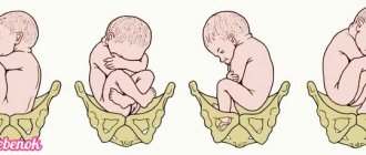
What position is called cephalic presentation of the fetus?
If the baby lies head down before birth, his body is located in a longitudinal position relative to the axis of the uterus - this is called cephalic presentation of the fetus in obstetrics. Nature arranges it in such a way that the parts of the skull close together and even overlap each other, which helps the baby overcome the birth canal. The deformation of the skull is ensured by the presence of elastic areas that allow the bones to move within certain limits. However, the baby’s body can be located differently in the womb.
By the end of pregnancy, the baby can take one of 3 main positions, determined by the relationship between the axes of the uterus and the fetus:
- along the axis of the uterus - longitudinal;
- across the axis of the uterus - transverse;
- oblique - this means that an acute angle appears between the axis of the uterus and the longitudinal axis of the fetus.
If the fetal position is longitudinal, the baby does not necessarily lie head down. Presentation can also be breech.
Oblique and transverse locations are less common; the possibility of self-resolution in such cases is determined by the doctor. Most often, before birth, the baby is in a longitudinal position - along the mother's body, head down.
Positions of the fetus in the womb
By the time of birth, 95% of women arrive with a longitudinal position of the fetus and a cephalic presentation. It is most favorable for successful resolution of pregnancy.
However, this position also has several options. There are 4 main types of cephalic presentation of a child:
- occipital - the baby passes through the birth canal with the back of the head forward, the head is pressed against the chest - this is the most successful version of the cephalic presentation of the fetus;
- anterior cephalic – the fetal head is not pressed tightly enough to the chest, in this case careful monitoring of the process by an obstetrician-gynecologist is required to avoid injury;
- frontal is rare, but poses a danger to the baby, it comes out forehead forward, so surgery is necessary for delivery;
- facial - the head is strongly tilted back, childbirth can be either natural or performed surgically, this is determined by the size of the mother’s pelvis and the build of the child.
If the baby is located along the uterus and the head is directed downward, they also talk about the type of fetal position:
- the most common first position, when the baby’s back is facing the left wall of the uterine cavity - this is the most physiological and favorable location;
- the second position is observed less frequently; it means that the baby’s back is facing the right wall of the pregnant woman’s uterus.
The relationship of the fetal back to the anterior or posterior wall of the uterus determines 2 more types of cephalic presentation. In this case, they talk about the front or rear version.
Reasons for the location of the placenta on the anterior wall
Pregnancy does not always proceed according to the ideal scenario. The embryonic organ can attach to the side or front, which often happens recently. Some doctors believe that chorion along the anterior wall of the uterus is a normal variant that requires special monitoring. There are many reasons for this condition. The mechanism of attachment of the placenta to the front is not well understood, but the following can provoke its disturbances:
- changes in the endometrium;
- multiple pregnancy;
- myoma;
- physiological characteristics of the ovum;
- inflammatory diseases, infections of the reproductive system;
- adhesions, scars on the walls of the uterus;
- multiple curettages (abortions);
- previous operation - caesarean section.
What is fetal articulation?
To predict the course of labor, the position of the fetus is also important. This obstetric term refers to the location of the baby's arms, legs, and head in relation to his body. The physiological position of the fetus is considered to be when the body is bent, the head is lowered to the chest, the legs are bent at both the knee and hip joints, pressed to the stomach, and the arms are crossed on the chest.
When the baby is not sleeping, he constantly makes movements that change his position. However, then the limbs are again located in their proper places. Only in 1–2% of cases can the fetal head be raised, and the arms and legs be in an incorrect position.
Various types of articulation are also possible with breech presentation. These include:
- Breech presentation, when the baby’s buttocks are at the bottom, the arms are folded on the chest, and the legs are extended along the body, with the feet touching the head. A complete or incomplete view is also possible, when one or both legs look down, while they are bent at both the knee and hip joints. The mixed type is distinguished by the correct position of the penis, but below is not the head, but the butt.
- Leg presentation is very rare, when the baby seems to be sitting above the opening to the small pelvis with crossed legs. In this case, the position of the head may vary.
- Knee means that the baby’s knees are directed towards the exit to the small pelvis.
Causes
In most precedents, the causes of this disease are associated with improper attachment of the embryo during implantation, as well as myometrial abnormalities. Disruption of the structure of the muscle fibers of the uterus occurs as a result of inflammation and sexually transmitted infections.
Marginal placentation during pregnancy is diagnosed when the myometrium is thinned. This is often due to abortions and sexually transmitted infections.
Sometimes pathology manifests itself when the uterus is deformed. They can be congenital and occur in the presence of benign formations. The anomaly is not uncommon in patients who have chronic diseases of the internal organs, cardiovascular system, and genitourinary organs.
Due to disruptions in blood circulation in the pelvis, there is a danger of incomplete or incorrect fixation of the fertilized egg. Sometimes marginal placenta previa is due to the fact that it attaches to the myometrium for a longer time.
Why might fetal malposition occur?
The baby's mobility is limited by size only in the third trimester. Sometimes mothers try to find out the position of the fetus after 20 weeks, but this is pointless. The baby is still very mobile and rotates freely in the uterus. Only after 26 weeks there are no changes in the position of the fetus in most pregnant women. However, the doctor makes conclusions about the presentation of the fetus after 30 weeks, usually in the interval from 32 to 34 weeks.
Medicine knows a number of reasons why the fetus may take an incorrect position. These include:
- polyhydramnios, which allows the baby to move freely in the later stages;
- oligohydramnios, preventing the fetus from taking the desired position;
- carrying two or more babies at the same time;
- narrow pelvis of the expectant mother;
- prematurity, when the fetal weight is less than 2.5 kg;
- various pathologies of the uterus;
- heredity;
- stretching of the abdominal muscles when carrying previous children.
Symptoms
Regional placenta previa along the posterior or anterior wall is accompanied by characteristic signs. The main one is bleeding. At the same time, the pregnant woman’s health does not deteriorate. Appear at rest or at night.
They may appear in the 1st trimester. It is also often observed at 28-32 weeks. At this time, the uterine muscles are more active as the organ prepares for childbirth.
Rarely, bloody lesions appear in the 2nd trimester. Their volume varies depending on the degree of damage to the blood vessels.
Bleeding at this stage occurs after sexual intercourse, active movements of the child, and physical activity. They may appear during or after a gynecological examination.
Often this condition provokes the threat of miscarriage. A pregnant woman experiences discomfort in the lower abdomen and hypertonicity of the uterus.
Since systematic hemorrhages appear against this background, iron deficiency anemia develops. The condition provokes increased fatigue and weakness. Due to insufficient amounts of nutrients, the risk of hypoxia and intrauterine growth retardation increases.
Diagnostic measures
An experienced doctor can diagnose cephalic or breech presentation during a routine gynecological examination. Probing the fetus through the vagina, he can touch the head or buttocks of the baby with his fingers. It makes sense to conduct such a study no earlier than 30–32 weeks, since before this the child does not yet have time to take the prenatal position.
You can try to determine the position of the child yourself. Lying on her stomach and slightly spreading her legs bent at the knees, mommy feels her stomach and is able to understand how the body is located and where the head is located.
However, it is better to focus on the results of ultrasound. The doctor will take a photo of the baby in the womb to reassure the mother.
Diagnostic features
As doctors note, central placenta previa during pregnancy is a rather dangerous disease, which is characterized by a high level of secrecy. The first striking symptom is bleeding, so before it appears it is quite difficult to determine the presence of presentation; it, of course, can be suspected, but only professional doctors can confirm the diagnosis.
Initially, the doctor carries out an external examination, as a result of which the height of the uterine fundus is measured (with central presentation, the height of the fundus is greater than it should be at the current stage of pregnancy) and an examination of the location of the fetus. Palpation does not make sense, since due to the placenta it is impossible to get precise sensations.
If bleeding has already occurred, the woman must be hospitalized and taken to the hospital, where an ultrasound scan is performed (it is advisable to use a vaginal sensor during this procedure). An examination is carried out in the mirrors to identify exactly where the source of bleeding is located, most often it is the cervix or varicose veins located in the vagina.
The peculiarity of the examination procedure with mirrors is that these manipulations are carried out in an operating room and using heated mirrors. This must be done so that if bleeding increases, surgery can be started immediately.
An ultrasound examination is also mandatory, with the help of which you can identify the presentation of the choreon, its appearance, as well as the area occupied by the placenta.
In the presence of such a pathology as central presentation, the timing of the ultrasound is slightly shifted, the procedure is performed at 16, 24 and 34 weeks of pregnancy.
Exercises and recommendations for different presentations of the baby
If the baby has not taken the desired position by 34 weeks, you should do simple exercises. Below are a few exercises to help your baby turn:
- Lie on your side on a hard surface, lie there for 10 minutes, then turn over and repeat the exercise on the other side.
- Get on all fours, leaning on your elbows and knees, stay in this position for up to 15 minutes.
- It is useful to swim during this period. In addition to light physical activity, swimming helps the baby take the right position.
- Quickly, sometimes the first time, the baby turns over if, lying on his back with his knees bent, he raises his pelvis to a height of up to 40 cm. To fix the pelvis, you need to place a pillow under it. Shoulders, pelvis and knees should be on the same straight line.
How is childbirth carried out with different presentations?
Head presentation is the most preferable, it allows the mother to give birth on her own. If the exit from the small pelvis is the occiput, childbirth occurs naturally. Frontal presentation poses a threat if the mother has a narrow pelvis, so a caesarean section is required. With anterior facial presentation and normal pelvic width, labor proceeds naturally. However, in the posterior position, to pass through the birth canal, the position of the child is changed manually or surgical intervention is used.
If the fetus is positioned longitudinally with its head down, forceps may be required. This rare type of obstetric care is used if the baby’s position in the birth canal is already low, but there is a danger of baby hypoxia or the mother’s labor has weakened.
With breech presentation, external rotation of the fetus onto its head is practiced. The manipulation is controlled using ultrasound. The effectiveness of the procedure is 60 – 70%.
Pure breech presentation, normal pelvic size, and baby weight up to 3800 g allow you to give birth to a baby naturally. Any deviations are indications for a cesarean section.
Transverse presentation can also be corrected by external rotation. Otherwise, as well as with mixed presentations, a caesarean section cannot be avoided.
Placenta previa
Ultrasound scanner HS50
Affordable efficiency.
A versatile ultrasound scanner with compact design and innovative capabilities.
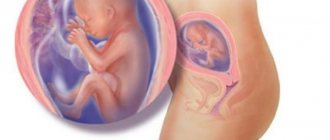
During the normal course of pregnancy, the placenta is usually located in the area of the fundus or body of the uterus, along the posterior wall, with a transition to the side walls, i.e. in those areas where the walls of the uterus are best supplied with blood. On the anterior wall, the placenta is located somewhat less frequently, since the anterior wall of the uterus undergoes much greater changes than the posterior one. In addition, the location of the placenta along the posterior wall protects it from accidental injury.
Placenta previa is a pathology in which the placenta is located in the lower parts of the uterus along any wall, partially or completely blocking the area of the internal os. The incidence of placenta previa averages from 0.1% to 1% of the total number of births.
If the placenta only partially covers the area of the internal os, then this is an incomplete presentation, which is noted with a frequency of 70-80% of the total number of presentations. If the placenta completely covers the area of the internal os, this is complete placenta previa. This option occurs with a frequency of 20-30%.
There is also a low location of the placenta, when its edge is at a lower level than it should be normally, but does not overlap the area of the internal os.
Causes of low position or placenta previa
There are several reasons for the formation of low position or placenta previa. The most common causes are pathological changes in the inner layer of the uterus (endometrium) due to inflammation, surgical interventions (curettage, cesarean section, removal of fibroids, etc.), and multiple complicated births. In addition, disturbances in placental attachment can be caused by uterine fibroids, endometriosis, underdevelopment of the uterus, isthmicocervical insufficiency, cervical inflammation, and multiple pregnancies. It should be noted that placenta previa is more common in repeatedly pregnant women than in first-time mothers. Due to these factors, the fertilized egg entering the uterine cavity after fertilization cannot be implanted in a timely manner in the upper parts of the uterus, and this process occurs only when the fertilized egg has already descended into its lower parts.
The most common manifestation of placenta previa is repeated bleeding from the genital tract. Bleeding can occur during various periods of pregnancy, starting from the earliest stages. However, most often they are observed already in the second half of pregnancy due to the formation of the lower segment of the uterus. In the last weeks of pregnancy, when uterine contractions become more intense, bleeding may increase.
The cause of bleeding is the repeated abruption of the placenta, which is unable to stretch following the stretching of the uterine wall as pregnancy progresses or labor begins. In this case, the placenta partially detaches, and bleeding occurs from the vessels of the uterus. The fetus does not lose blood. However, he is at risk of oxygen starvation, since the detached part of the placenta does not participate in gas exchange.
Provoking factors for bleeding during pregnancy can be: physical activity, sudden coughing, vaginal examination, sexual intercourse, increased intra-abdominal pressure during constipation, thermal procedures (hot bath, sauna).
With complete placenta previa, bleeding often appears suddenly, without pain, and can be very heavy. Bleeding may stop, but after some time it may recur, or it may continue in the form of scanty discharge. In the last weeks of pregnancy, bleeding resumes and/or intensifies.
With incomplete placenta previa, bleeding may begin at the very end of pregnancy. However, more often this occurs at the beginning of labor. The severity of bleeding depends on the size of the presenting area of the placenta. The more placental tissue is present, the earlier and more severe the bleeding begins.
Repeated bleeding during pregnancy complicated by placenta previa in most cases leads to the development of anemia.
Pregnancy with placenta previa is often complicated by the threat of miscarriage, which is due to the same reasons as the occurrence of abnormal location of the placenta. Preterm birth most often occurs in patients with complete placenta previa.
Pregnant women with placenta previa are characterized by low blood pressure, which occurs in 25%-34% of cases.
Preeclampsia (nephropathy, late toxicosis) is also no exception for pregnant women with placenta previa. This complication, occurring against the background of dysfunction of a number of organs and systems, as well as with the phenomena of blood clotting disorders, significantly worsens the nature of repeated bleeding.
Placenta previa is often accompanied by fetoplacental insufficiency, lack of oxygen for the fetus and delayed development. The detached part of the placenta is excluded from the general system of uteroplacental circulation and does not participate in gas exchange. When placenta previa often occurs, an abnormal position of the fetus (oblique, transverse) or pelvic presentation is formed, which in turn is accompanied by certain complications.
In obstetric practice, the term “placental migration” has become widespread, which, in fact, does not reflect the real essence of what is happening. The location of the placenta changes due to changes in the structure of the lower segment of the uterus during pregnancy and the direction of placental growth towards better blood supply to areas of the uterine wall (towards the fundus of the uterus) compared to its lower sections. A more favorable prognosis in terms of placental migration is observed when it is located on the anterior wall of the uterus. Typically, the process of placental migration occurs within 6-10 weeks and is completed by the middle of 33-34 weeks of pregnancy.
Diagnosis of placenta previa
Detecting placenta previa is not particularly difficult. The presence of placenta previa may be indicated by a pregnant woman's complaints of bleeding. In this case, repeated bleeding in the second half of pregnancy is usually associated with complete placenta previa. Bleeding at the end of pregnancy or at the beginning of labor is most often associated with incomplete placenta previa.
If there is bleeding, you should carefully examine the walls of the vagina and cervix using speculum to exclude trauma or pathology of the cervix, which may also be accompanied by the presence of bleeding.
A vaginal examination of a pregnant woman also easily reveals clear diagnostic signs indicating an abnormal location of the placenta. However, such a study must be performed as carefully as possible, in compliance with all the necessary rules to prevent possible bleeding.
Currently, the most objective and safe method for diagnosing placenta previa is ultrasound, which allows you to establish the fact of placenta previa and the variant of presentation (complete, incomplete), determine the size, structure and area of the placenta, assess the degree of abruption, and also obtain an accurate idea of placental migration.
If an ultrasound reveals complete placenta previa, then a vaginal examination should not be performed at all. The criterion for a low location of the placenta in the third trimester of pregnancy (28 - 40 weeks) is the distance from the edge of the placenta to the area of the internal os of 5 cm or less. Placenta previa is indicated by the detection of placental tissue in the area of the internal os.
The nature of the localization of the placenta in the second and third trimesters of pregnancy (up to 27 weeks) is judged by the ratio of the distance from the edge of the placenta to the area of the internal os, with the diameter (BDP) of the fetal head.
If an abnormal location of the placenta is detected, a dynamic study should be carried out to monitor its “migration”. For these purposes, it is advisable to perform at least three echographic controls throughout pregnancy at 16, 24-26 and 34-36 weeks.
Ultrasound should be performed when the bladder is moderately full. Using ultrasound, it is also possible to determine the presence of blood accumulation (hematoma) between the placenta and the wall of the uterus during placental abruption (in the event that there is no bleeding from the uterine cavity). If the area of placental abruption occupies no more than 1/4 of the area of the placenta, then the prognosis for the fetus is relatively favorable. If the hematoma occupies more than 1/3 of the area of the placenta, then most often this leads to the death of the fetus.
Medical support for pregnant women with placenta previa
The nature of the management and treatment of pregnant women with placenta previa depends on the severity of bleeding and the amount of blood loss.
In the first half of pregnancy, if there is no bleeding, the pregnant woman can be at home under outpatient monitoring, following a regimen that excludes the action of provoking factors that can cause bleeding (limitation of physical activity, sexual activity, stressful situations, etc.)
Observation and treatment for pregnancy over 24 weeks is carried out only in an obstetric hospital.
Treatment aimed at continuing pregnancy up to 37-38 weeks is possible if the bleeding is not heavy and the general condition of the pregnant woman and the fetus is satisfactory. Even despite the cessation of bleeding from the genital tract, pregnant women with placenta previa under no circumstances can be discharged from the hospital before delivery.
Management of pregnant women in an obstetric hospital includes: strict bed rest; the use of drugs that ensure optimization and normalization of contractile activity; treatment of anemia and fetal-placental insufficiency.
Indications for emergency caesarean section, regardless of the stage of pregnancy, are: repeated bleeding; a combination of small blood losses with anemia and decreased blood pressure; immediate heavy blood loss; complete placenta previa and bleeding.
The operation is performed according to vital indications on the part of the mother, regardless of the duration of pregnancy and the condition of the fetus.
If the pregnancy was carried to 37-38 weeks and placenta previa persists, depending on the current situation, the most optimal method of delivery is chosen individually.
The absolute indication for elective caesarean section is complete placenta previa. Childbirth through the vaginal birth canal is impossible in this situation, since the placenta blocking the internal os does not allow the presenting part of the fetus (fetal head or pelvic end) to be inserted into the pelvic inlet. In addition, as the uterine contractions increase, the placenta will peel off more and more, and the bleeding will increase significantly.
In case of incomplete placenta previa and in the presence of associated complications (breech presentation, abnormal position of the fetus, uterine scar, multiple pregnancy, severe polyhydramnios, narrow pelvis, primigravida over 30 years of age, etc.), a caesarean section should also be performed as planned.
If the above associated complications are absent and there is no bleeding, then you can wait until spontaneous labor begins, followed by early opening of the amniotic sac. If, after opening the amniotic sac, bleeding still begins, then it is necessary to resolve the issue of performing a cesarean section.
If, with incomplete placenta previa, bleeding occurs before the onset of labor, then the amniotic sac is opened. The necessity and expediency of this procedure is due to the fact that when the membranes are opened, the fetal head is inserted into the entrance to the pelvis and presses the detached part of the placenta against the wall of the uterus and pelvis, which helps to stop further placental abruption and stop bleeding. If bleeding continues after opening of the membranes and/or the cervix is immature, then a cesarean section is performed. If the bleeding stops, it is possible to conduct labor through the natural birth canal (if the obstetric situation is favorable).
Bleeding can begin in the early stages of labor, from the moment of the first contractions. In this case, early opening of the amniotic sac is also indicated.
Thus, management of childbirth with incomplete placenta previa through the natural birth canal is possible if: the bleeding stopped after opening the membranes; the cervix is mature; labor activity is good; there is a cephalic presentation of the fetus.
However, cesarean section is one of the most frequently chosen methods of delivery by obstetricians for placenta previa and is performed with a frequency of 70% -80% for this pathology.
Other typical complications during childbirth with incomplete placenta previa are weakness of labor and insufficient oxygen supply to the fetus (fetal hypoxia). A mandatory condition for conducting childbirth through the natural birth canal is constant monitoring of the condition of the fetus and the contractile activity of the uterus.
After the birth of the child, bleeding may resume due to a disruption in the process of separation of the placenta, since the placental site is located in the lower parts of the uterus, the contractility of which is reduced.
Heavy bleeding often occurs in the early postpartum period due to decreased uterine tone and damage to the extensive vascular network of the cervix.
Prevention of placenta previa
Prevention of placenta previa involves reducing the number of abortions, early detection and treatment of various inflammatory diseases of the reproductive system and hormonal disorders.
Ultrasound scanner HS50
Affordable efficiency.
A versatile ultrasound scanner with compact design and innovative capabilities.
Prevention of complications
Childbirth is a complex and responsible process. Only 37% of women in labor cope with it on their own. To avoid complications, you should regularly consult with a gynecologist, and if necessary, go to the hospital for conservation.
Even if the child is in the most physiological position, one must remember the possibility of complications. Attenuation of labor, narrow pelvis, pathology of the membranes, injuries in previous births, chronic diseases can lead to disruption of the process. The low probability of giving birth safely without medical assistance is a reason to think in advance about where and how to give birth. The health of both mother and baby depends on how successfully the pregnancy is resolved.
Loading…
Share with friends!