As you know, all pregnant women must undergo three mandatory screenings, including ultrasound, during their pleasant wait. 19 weeks is exactly the period at which the second screening most often occurs. It is carried out from 16 to 20 weeks of pregnancy, but preferably no later: after this, the increased size of the fetus may interfere with the examination, and the results of a biochemical blood test are no longer informative.
Ultrasound examination is the most significant part of the second screening, which absolutely all women who register for pregnancy at the antenatal clinic undergo . Therefore, if the doctor prescribed you an ultrasound at 19 weeks, there is no doubt about its necessity.
Pregnancy at 19 weeks
Even if the previous weeks of pregnancy passed quickly for the expectant mother, and until the 19th week she did not notice her position, at such a significant period almost any pregnant woman understands that childbirth is just around the corner. An enlarged belly will remind you of the “equator” - almost all women notice it by this time. Even the not-so-pleasant signs of the second trimester—shortness of breath and swelling in the legs, possible weakness—are the norm at 19 weeks . But there is also an inspiring side: toxicosis is behind us, maternity leave is approaching, half of the pregnancy is over.
The best news: most pregnant women at this stage clearly feel the baby’s movements! Some mothers, even at this considerable stage, are afraid of harming the baby during an ultrasound. In fact, the risks of performing the procedure significantly exceed the impact of ultrasonic waves on a baby, especially one as strong and formed as at 19 weeks of pregnancy.
Why is the study done and to whom is it recommended?
Ultrasound diagnostics in accordance with the second screening program is prescribed to all pregnant women.
For a certain category of patients, the risk of developing genomic pathologies of the fetus increases:
- Age over 35 years.
- Patients with children or relatives with genetic disorders.
- Age up to 17 years.
- Taking medications contraindicated for pregnant women.
- Poor results of the first screening.
- History of miscarriages, threat of miscarriage during current pregnancy.
Week 19 is considered the most informative period for determining anatomical pathologies accompanying congenital syndromes and diseases.
Important! Patients whose first screening results indicate a high probability of genetic diseases in the child are subject to mandatory examination. Diagnostics will restore calm and confidence to a woman who is worried about the health of her baby.
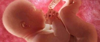
How does the baby develop by the 19th week of pregnancy?
Fetus at 19 weeks of gestation
The nineteenth week is almost the middle of pregnancy. What happens to the fetus at this time?
The norm may vary, but almost all children develop the following signs in utero:
- the baby becomes fully proportional, his body will remain this way until childbirth;
- The sex of the child is clearly visible on ultrasound, the likelihood of error is extremely low;
- the child hears sounds outside well and distinguishes the mother’s voice; according to the results of observations, if the future dad talks to the baby or if poetry is read to the tummy, he remembers the sounds and subsequently reacts to them;
- the formation of original feces - meconium - begins in the intestines;
- the nervous, immune, and cardiovascular systems of the fetus are actively developing;
- already from the 22nd week (less than a month!), a child born prematurely has a chance of surviving in a hospital;
- the content of subcutaneous fat increases, folds form on the skin, and a unique papillary pattern appears on the baby’s fingers;
- by the 19th week, the molars and milk teeth of the unborn child are even formed;
Mother's condition and fetal development
For the expectant mother, pain in the back and lower back becomes more intense. The joints gradually diverge, ensuring the unhindered passage of the fetus in the upcoming birth.
During this period, the movements of the child in the woman’s womb become intense. The baby begins to react to the mother’s voice, perceives light, and becomes anxious at loud sounds. The immune system is being developed.
Parts of the brain and the central nervous system are intensively developing. The lungs and bronchi pass through a critical stage of formation. The child’s movements acquire a meaningful character as a response to external stimuli.
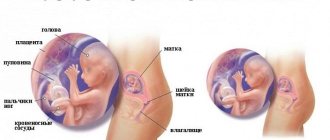
Why is ultrasound performed?
Ultrasound of a 19-week pregnancy is a very important study, which is not recommended to be neglected even by an absolutely healthy woman. Based on the size of the fetus and its parts, a specialist can diagnose delayed or advanced development, which affects the management of pregnancy and the expected date of birth. By assessing the structure of internal organs, including the brain, the doctor will report the presence of possible developmental anomalies.
During an ultrasound at 19 weeks of pregnancy, the fetal face is clearly visualized, on which the so-called markers of intrauterine pathology are easily distinguishable. The amount of amniotic fluid, the structure of the umbilical cord and the blood vessels of the placenta must be assessed - this is an important sign characterizing the condition of the baby in the womb. Additionally, the condition of the cervix and its appendages is examined.
If a pregnant woman is at risk, in addition to an ultrasound, she is recommended to undergo a so-called triple test . In this case, the level of three hormones is determined in the woman’s venous blood: hCG, estriol and alpha-fetoprotein. In rare cases, the concentration of inhibin is also measured. Such tests are prescribed only if pathology is suspected based on the results of the first screening at 10-14 weeks or if there is an increased risk of chromosomal abnormalities of the fetus.
What is the size of the fetus at 19 weeks of pregnancy?
Ultrasound at 19 weeks of pregnancy
At this stage, the baby’s size from head to toe is 13-15 cm, weight is about 200 grams. It is no coincidence that one of the first indicators assessed by ultrasonography at 19 weeks is fetometry data - the size of the fetus. The doctor measures the circumference of the child's head, chest and abdomen, as well as the length of the limb bones and the diameter of the head. The conclusion is given only on the totality of sizes for a given period. Those. If any indicator is below the recommended values for the term, the expectant mother should not worry too much: this may be the baby’s individual norm.
Stomach
Abdominal pain may indicate the possibility of miscarriage. If they are pronounced, have a cramping nature, if the lower abdomen really hurts and is very tight, and also if these pains are accompanied by bloody discharge, then you need to tell the doctor about it.
At the 19th week of pregnancy, the belly should have grown noticeably. Most likely, you can no longer sleep on your stomach, and you don’t need to: in this position, the uterus is compressed, and this is undesirable and unpleasant. It is also not recommended to sleep on your back so that the weight of the uterus does not compress the vena cava, blocking the access of blood and oxygen to the fetus.
Is it possible to determine the sex of an unborn child at 19 weeks?
Boy or girl? This is one of the first questions that worries future parents when they find out they are pregnant. In fact, the gender of the baby is determined already at the moment of conception. Already from the 8th week of pregnancy you can find out quite accurately if you do a DNA blood test. But this is a rather expensive study.
On ultrasound, the sex of the unborn baby is visible from about 14 weeks, but may be difficult to distinguish at the beginning. And it is at 19 weeks that gender determination is the norm rather than the exception to the rule. The likelihood of an error at this time is very small. You can see the differences in gender characteristics of boys and girls on an ultrasound at 19 weeks in the picture.
Normal values of various indicators on ultrasound
If you undergo an ultrasound at 19 weeks of pregnancy, the doctor will definitely make a conclusion and report the established diagnosis. But expectant mothers are always interested in figuring out all the symbols and numbers themselves!
We recommend trusting the opinion of a specialist and not panic if some values do not correspond to the table: this may be an individual norm for your baby!
Below are a few abbreviations that you can see in the conclusion of sonography.
The norms of the fetometry values below are given in the table.
KTP - coccygeal-parietal size, fetal height is normally 130-150 mm
BPR – biparietal head size
LZR - fronto-occipital size
DB – length of the femur
DgrK – chest diameter
The length of the nasal bone by week 19 is about 5 mm. Its shortening may indicate a serious chromosomal abnormality - Down syndrome. But an incorrectly determined gestational age can affect the measurement result, therefore, if shortening of the nasal bone is suspected, additional biochemical screening must be prescribed.
The thickness of the collar zone/space (collar fold) should be no more than 3 mm when examined transabdominally. An increase in this indicator indicates serious disorders such as Edwards syndrome.
Another important digital index relates not to the fetus, but to the amniotic fluid. This is the so-called amniotic index (AI) - the sum of the distance from the fetus to the uterine wall at three measurement points. The norm of AI by the 19th week of pregnancy is from 83 to 225 mm. If the indicator is higher than normal, they speak of polyhydramnios, if lower, a diagnosis of oligohydramnios is possible. Both conditions require careful investigation, because may indicate gestosis, infection of the birth canal and other serious pathologies of pregnancy.
The degree of maturity of the placenta and the number of vessels in the umbilical cord indirectly characterize the condition of the fetus in the womb. Normally, at week 19, the degree of maturity of the placenta should be zero, and there are normally three vessels. Deviations in indicators require additional studies - Dopplerography and CTG - to make an accurate diagnosis.