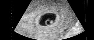
An ultrasound at 30 weeks of pregnancy visualizes the fetus, which in general is already a human being. At this stage, the cardiovascular, skeletal and nervous systems of the fetus are almost completely formed.
At 28-30 weeks, doctors look at hormone levels in the mother’s body, deviations from the norm in the baby’s development, and its growth rate. Particular attention is paid to assessing the performance of the fetal heart (indicators of its contractility) and large vessels extending from it.
In this article we will talk about what an ultrasound shows at 28–30 weeks of pregnancy. We will also talk about what diseases can be prevented during this period of pregnancy.
Ultrasound at 30 weeks of pregnancy
Ultrasound of a child at 30 weeks of pregnancy does not include a number of mandatory diagnostic measures. In most cases, ultrasound diagnostics at this stage are carried out according to indications or if any complaints arise in the pregnant woman. In some cases, the woman herself expresses a desire to undergo diagnostics to confirm the normal development of the fetus. But still, most often an ultrasound at 30 weeks of pregnancy is performed if indicated. These include:
- a decrease in the activity of fetal movement or, conversely, when excessively active movement appears;
- during multiple pregnancy;
- if pregnancy occurs as a result of the use of assisted reproduction technologies, namely the method of in vitro fertilization;
- the appearance of painful sensations accompanying discharge atypical for pregnancy;
- to evaluate ongoing drug therapy for various pathological conditions, both on the part of the fetus and mother;
- eliminating possible entanglement with the umbilical cord, which can lead to fetal hypoxia, which is a threat to the life of the fetus;
- suspicion of intrauterine pathologies of the fetus.
Also, an ultrasound at 30-32 weeks of pregnancy is a routine examination in case of carrying twins. Managing a multiple pregnancy has its own characteristics, one of which is the timing of routine ultrasounds. The development of the fetus during such a pregnancy is somewhat different from a normal pregnancy. Such a pregnancy requires special attention and monitoring of the development of both fetuses. Often the reason for ultrasound diagnosis at this stage is a discrepancy between the height of the uterine fundus and the height of the uterine fundus. The cause of the discrepancy may be a developmental delay of the fetus or a frozen pregnancy.
Ultrasound 30-31 weeks of pregnancy
As a rule, pregnant women experience an increased sense of fear and anxiety for their baby. The vast majority of women have the erroneous opinion that ultrasonic waves can harm the unborn child. This is a myth that makes many women afraid of undergoing routine ultrasounds. But to date there is no confirmed data on the dangers of ultrasound diagnostics. Ultrasound after 30 weeks of pregnancy is performed transabdominally. At this time, transvaginal examination should not be performed, not because such a method can harm the child or mother, but because it is not necessary. The fetus is already quite large, and the uterus has risen so much that it has pushed back the intestinal loops. In this regard, at this stage of pregnancy, preparation for the study is not required. There is also no need to fill the bladder, because the fetus is much higher and the amniotic fluid now serves as a conductor. In addition to the standard two-dimensional examination, the method of four-dimensional and three-dimensional examination can be used. These methods show a three-dimensional image, which allows you to see what the fetus looks like. And with 4D ultrasound, you can not only see its external data, but also its activity.
Ultrasound at 30 weeks of pregnancy: what they look for
At 30 weeks of age, the studied parameters in one fetus may differ significantly from the data of its “peers”. This is due to the fact that at this time a hereditary predisposition comes into force. Accordingly, large parents will have a larger baby than the average couple at the same age. It is important that the fetal parameters are not below or, conversely, significantly above the acceptable limits at this period. At this time, the doctor still evaluates the parameters of the fronto-occipital, biparietal size of the head, and measures the length of the limbs. Also, much attention is paid to determining the circumference of the chest, abdomen, and head.
More and more attention is being paid to the presentation of the fetus, but there is no need to focus on this, since the fetus can change its position before birth.
Normally, according to an ultrasound scan of 30 weeks of pregnancy, the weight of the fetus is on average 1500 g, and its height reaches 35-37 centimeters. The fronto-occipital size is 8.9-10 cm, and the transverse size is 8.5 cm. When assessing the growth and development of the fetus, the doctor is guided by the following data: forearm bone - 4.2 -5 cm, humerus - 5-5.7 cm. The lower limbs have the following dimensions: the femur is 5.2-6 cm, and the lower leg is about 4.9 - 5.7 cm.
Doppler ultrasound at 30 weeks of pregnancy allows you to assess the state of blood flow in the umbilical cord and placenta. During the procedure, the doctor assesses the oxygen saturation of the fetus and whether it suffers from hypoxia due to a possible blood flow disorder. The data obtained also make it possible to assess the functioning of the circulatory system and how well it copes with the load during pregnancy.
Results of Doppler study
Doppler data at 31 weeks are normal:
- The resistance index inside the uterine artery is 0.34 – 0.61;
- IR inside the umbilical cord arteries is approximately 0.53 – 0.76;
- Systole-diastolic ratio of the uterine artery 1.4 – 3.5;
- The SDO of blood flow inside the umbilical cord artery remained the same as at week 30 – 2.88 – 2.96;
- SDO inside the baby's middle cerebral artery is about 4.4;
- IR inside the middle cerebral artery – approximately 0.8;
- IR VSA reaches 0.80 – 0.85.
Decoding the norm on ultrasound at 30 weeks
As a rule, the parameters examined during ultrasound diagnostics are individual and depend on many factors. To determine deviations in fetal development at a given time, it is customary to compare the actual results with the proper results of the norm.
Ultrasound at 30 weeks of pregnancy: normal
During a normal pregnancy, parameters such as the height and weight of the fetus are determined using ultrasound. Normally, at this stage, the embryo has long become a fetus, and its weight can be from 1200 g to 1600 g. During this period, the baby’s height is on average about 38 - 40 centimeters. Also during an ultrasound examination, fetal parameters such as head size, length of the upper and lower limbs, chest and abdominal circumference are measured.
Decoding the results obtained allows us to determine possible deviations from the norm using ultrasound at 30 weeks of pregnancy. Thus, normally the transverse size of the fetal head is on average 7.5 cm, and the size from the frontal bone to the occipital bone is 9.5 cm. The permissible head circumference is 26.5 cm. The thigh length in a healthy fetus is 5.5 cm to 6 cm. But it is worth noting that normal indicators depend on the anthropometric data of the parents.
More on the topic First, second, third ultrasound by week
Well, I went for an ultrasound! I found a high-quality doctor with us and without even looking, made an appointment for the near future. According to the cycle 5.4 days, and according to ultrasound 6.4, according to KTR - 6.1