- Dates
- What does a fetus look like in the eighth month?
- Preparation
- How is it done?
- Normal indicators for deciphering ultrasound results
The period of 32 weeks of pregnancy is decisive for determining the tactics of childbirth. All important indicators, the degree of maturity of the lungs, the growth and parameters of the fetus, the exact date of its birth can be assessed using ultrasound. By conducting this study in time, you can avoid natural births that are dangerous for the fetus and reduce the consequences of placental insufficiency.
Timing of the third planned ultrasound
Sonography in the third trimester is prescribed to:
- accurately determine whether it is possible to give birth naturally, or whether only a caesarean section is possible;
- timely improve the supply of oxygen to fetal tissues, especially brain tissues;
- take timely measures in case of premature aging of the placenta;
- attempt to rotate the fetus from a breech presentation to a cephalic presentation (only if a natural birth is planned);
- perform surgery in utero to correct life-threatening malformations or be prepared for it immediately after birth.
It is important to comply with the timing of the ultrasound examination. This is 32-34 weeks of pregnancy.
If it is necessary to turn the fetus into a cephalic presentation, perform intrauterine surgery or invasive intervention (cordocentesis or blood transfusion), the results of an ultrasound examination determine how mature (that is, ready for “extrauterine” breathing) the lungs are. If a conclusion is given about zero or first degree of maturity, before carrying out the above manipulations, a course of injections is prescribed that will help the lungs mature faster. Only after this the planned interventions begin.
This algorithm is followed due to the high risk of premature birth, which can be provoked by any intervention in the pregnant uterus. The fetal brain and heart are already fully mature by this time. The only thing that needs to be prepared for extrauterine conditions is the lungs, and they are assessed during this ultrasound.
Indicators of the mother's body
The ultrasound method also allows you to monitor the condition of the mother’s body, determining its readiness for a less painful and safe childbirth.
Cervical length
The lower narrowed part of the uterus should exceed 29 mm. The optimal value is considered to be 30–36 mm. A patient whose cervical length was insufficient (up to 26–27 mm) during a routine ultrasound examination will most likely have to be kept in hospital until delivery. Otherwise, premature delivery may be required.
Condition of pharynx
The pharynx is the opening of the upper and lower part of the cervix. One is called internal, and the other is called external. The normal ultrasound scan at 33 weeks of pregnancy suggests that there is no dilation of the pharynx - they should both be closed.
Amount of amniotic fluid
The amount of amniotic fluid must be determined. Its minimum is approximately 72–77 mm, its maximum reaches 245–265 mm. During the examination, neither high water nor low water should be observed.
Location and thickness of the placenta
A female organ such as the placenta in the third trimester has a thickness of 35–43 mm. A deviation of 2 units to the left or right is not considered a deviation. When an ultrasound is performed, the uterine position of the structure is established - it should be located in the upper part of the hollow organ. Placenta adherence signals problems that require specialist intervention.
Placental maturity
Readiness for a full birth of the placenta is determined by its maturity. There are 4 main stages - 0, 1, 2 and 3. At the 33rd week of gestation, the mark is located between 1 and 2.
Umbilical cord length
The umbilical cord in each individual case has an individual indicator. The main thing to remember is that exceeding 70 cm can provoke the formation of loops around the baby.
The degree of supply of blood vessels to the umbilical cord
A healthy umbilical cord provides adequate blood supply and nutrition to the developing body. If the ultrasound results show 3 umbilical vessels, there is no need to talk about violations.
What does a fetus look like in the eighth month?
The main organs have already been formed: some of them are undergoing additional development, as well as active weight gain and lengthening of the body.
Almost all internal organs function:
- the kidneys filter ingested amniotic fluid, secreting urine;
- the heart contracts 140-160 beats per minute, but the blood does not circulate quite as much as after birth;
- feces appear in the intestines;
- all senses work;
- brain neurons actively connect with each other, forming strong connections (this allows the fetus to form a reaction to a certain impact);
- the ovaries are filled with eggs, and in most boys the testicles are already in the scrotum (4% complete the process within the first 12 months).
Only the lungs do not work, which at 32-34 weeks still resemble the liver in density. They already have the necessary “bags” where gas exchange occurs (alveoli), but there is still insufficient surfactant - a substance that will not allow these “bags” to subside. The immune system is also underdeveloped.
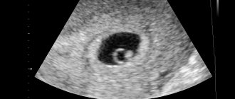
Ultrasound photo at 32 weeks of pregnancy
The period of pregnancy itself, during which the third ultrasound is performed, is considered a critical period due to the likelihood of premature birth. Right now it may develop:
- premature abruption of the placenta, both presenting and normally located;
- isthmic-cervical insufficiency;
- worsening of gestosis, up to the development of eclampsia.
At 30-34 weeks, twins or triplets are most often born. And although the born children are already viable, their appearance at this stage will often require subsequent treatment of varying durations.
Ultrasound 32 weeks pregnant
With the introduction of ultrasound diagnostics into medical practice, the determination of most pathologies has become more accessible and, moreover, their identification in the early stages. In obstetric practice, ultrasound diagnostics allows early confirmation of pregnancy. In the future, with the growth and development of the fetus, this diagnostic method makes it possible to identify chromosomal abnormalities and fetal malformations.
Ultrasound of the fetus at 32 weeks of pregnancy is a mandatory diagnostic measure. As a rule, the third mandatory ultrasound examination is performed at this time. The study at this stage is quite important, because deciphering the results will allow us to assess the growth of the fetus and its compliance with the gestational age.
Ultrasound at 32 weeks of pregnancy: what they look for
The main goal of the study at this time is to determine a number of indicators:
- identification of fetal anomalies that were not detected during the second planned ultrasound examination;
- analysis of fetometric data, assessment of fetal size. Determination of limb length, head circumference, abdomen and chest;
- assessment of the development of internal organs, identification of possible somatic pathologies;
- determination of fetal activity, passive position or excessive activity;
- confirmation of the gender of the fetus. It may not have been possible to determine gender at previous ultrasounds. An ultrasound at 32 weeks of pregnancy accurately determines the sex of the child;
- identifying the possible presence of a loop on the fetal neck, the so-called umbilical cord entanglement. This is necessary for the further management of pregnancy and is important for the management of childbirth;
- a preliminary determination of the presentation of the fetus, but this does not indicate its final position; it may turn around by the due date.
In addition to all of the above, an ultrasound examination at this period is necessary in order to determine the condition of the placenta, its structure and measure its thickness. At 32 weeks, the fetus is still completely dependent on the placenta, so pathological changes on its part at this period are fraught with premature birth. At the time of diagnosis, the doctor assesses the condition of the uterus and determines the length of the cervix. In addition, during the 3rd ultrasound at 32 weeks of pregnancy, the doctor predicts more accurately the preliminary date of birth.
Ultrasound at 32 weeks of pregnancy with Doppler
At 32 weeks of gestation, the doctor quite often, or rather almost always, conducts, in addition to a standard ultrasound examination, a more detailed examination of the condition of the blood vessels. Using Doppler, the doctor can evaluate the blood flow in the uterine blood vessels, as well as in the umbilical cord. Determine the ratio of blood circulation speed during the period of contraction of the heart muscle and at the time of its relaxation. If blood flow is disrupted, the fetus shows signs of oxygen starvation, which can lead to serious problems in the development of the fetus. If a circulatory abnormality is detected, depending on its degree, the doctor determines the tactics for further pregnancy management. In the third degree of violation, a decision is made to perform a caesarean section.
An ultrasound of a child at 32 weeks of pregnancy is performed exclusively using the transabdominal ultrasound diagnostic method. Thanks to the amniotic fluid, the doctor receives more accurate information during the examination. Transvaginal examination is not used in late pregnancy. The fetus has parameters large enough to be fully assessed during a transabdominal examination.
32 weeks of pregnancy: ultrasound showed
During ultrasound diagnostics, head size, chest and abdominal circumference are measured. The length of the tubular bones is also measured: femur, tibia, shoulder and forearm.
The transverse size of the head at the 32nd week of pregnancy is 8.5 cm, the frontal-occipital size of the head is 10 cm.
When measuring the abdominal circumference, the normal values are 28.6 cm. The length of the femur bones is 6.5 cm, the lower leg is 5.6 cm-6 cm, the humerus is 5.6-5.8, and the forearm is 5.2. At 32 weeks of pregnancy, according to ultrasound, the baby’s weight is normally 1700-1900 g, and the baby’s height at this stage is on average 42 centimeters.
The normal thickness of the placenta at 32 weeks of pregnancy should not exceed 4.5 centimeters. Also an important indicator of the normal state of the placenta is its homogeneous structure. If there is a deviation from the norm, there is a risk of a detrimental effect on the fetus. According to the degree of maturity, the placenta is normally in the 1st degree of maturity until 34-35 weeks of pregnancy.
Another parameter being studied is amniotic fluid, the amount of which is normally clearly determined. During ultrasound diagnostics, the doctor carefully observes the baby's activity and contractile actions to assess his biophysical profile.
Preparation
The study is carried out without preliminary preparation: there is no need to diet or fill the bladder during these periods. The amniotic fluid will serve as a liquid “window” that allows you to look into the pregnant uterus.
The only caveat concerns expectant mothers who have not quit smoking. Since ultrasound in this period is almost always performed with Doppler measurements (it allows you to assess blood flow through the vessels), you need to take a break of at least 2 hours before the study, otherwise the data obtained will be uninformative due to generalized spasm of the arteries.
Preparation and performance of ultrasound
Increased gas formation in the intestines reduces the information content of ultrasound. Therefore, the day before the examination, it is recommended to reduce the consumption of the following foods:
- peas, legumes;
- black bread;
- cabbage;
- carbonated drinks;
- baking.
Ultrasound is performed transabdominally, through the abdominal wall. The woman lies on the couch, on her back or side, puts her legs horizontally, less often, keeps her knees bent. The examination is carried out using a sensor that the doctor moves over the abdomen. The procedure takes 10–20 minutes.
How is it done?
The pregnant woman will need to lie on her back and place a small cushion or her own fist under her right side. This will displace the uterus from the inferior vena cava, which will help her avoid a sharp decrease in blood pressure and disruption of the blood supply to the fetus.
At this stage, ultrasound is performed transabdominally, that is, percutaneously. An examination is not carried out with a vaginal sensor, even if there was an ICI and a pessary is in place (or the cervix is sutured). This is due to the risk of causing premature birth during this critical period for pregnancy.
The duration of “conventional” (two-dimensional) sonography with Doppler examination is about 20 minutes. If necessary (if the question is about umbilical cord entanglement) or if you want to perform three- or four-dimensional ultrasound imaging in the office, you will have to spend at least 40 minutes.
Indications and contraindications for examination
Typically, a woman undergoes a third screening at –33 weeks. If for some reason this does not happen, then an ultrasound is performed at 34 weeks of gestation. It is recommended for absolutely all pregnant women according to the standards.
There are no contraindications to ultrasound. If desired, a woman can refuse the examination by issuing an official written refusal.
Please watch a video review from a medical consultant on the topic:
Normal indicators for deciphering ultrasound results
Let's look at everything that a sonologist examines, point by point.
- Presentation
Normally, at 33 weeks of pregnancy, presentation should be cephalic. In case of breech presentation, the pregnant woman is recommended to carry out external rotation of the fetus. The manipulation is performed under ultrasound guidance.
If detected at 34 weeks or later, breech presentation cannot be corrected.
If at this stage an oblique or transverse position of the fetus is diagnosed, then, with rare exceptions (for example, when a pregnant woman has abnormalities in the structure of the uterus), this indicates fetal hypoxia. This condition requires hospitalization and treatment.
- Umbilical cord
It should contain 3 vessels: two arteries and a vein. A condition where 2 vessels are detected requires hospitalization in order to have time to provide assistance to the pregnant woman and the fetus.
If the question is about umbilical cord entanglement, an additional three- or four-dimensional ultrasound is recommended.
- Placenta
Normally, it is most often located on the back, but can also be on the anterior wall of the uterus. The main thing is that it does not cover the exit from the uterus (central placenta previa) and is not located lower than 70 mm from the edge of the internal os.
The maturity of the placenta at 32 weeks is “1”, at 33 – “1”, at 34 – “2”. The thickness of the placenta at 32 weeks is 2.53-4.2 cm.
- Cervix
At any stage of pregnancy, with the exception of the period of labor, it is at least 2.9 cm. Both pharynxes must be closed until the moment of birth. An open pharynx and shortening of the cervical ridge indicate a diagnosis of isthmic-cervical insufficiency.
- Uterine muscle
She is examined for tone. Normally, during the study, a short-term increase in tone may be recorded when stimulated by the sensor, but it quickly passes.
If the pregnant woman had a cesarean section, the condition of the scar must be assessed. At the slightest suspicion of its failure, the woman should go to the hospital for possible emergency surgical delivery in the interests of the fetus.
Is everything okay?!
Often, expectant mothers try to independently determine whether their pregnancy indicators correspond to the norm. They post data and photos of the child on various forums, asking advice from more experienced mothers. And although support from other women is of great importance during pregnancy, it is better to rely on the opinion of a specialist!
Only a qualified doctor can determine that there is intrauterine growth restriction in the fetus or determine the presence of other pathologies based on the results of an ultrasound scan at 34 weeks. And it’s better for the doctor leading the pregnancy to be like that.
When making a serious diagnosis, he will rely not only on the indicators of one ultrasound, but will prescribe a number of certain studies. It will be necessary to establish several additional parameters: what is the situation with the blood flow in the vessels of the umbilical cord, fetus and uterus, measure the heartbeat of the unborn child, etc. Despite all sorts of concerns and numerous tests and diagnostics, the likelihood of identifying significant deviations in the development of the raft is quite small.
Child indicators
Child indicators
The following parameters are assessed: the size of the arms (the length of the shoulder and forearm), the legs (the length of the thigh and lower leg), the head (biparietal size - transverse size (distance between the parietal bones), fronto-occipital size and head circumference), chest circumference.
For a normally developing child, these indicators at 32 weeks of pregnancy should be:
Table of normal somatic indicators of a child:
| index | norm |
| Shoulder length | 52-62 mm |
| Forearm length | 45-55 mm |
| Thigh length | 55-65mm |
| Calf length | 50-60 mm |
| Biparietal size | 75-90 mm |
| Fronto-occipital size | 95-110 mm |
| Head circumference | 280-330 mm |
| Abdominal girth | 255-315 mm |
If the indicators are normal, a conclusion is written that the pregnancy corresponds to 32 weeks . The indicators, of course, are not absolute; small deviations to the sides are acceptable; it is especially important to take into account the family predisposition to have certain somatic dimensions. If the size of the fetus is increased, then they speak of fetal hypertrophy. Fetal hypertrophy is an indicator of a possible disturbance in the development of the placenta: its hypertrophy or high maturity. Both can disrupt the development of the fetus. Also, the thickness of the placenta is an indirect sign of a possible infection.