Last updated - January 19, 2020 at 14:34
Reading time: 5 min
Enterobiasis is an infection of a person with a special type of helminths that parasitize the large and small intestines.
The female pinworm lays eggs directly in the anus. If personal hygiene is not observed, they get onto household items through dirty hands, thereby infecting others and reintroducing the infection into their body.
This is a helminthic disease that most often affects children under 12 years of age.
The causative agent of enterobiasis
The pinworm is a nematode of white color, small in size, the female reaches 5 mm, the male 12 mm.
The male has a curled end, the female has a pointed end. A vesicle is visible in front - the oral part of the parasite, with the help of which the pinworm is attached to the walls of the body
The vital activity of the parasite is carried out due to the contents of the intestine; the main component of the diet is beneficial human bacteria. They can also feed on blood. As a result, sick people experience indigestion and diarrhea.
The laid egg of the parasite contains a not quite mature larva. Final ripening occurs within 4–5 hours, reaching adult size in a month.
Then the ripening cycle repeats again. In this case, favorable conditions are necessary, a certain human body temperature of not lower than 36.5 degrees, increased humidity up to 70% - 100% (the optimal place is the human perineum).
Consequences of invasion
Why are pinworms dangerous? The disease caused by this type of worm is called enterobiasis. Its pathogenesis consists of several points:
- mechanical damage to the intestinal mucosa;
- nutrition with intestinal contents;
- release and absorption into the blood of waste products of worms and toxins after their death.
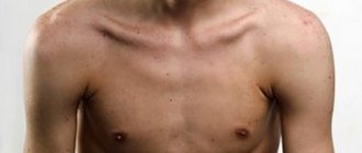
Patients may complain of general weakness, increased body temperature, pain in muscles and joints. As a result of nutritional deficiencies, body weight is lost. There may be deterioration in the condition of hair, skin, nails, and allergic manifestations. The level of hemoglobin in the blood is reduced. Colitis forms due to damage to the mucous membrane. Diarrhea, constipation, bloating, and abdominal pain in the navel or diffuse localization appear. With a small number of worms, they do not pose a threat to the life and health of the patient. Over time, as the population of parasitic individuals grows, health may deteriorate, which is especially noticeable in children.
Life cycle of pinworms
The life cycle of a pinworm is not long. The individual lays eggs with an embryo, which transforms into a larva within 5 hours. In a favorable environment, eggs live for about one month. Infection occurs through contact with a sick person or close contact with things or utensils of an infected person.
The larvae penetrate and hatch in the small intestine, then move into the large intestine.
After two weeks, the larva becomes an adult, capable of laying eggs on its own, but it retains them in its body, gradually migrating down the large intestine.
During the night's rest, the female exits through the anus and lays up to 20 thousand eggs around the anus. The development cycle of pinworms in the body does not exceed one month. The male dies immediately after fertilization of the female.
When does a female die?
- after laying eggs, the patient can crush the individual while scratching;
- dies even before the laying of eggs begins, but after the decomposition of the helminth, the larvae are released and begin their life activities.
For the infected person, frequent diarrhea is not so much a discomfort as irritability and itching in the anal area. Since helminths more often emerge and lay eggs at night, a person’s sleep may be disturbed, fatigue may develop, the nervous system may experience nervousness and unpredictable aggression towards others.
With the loss of nutrients in the body, people noticeably lose weight, and the level of hemoglobin in the blood drops sharply.
Children, due to their age, do not understand what is happening to them; they can scratch the itchy area, thereby ending up with eggs on their hands or under their nails. In girls, eggs often enter the vagina and cause inflammation - vulvovaginitis. The girl is taken to a female doctor, while the cause of the disease itself often remains unknown.
The patient becomes a carrier of helminths by touching toys, things, and dishes with infected hands. Then the parasite quietly emigrates back into the body through dirty hands and the pinworm development cycle repeats.
Important! It is known that if hygiene is observed, helminths die in the body and come out within three months!
The life cycle of this parasite is quite simple, it reproduces only in a living organism, and does not even require another host. The infected person himself may be the sole or intermediate carrier of helminths.
Main routes of infection
Drits Irina Alexandrovna. Parasitologist Helminth infections can lead to numerous health problems, shortening life by 15-25 years. Many parasites are extremely difficult to detect. They can be anywhere - in the blood, intestines, lungs, heart, brain. Symptoms of helminthic infestation can be confused with ARVI, gastrointestinal diseases and others. The main mistake in such cases is delay! If you suspect the presence of parasites, you need to contact a specialist. More information about modern methods of treating helminth infections is described in this interview with a doctor . If we talk about medications and self-treatment, then this antiparasitic complex .
Humans are the main source of invasion. Due to severe itching, the infected person combs the clutches, transfers the eggs with their hands to the face, household objects, and into the mouth. They also get on underwear and bedding. They can be found on the following items:
- dishes;
- toys;
- table;
- chairs;
- money;
- Food;
- hygiene supplies.
Pinworm eggs can be carried by insects, travel with dust, or on animal fur. They can maintain vital functions at temperatures of -15-55 degrees Celsius. Typically infection occurs:
- through hands;
- household items;
- unwashed and poorly processed food products;
- dust;
- ingestion of water from contaminated reservoirs and swimming pools.
Detection of enterobiasis
Detecting the parasite is quite simple; you need to go through two stages.
The first stage involves a thorough interview:
- what is he complaining about?
- appearance of the first symptoms
- Does anyone in your immediate circle complain of similar symptoms?
- Is the rule of personal hygiene observed (constantly clean hands).
At the second stage:
- laboratory examination of stool is carried out;
- scrapings are taken near the anus using adhesive tape;
- examination of a general blood test from a finger (there are characteristic indicators of the presence of parasites).
The main indicator is a low level of hemoglobin, the normal level is 120 - 140 g/l, red blood cells - 3.5 - 5.3, with a low level anemia occurs. Anemia appears when the disease is advanced, with a large presence of parasites and during an inflammatory process in the body.
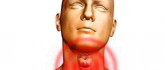
Inflammation of the genital organs, mainly in girls. Appendix - there is no reliable evidence that helminths entering it lead to inflammation of the appendix. The gallbladder is involved in the digestion process.
Active pinworms can move through the intestines not to the exit to the anus, but rather in the other direction to the anterior sections of the gastrointestinal tract. The bile ducts penetrate into the gallbladder itself and, irritating the mucous membrane of the wall, cause inflammation - cholecystitis.
Diagnosis of the disease
Female pinworms lay their eggs in the anal area, so to detect them, a smear is taken for analysis in this place.
In addition, the patient may be prescribed a blood test. When the body is infected with parasites, the level of eosinophils in the blood will be exceeded.
Stool analysis can show the presence of certain types of helminths in the body, but pinworms cannot be detected in this way. These parasites lay eggs outside the rectum.
Treatment
All contacts, including family members without symptoms, must be treated for prevention. More often, adults do not feel any abnormalities in their health and have no signs of illness.
But it is still necessary to undergo examination and proper treatment. Otherwise, you can infect a re-cured family member. After all, if one person is a carrier of parasites, then the likelihood of infecting other members of society increases significantly.
We come to the conclusion that every carrier of helminthiasis needs treatment!
Deworming is the main method of treating enterobiasis with the help of medicinal drugs.
The most widely used drugs are:
- Vermox , white tablets 0.1 g. They have a paralyzing effect on the parasite. After ingestion, the helminths die and are excreted in the feces. Children from 3 to 10 years old are given 0.5 g of the drug. Adults are given 0.1 g of the drug. Doctors recommend treating the whole family at once, under the supervision of the attending infectious disease specialist or therapist.
- Medamin - tablets No. 10, penetrating orally into the body, paralyzes pinworms, which in turn cannot hold on to the mucous membrane and are expelled. A dose of 10 mg per 1 kg of body weight is applied once 3 times a day.
- Piperazine – tablets No. 0.2, 0.5g. It has a detrimental effect on the nerve impulse of the muscles, the parasites come out on their own.
The dose is selected individually by the doctor, depending on the age and weight of the patient. The drug is taken for 5 days.
For infants under three months of age, it is prohibited to prescribe medications.
It is necessary to limit ourselves only to preventive measures. The drug does not contain a dosage regimen in the annotation for infants!
How does the parasite work?
The appearance is unremarkable: light body color, small size (up to 13 mm). The pinworm belongs to the class of round parasites (a group of thin worms). Females and males differ somewhat in structure: in the former the body is pointed on one side, in the latter it twists like a snail. In addition, the sizes differ: the female is twice as long as the male in both length and width.
For attachment, parasites use a vesicle - it is located on the front of the body and is a small swelling. The parasite egg is also unremarkable from the outside: light in color, oval in shape and microscopic in size. On one side it is flattened. The offspring at this stage of development are not exposed to many external factors, but the protective shell dissolves in the intestines, which contributes to the appearance of small larval worms. The duration of their formation until sexually mature parasites is 2-4 weeks.
Females are large from 8-13 mm by 0.3-0.5 mm in cross-section, males are small from 2-5 mm by 0.1-0.2 mm in cross-section
After emerging from the eggs, the larvae move to different parts of the intestine: the lower small intestine, the upper large intestine, and move towards the cecum. With enterobiasis (helminthic infestation by pinworms), the female leaves eggs on the outer integument of the human body, or more precisely, in the folds of skin near the anus.
During pregnancy
During pregnancy during the first three months, drug treatment is prohibited. It is necessary to limit ourselves to preventive measures.
If hygiene rules are followed, the helminths will die on their own. But if a doctor considers it appropriate to prescribe a drug, then Mebendazole, which he recommends, also does not have a license for use by pregnant women.
When breastfeeding, treatment with anthelmintic drugs is also prohibited. It is enough to limit yourself to compliance with preventive measures.
Appearance and structure
The pinworm is a grayish-white round worm, convex on one side and flattened on the other, looking like a thin thread. The appearance of females differs from males. The first ones are 2-3 times longer in length - 8-13 mm and 2-5 mm.
In females, the rear end is pointed and resembles an awl; in males, this part is twisted. At the opposite end, pinworms of both sexes have a mouth opening, followed by three folds. Behind the folds is a vesicle, with the help of which parasites attach to the intestinal wall. Pinworms feed on intestinal contents and intestinal bacteria.
Pinworm eggs cannot be seen with the naked eye, but under a microscope they are clearly visible. The shape of the egg is a colorless asymmetrical oval clot surrounded by a transparent shell. Egg size is 0.02 – 0.04 mm.
Products useful for enterobiasis
It is recommended to eat spicy and bitter foods. Walnuts, pumpkin seeds, flax seeds. Vegetable oils have a beneficial effect on irritation and inflammation of the intestinal walls. All sour berries, fruits and vegetables.
Preventive actions
- If enterobiasis is detected, immediately begin treatment to prevent infection of others;
- compliance with hygiene rules;
- regular change of underwear and outerwear;
- hot processing of raw food;
- do not hesitate to tell adults about the presence of itching in the anus.
After treatment, general cleaning is carried out using disinfectants, washing agents, and quartz coating of the room.
When an enterobiasis epidemic is detected, persons in contact with patients use anthelmintic drugs for prevention.
Causes in adults and children
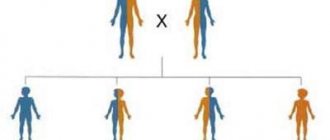
Most people believe that under no circumstances are they at risk of becoming infected with worms. All causes of pinworms in adults and children ultimately come down to non-compliance with personal hygiene rules. People at increased risk of infection include:
- children of preschool and younger age attending kindergarten or school;
- neglecting the rules of personal hygiene;
- consuming unwashed vegetables and fruits, undercooked foods;
- using other people's hygiene products;
- eating in places not designated for this (in bed, at the desk, etc.);
- wearing long nails;
- having the habit of biting their nails and sucking their fingers.
In the environment
One of the life stages of pinworms is that they enter the environment through bedding or underwear, unwashed hands or objects. Their lifespan directly depends on the climate in which they are located. Under favorable conditions, larvae can survive from 30 to 60 days if the air temperature is from 20 to 30 ° C and the humidity is at least 70%. In other cases, the viability of eggs is noticeably reduced, most often this happens:
- At low humidity levels. The larvae slow down in development and then die completely. Therefore, pinworm infection is a rare occurrence in countries with hot climates with low air humidity.
- At high temperatures, namely more than 35-40°C, and when the larvae are exposed to sunlight. As a result of a decrease in air temperature to -15°C, the larvae die within 40 minutes.
- When exposed to strong detergents.
- When washed in water above 55 °C, the parasite eggs die within 10 minutes.
Parasites are quite resistant to natural conditions, so they can easily retain their properties before they enter the human body. When infected, it is very important to take all possible measures to prevent the spread of larvae.
In summer or winter, you can take household items that cannot be cleaned to the balcony. This will help kill the larvae due to solar radiation or temperature. The remaining items should be washed at the desired temperature and ironed thoroughly. It is necessary to take into account everything that the patient could touch, including the carpet, curtains, pillows. After all, eggs can be located anywhere and cause re-infection.
Dienthamoeba. Dientamoeba fragilis.
Species: Intestinal amoeba (Entamoeba coli)
Habitat: upper large intestine and lower small intestine.
Invasive form: vegetative form, is a commensal, facultative parasite.
Method of infection: fecal-oral. Anthropogenic invasion.
Morphology of the parasite: division of the cytoplasm into 2 layers is visible in a dead amoeba or during the formation of pseudopods. The cytoplasm contains bacteria, fungi, protozoa, and food debris. There are NO formed elements of blood in the cytoplasm. The nucleus of a living amoeba is clearly visible (unlike e.histolytica) in the form of a ring formation of shiny chromatin grains. The pseudopods are short, wide and there are several of them. Movement - marking time.
Life cycle: lives in the large intestine, non-pathogenic.
Laboratory diagnostics: microscopy of stool smears.
Type: sarcoflagellates
Class: sarcodina
Order: amoebas
Genus: dientamoeba Jepps
Species: dientamoeba fragilis
Disease: dienthamoeba diarrhea.
Invasive form: vegetative form, pathogenic.
Method of infection: due to extreme instability in the external environment, the human body enters the human body with the eggs of roundworms (symbiosis with children's pinworms), into which the amoeba penetrates in their early stages of formation.
Small. It lives in the lumen of the colon and feeds on bacteria, fungi, and red blood cells. Only vegetative forms of this amoeba are known. Ectoplasm and endoplasm are clearly distinguishable. It has 2 nuclei (rarely 3), visible only after staining. Found only in loose stools, usually in various intestinal disorders. Can be found in appendicitis.
Life cycle: the vegetative form, through the mouth with pinworm eggs, enters the large intestine, where it multiplies and parasitizes. Does not form cysts. In the external environment it quickly dies.
Laboratory diagnostics: microscopy of smears from fresh (warm) feces.
Order: amoebas (amoebina)
Genus: entamoeba
Species: dysentery amoeba (entamoeba histolytica)
Medical significance: amoebiasis (amebic dysentery)
Invasive form: large vegetative and tissue form.
Form of infection: mature 4-core cyst.
Epidemiology: anthropogenic invasion. Infection is fecal-oral. The source of invasion is cyst carriers and patients.
Morphology of the parasite: exists in the form of several forms (large vegetative form, small vegetative, tissue, luminal, pre-cystic form and cysts).
- Large vegetative form: the cytoplasm is divided into 2 layers (ectoplasm - like crushed glass, and endoplasm - a glassy mass). In a living amoeba the nucleus is not visible; in a dead amoeba it is in the form of a ring-shaped cluster of grains. The endoplasm contains several red blood cells. It differs from other forms in its forward movement - an outgrowth of ectoplasm is formed in a jerky manner, into which the endoplasm flows with a swirl.
- Cyst: formed from a luminal form in a thick body, motionless, round, colorless, sometimes shiny rods are visible in them - chromatoid bodies (RNA and protein). When stained with Lugol's solution, 4 nuclei are visible.
Each cyst enters the gastrointestinal tract, where in the large intestine it produces 8 cells that transform into a small vegetative form (non-pathogenic, feeds on bacteria and food debris). When the immune system is weakened, it transforms into a large vegetative form, which lives in the lumen of the descending and sigmoid colon (pathogenic, feeds on mucous membranes and red blood cells).
Cysts f.minuta→f.magna→ luminal form → cysts
Pathogenesis.
f.magna lives in the lumen of the lower parts of the large intestine (descending and sigmoid colon), secretes an enzyme that destroys tissue (necrosis of the mucosa) and the formation of bleeding ulcers (ulcerative colitis) and the addition of a secondary infection. With a decrease in immunity, the tissue form of the amoeba penetrates the bloodstream (generalization of the process) and enters the liver... where abscesses can develop, which in 5% of cases break into the abdominal cavity with the development of peritonitis. Which also develop during perforation (perforation).
Clinic:
- Tenesmus - false urge to defecate
- The stool is raspberry jelly (mucus with red blood cells), often watery.
- Lower abdominal pain
- Symptoms of intoxication: weakness, low-grade fever, headache, nausea.
- Symptoms of anemia, exhaustion and hypovolemia (dehydration)
Laboratory diagnostics:
- When carrying cysts: in formed or semi-formed feces, cysts can be found, which are distinguished by size and number of nuclei. The smear is examined microscopically with Lugol's solution.
- In acute or subacute cases: a native smear is prepared from fresh liquid feces and mobile vegetative forms of amoebas with red blood cells in the cytoplasm are observed. Stool is examined within 10-20 minutes after excretion.
Prevention:
- Personal: boiling water, breaking the chain of fecal-oral infection, washing hands, washing vegetables, fruits, destroying vectors (cockroaches, flies).
- Public: identification and isolation of patients and carriers, prevention of fecal contamination of the environment (disinfection of feces), sanitary education work.