A skin nevus is a benign formation that appears on its surface. It can be congenital (from birth) or acquired (appear at a certain stage of life) in nature. Popularly, such growths are called birthmarks. In medical practice, they are all similar in their structure and mechanisms of formation.
In the lion's share of clinical situations, nevus does not require a therapeutic process, since it does not have any effect on the quality of human life. However, doctors remove some types of moles because they believe that they run the risk of developing into an oncological condition. By the way, this is their key negative consequence.
Etiology
Nevus cells are born during the formation of the embryo. The development of various anatomical objects requires a neural crest. This is a collection of cells from which nerve ganglia arise, elements that form the skin (pigment cells, melanocytes), membranes that envelop the medulla, and parts of the adrenal glands. Nevus cells develop from 40 to 180 days of embryo formation. If they fail to mature into melanocytes, they remain in the dermis and cannot move into the deeper layers of the epidermis. The reasons for this phenomenon have not yet been established. This skin anomaly is not acquired, but has a congenital etiology.
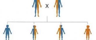
Hereditary nevi are:
- pigmented;
- vascular;
- hyperkeratotic;
- cystomatous;
- dermoid.
Types of moles by continuity:
- hereditary;
- non-hereditary.
Provoking factors for the development of congenital nevi in embryos:
- genetic failure;
- heredity;
- hormonal imbalances in the expectant mother;
- pathological course of pregnancy (toxicosis, threat of miscarriage);
- infectious diseases of the genitourinary system in a pregnant woman;
- the influence of toxins and allergens on the expectant mother;
- chemical, ionizing, radioactive, toxic, radiation effects on the pregnant woman’s body.
Factors influencing the manifestation of congenital nevi in people of different ages:
- ultraviolet action;
- skin diseases of infectious and allergic nature;
- hormonal surges (especially during puberty and pregnancy);
- taking contraceptives;
- mechanical damage to the skin.
Causes
Pigment spots can form due to congenital or acquired reasons.
Congenital nevi are a defect of embryonic development. Their occurrence is caused by a disruption in the process of movement of melanocyte precursor cells (melanoblasts) from the neuroectodermal tube into the skin. The accumulation of these cells in the skin causes the appearance of pigment spots.
Acquired age spots are formed under the influence of the following factors:
- significant changes in hormonal levels (puberty, pregnancy);
- skin infections;
- excessive skin exposure.
Symptoms of congenital nevus
Hereditary nevi can appear from birth, a couple of months after birth, or at a more mature age.
Stages of nevus development:
- intraepithelial;
- border;
- intradermal (about 30 years);
- reverse development (in the elderly).
Development time:
- immediately after birth;
- in early childhood (1−24 months);
- at a later age (develop along with the child’s growth).
If a person has many congenital moles (this occurs in 5% of people), then one of them will be large in size, and over time they may increase in size.
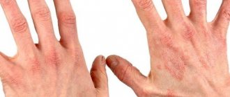
Hereditary nevi have different clinical manifestations:
- quantity (from 1 to many);
- size (small - 0.5-1.5 cm, medium - 1.5-10, large - 10-20, giant - more than 20 cm);
- color (different shades of brown from light to dark);
- convexity (merges with the surface of the skin or protrudes above it);
- shape (various - nodules, spots, plaques);
- boundaries (clear or blurred);
- surface (lumpy, smooth, lobed);
- skin pattern (sometimes preserved);
- consistency (small ones do not differ from the skin, large ones are soft-elastic);
- hair (appears on the surface with age);
- cellular location (subcutaneous tissue, lower layer of dermis);
- localization (can be on different parts of the skin and mucous membranes, but more often on the lower torso, upper back, chest, limbs, forearms).
Risk of degeneration
Nevi can cause emotional discomfort and become damaged.
In any case, the presence of nevi in itself is an increased risk of developing cancer. Risk factors for developing melanoma (the most aggressive skin cancer):
- a significant number of moles (more than 20);
- mechanical damage;
- the presence of moles protruding above the skin;
- diameter more than 2 cm;
- large total area on the body surface (more than 2.5%);
- moles that are localized on the face;
- congenital pigment spots;
- giant nevi;
- change in color, shape, size;
- the appearance of nodes or ulcers on the nevus.
Differences in hereditary nevi according to histological structure:
- melanoma-dangerous - with a high probability they can degenerate into malignant formations (most often into melanoma), require increased attention and control (dysplastic, verrucous, borderline pigmented, giant pigmented, blue nevus, nevus of Ota);
- melanoma-monogenic - very rarely become malignant, they interfere only from an aesthetic point of view (halonevus (Setton's nevus), Mongolian spot, birthmark, Sturge-Weber syndrome, nevus of the sebaceous glands, vascular (hemangiomas), papillomatous, intradermal pigmented, epidermal, fibroepithelial nevi) .
Congenital melanocytic moles pose the greatest danger in childhood and puberty, since at this time they grow and develop along with the area in which they are located. In adults, they usually remain stable unless changes are provoked (excessive exposure to ultraviolet radiation, mechanical damage).
In 6−10% of cases, giant nevi degenerate into melanoma.
Diagnostic complex
A doctor performing diagnostic procedures undertakes to set a set of goals:
- establishing the type and class of moles,
- identifying treatment options,
- timely recognition of the onset of cancer,
- identification of indications for additional implementation. examination methods,
- taking measures of the therapeutic process.
During the initial examination, the medical specialist undertakes to establish several answers to the most important questions:
- at what time did the growth appear?
- it is present from birth or from any age,
- how has he behaved over the past days, weeks, months,
- whether there were changes in dimensional indicators,
- whether the color characteristics, contour parameters,
- whether diagnostics were carried out,
- whether healing measures were taken.
So, the first stage is communication with the patient. This is followed by an examination, during which the physician assesses the shape, size, and location. If the diagnostic goal is not achieved, auxiliary measures are prescribed:
- smear from the surface (subsequent examination of the material through a microscope) with results obtained the next day,
- examination on the patient’s body (a microscope is used, a smear is not taken, the procedure is performed directly on the body),
- computer diagnostics (provides an image of a pigment spot saved on a computer, is expensive, but effective),
- laboratory examination (determines the fact of degeneration into melanoma),
- histology (shows the structure of the formation, reveals the degree of its danger and stage of development),
- biopsy (an impressive examination in all directions, making it clear what should be done for the further therapeutic process).
How is diagnosis carried out?
First of all, a dermatologist-oncologist sets himself the task of distinguishing a benign disease from a malignant one. To do this, he carries out a number of manipulations:
- collects anamnesis (finds out how long the patient has had a nevus, what changes it has undergone and over what period of time, whether it is hereditary, etc.);
- conducts an inspection;
- performs dermatoscopy, thermometry, echography, and phosphorus isotope studies;
- performs epiluminescent microscopy - a non-traumatic diagnostic method (allows you to examine the structure of the mole);
- takes a histological analysis using a biopsy (this manipulation is carried out in extreme cases, since the procedure itself can provoke malignant degeneration).
Treatment and removal
The decision whether a mole needs to be treated (by removal) or whether it can simply be monitored is made by the doctor based on the overall clinical picture and diagnostic results. In this case, several indicators are taken into account - aesthetic defect and the risk of developing cancer. Mole removal methods:
Elimination of hereditary nevi is prescribed when the patient experiences physical and emotional discomfort.
- surgical - not only the nevus is excised, but also nearby tissues (3-5 cm), the operation is performed under local anesthesia and leaves scars;
- electrocoagulation - removal by burning is painful and can leave scars, suitable for small and medium-sized moles;
- laser - the most common and painless;
- cryodestruction - removal with liquid nitrogen is painless and leaves no scars, suitable for small formations.
Large melanoma-dangerous moles are recommended to be removed:
- giant - in early childhood;
- small (with a suspicious shape or color) - up to 12 years.
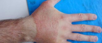
There are a number of indications in which a mole must be removed immediately:
- increase in size;
- height change;
- strong darker;
- color unevenness;
- the appearance of erosion;
- itching;
- inflammation of the halo;
- pain;
- bleeding
The most reliable method is surgical; for oncological indications, only this is used, and for cosmetic indications - laser, nitrogen or current.
Therapeutic process
Treatment is carried out in various ways. Surgery is common. The practice of removal by other means is used. Indications for choosing one or another option are determined by the treating specialist. Several key factors are taken into account.
- Key features of age spots. These include its type, size, and risks of transformation into cancer.
- Availability of the necessary equipment in the clinic. Many establishments use only a scalpel and do not use other methods due to lack of equipment.
- Individual characteristics of the body. Some patients cannot bear the pain, so painless techniques are preferred. The degree of sensitivity of the skin is also taken into account.
Surgical excision
The main tool of a doctor is a scalpel. Due to the undemanding nature of the technique, the method is considered the most common. Doctors give it preference when it is necessary to remove large growths. The disadvantages are in the following aspects:
- the presence of scars after surgery,
- the need to remove not only the spot itself, but also the surrounding skin in the area of 3-5 cm),
- the need for anesthesia (local in adults and mainly general in children).
Recently, this type of excision is rarely used, because there is a possibility of the spot degenerating into a tumor process. Along with surgical techniques, there are other common types of birthmark removal.
- A liquid nitrogen. Provides treatment for nevus through freezing. Low temperatures cause the spot to die, which then turns into a scab. After healing, natural skin begins to grow in its place. There are no scars or cicatrices, no pain.
- Electrocoagulation. The method is the opposite of the previous one, since high temperatures are used for excision. Compared to the classical operation, the method has a number of advantages: no blood, no need to remove large areas. But the method is not used for excision of large moles.
- Laser removal. The technique has become widespread in modern cosmetology salons. It helps to instantly remove small-sized moles on the face and neck. The laser penetrates deeply, and there are no scars or burn consequences. The patient does not feel pain.
- Radiotherapy. Has good recommendations within the framework of modern world medicine. The essence is to use a special knife capable of generating a beam of radiation concentrated in the area of the pathology.
If there is a suspicion of a malignant nature of the process, surgical intervention is traditionally used. In this case, surrounding tissue is removed to avoid subsequent growth of the tumor process.
Prevention
There are no preventive measures that would guarantee that congenital nevi will not appear. Therefore, it is necessary to monitor existing formations and prevent the degeneration of moles into melanoma:
- find out which moles are melanoma-hazardous;
- do not spend a lot of time under active ultraviolet radiation (from 11 a.m. to 5 p.m.);
- avoid injury;
- know how to distinguish an ordinary mole from a malignant one;
- take photographs of existing moles so that you can monitor their condition over time;
- know about your heredity;
- use highly effective sunscreens;
- monitor beta-carotene intake and avoid carcinogens;
- remove atypical formations.
The prognosis for this phenomenon directly depends on the clinical picture and accompanying factors. If the moles are not large, there are few of them, and they do not have pathological signs, then you don’t have to worry about the risk of cancer. If there are a lot of nevi, they are huge, non-standard, and accompanied by abnormal symptoms - this is a reason to immediately consult a doctor.
Characteristic signs of hemangiomas
Cavernous hemangioma is a soft, nodular neoplasm that is purple or bright cherry in color. The composition of such a vascular nevus is pathologically enlarged blood vessels. The most common causes are congenital pathology.
Due to the abnormally enlarged size of the vessels, in some cases, cavernous hemangioma may bleed, after which it (rarely) undergoes partial, spontaneous disappearance. Treatment methods: surgical excision, electrocoagulation.
The second type of vascular nevus , capillary hemangioma, appears as a large, purplish-red spot. The size of such a benign neoplasm can vary from 1 to 10 cm in diameter. Also applies to congenital type pathology. Appears at birth and tends to grow rapidly during the first months of a child's life.
As a rule, capillary nevus does not require special treatment or surgical removal, since in most cases it disappears on its own by the age of seven. In addition, with a scalpel excision, there is a possibility of the formation of noticeable scars and extensive scarring on the skin. If necessary (if the nevus is located near the eyes), the doctor prescribes hormonal therapy as a treatment for capillary hemangioma.