Causes of anisocoria
In many cases, the presence of slight differences between the pupils is normal and is not considered to be an expression of any pathology or a consequence of injury. As a rule, if one pupil is larger than the other or smaller than 1.0 mm without an objective reason, then this is called physiological anisocoria, benign or simple. Its appearance is not affected by a person’s gender, age, or eye color; this phenomenon can be observed in approximately 20% of the population.
The causes of non-physiological anisocoria (exceeding 1.0 mm) may be the following:
- Trauma to the organs of vision, traumatic brain injury, which may damage the nerves or areas of the brain responsible for the tone of the sphincter and dilator of the pupil.
- The use of local or systemic medications that affect pupil width (pilocarpine eye drops, ipratropium bromide).
- Inflammation of the iris. Iritis (anterior uveitis) can cause anisocoria, which is usually accompanied by eye pain.
- Adi's syndrome is a benign dilatation of the pupil in which it stops responding to light. This may be due to eye trauma, eye surgery for cataracts, ocular ischemia, or an eye infection.
Neurological disorders in which anisocoria may occur:
- Strokes, usually hemorrhagic. Its additional signs include the sail sign (when breathing, swelling of the cheek on the side of the brain lesion), asymmetry of the palpebral fissures.
- Spontaneous hemorrhages or traumatic brain injury.
- Aneurysm.
- Abscess inside the skull.
- Excessive pressure in one eye caused by glaucoma.
- Increased intracranial pressure due to cerebral edema, intracranial hemorrhage, acute stroke, or intracranial tumor.
- Meningitis or encephalitis.
- Migraine.
- Diabetic nerve palsy.
Pupils of different sizes - what is it?
Many people wonder whether it is possible to influence the size of their pupils on command. For example, you need to narrow or dilate your pupils during a photo shoot. In fact, there is nothing complicated about it. Below are step-by-step instructions, following which will lead you to the desired result.
Step 1. Go into a dark room. As noted earlier, in a dark room the pupils dilate as they try to “catch” more light. If it is not possible to completely turn off the light in the room, then simply turn away from the windows, thereby protecting yourself from light sources.
Go into a dark room
Step 2. To constrict your pupils, turn towards a light source in your home and remain in this position for a few seconds. If you are on the street, then all you need to do is look up. Of course, looking at the sun is bad for your eyes, so it's best to focus your gaze on something else.
Turn towards the light source
Step 3: Another easy way to constrict your pupils. Just look at the object that is located next to you. When focusing the gaze, the pupils constrict. Alternatively, you can place your finger in front of your eye and focus your attention on it.
Focus your gaze on a nearby object
Step 4: Tighten your abdominal muscles. Many people manage to dilate their pupils by keeping their muscles in constant tension. To test this method, you need to tense your stomach and look at yourself in the mirror. If your pupils are dilated, then this method is suitable for you, but if not, then try another method.
Tighten your abdominal muscles
There are several environmental factors and a number of diseases that can affect the diameter of the hole in the iris.
In adults
Among the reasons that influence the development of anisocoria, there are a number of factors:
- Impact of medications. In this case, giving up medications and looking for alternative options will help get rid of the deviation;
- Optic nerve damage;
- Traumatic brain injury causing hemorrhage;
- Drug use (cocaine, tropicamide, etc.).
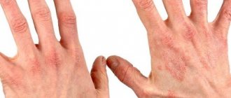
Anisocoria is a condition in which the pupils of the right and left eyes differ in size or diameter. The pupil is the round black area in the center of the iris. Depending on the lighting, it can have dimensions from 1mm to 6mm in diameter.
In the presence of general or ocular pathology, anisocoria is always combined with the following manifestations:
- restriction of eye movement, or the eye on which the pupil is larger
- drooping upper eyelid (ptosis)
- Pain in the eyes
- increased temperature or fever
- headache
- decreased vision
- double vision
When observing a person with different pupils, one can suspect pathology or a hereditary factor in the transmission of symptoms from parents. A condition in which the pupils become different in size is called anisocoria, one pupil of which can narrow and the other dilate.
Among doctors, such a symptom is not considered a disease, but is understood as a deviation from the norm. This pathology can be seen with the naked eye in poor lighting; the difference in size can be minimal one millimeter, and if you use drops to dilate the pupils, they can become almost identical for a short time. For people to have such a pathology means being more susceptible to eye diseases and inflammation.
A type of anisocoria is Horner's syndrome, which is directly related to damage to the sympathetic system, after which the pupil dilates. Visually, symptoms may look like a sunken eyeball, drooping eyelid, and decreased sweating.
Main symptoms of Horner's syndrome:
- miosis;
- slow pupil reaction
- anhidrosis;
- ptosis.
On this topic
- Ekaterina Nikolaevna Kislitsyna
- March 26, 2020
Cocaine or tropicamide tests can verify the accuracy of the data and confirm the pathology; the size of the pupils will be especially noticeable at dusk.
A similar syndrome associated with anisocoria is oculomotor nerve palsy caused by a tumor or other formation. Visually, the patient can see a narrowing of the affected pupil and drooping of the eyelid.
Types of anisocoria
In an adult, anisocoria is most often acquired as a consequence of one of the reasons mentioned above.
Congenital
Congenital anisocoria is often found in newborns. Most often it is caused by pathology of the iris or weak or defective development of the brain and nervous system.
However, if a child has different pupils from birth, just like adult family members, and no neurological symptoms are observed, then such anisocoria can be considered a genetic feature. In this case, there is nothing to worry about.
Some physiological difference in the size of the pupils in infants, as well as congenital nystagmus caused by an imperfection of the nervous system, can self-correct up to a year, according to the development and strengthening of the visual organs and the centers in the brain responsible for their innervation. They are eliminated naturally, and treatment is prescribed only if pathology is detected.
Acquired anisocoria in children is often a consequence of injury or infectious diseases of the brain.
Transient
The change in pupil size may not be permanent and is called transient anisocoria. It is very difficult to make this diagnosis because symptoms may not appear at the time of examination. The transient nature corresponds to the moment of onset of the underlying disease, for example, migraine, sympathetic or parasympathetic dysfunction.
Hyperactivity of sympathetic innervation is expressed in a normal or delayed reaction of the pupils to light, different widths of the palpebral fissures. She's more on the losing side.
Paresis of parasympathetic innervation leads to the absence of pupillary reactions, and the palpebral fissure on the affected side is much smaller.
Classification
All lesions of the pupillary opening can be divided into congenital and acquired. Variable pupil sizes can be persistent or transient; in the intermittent variant, the diameter is restored after the end of the influence of the trigger factor; in the persistent variant, it persists for a long time. There are two main forms of pathology:
- Physiological. It often occurs in healthy people and can be observed at rest. The difference in pupil diameter does not exceed 1 mm. Visual differences in the size of the pupillary opening persist regardless of lighting conditions.
- Pathological. This form of anisocoria is a symptom of a neurological or ophthalmological disease. Pupil differences vary widely. There is a relationship between the size of the pupil and the reaction of the eye to changes in light intensity.
Diagnostics
Often you may not even realize that your pupils are different sizes. If this is not due to the presence of pathology, then physiological anisocoria is not reflected in the quality of vision.
However, if anisocoria is caused by eye or nervous system health problems, there may be additional symptoms associated with these problems. These include:
- involuntary drooping of the eyelid (ptosis, partial ptosis);
- difficult or painful eye movement;
- pain in the eyeball at rest;
- headache;
- temperature;
- decreased sweating.
See a neurologist
A neurological examination is required. People with nervous system disorders that cause anisocoria often also have ptosis, diplopia, and/or strabismus.
Anisocoria is also included in the triad of classic Horner's syndrome: drooping of the eyelid (ptosis of 1-2 mm), miosis (constriction of the pupil less than 2 mm, causing anisocoria), facial anhidrosis (impaired sweating around the affected eye). These usually occur due to a brain injury, tumor, or spinal cord injury.
Horner's syndrome (oculosympathetic paresis) can be distinguished from physiological anisocoria by the speed of pupil dilation in dim lighting. Normal pupils (including normal pupils that are slightly unequal in size) dilate within five seconds after the light in the room dims. A pupil suffering from Horner's syndrome usually needs 10 to 20 seconds.
At the ophthalmologist
An examination by an ophthalmologist is carried out to determine the size of the pupils and their reaction to light in bright and dark conditions. In a dark room, the pathological pupil will be smaller. However, this will also be characteristic of physiological anisocoria and Horner's syndrome. Further differential diagnosis is carried out by instillation of mydriatics (medicines that dilate the pupil) into the eye. With pathology, a smaller pupil will still remain constricted and will not respond to the action of the medicine.
When the difference in pupil size is greater in a lit room, the larger pupil is considered abnormal. In this case, additional difficulty in eye movement may be detected, which indicates damage to the third pair of cranial nerves. If normal eye movement is maintained, a test is performed with miotic drugs, which should cause pupil constriction. If this does not happen, then the presence of tonic Adi syndrome is assumed; if there is no reaction to the drug, then damage to the iris can be suspected.
Accommodation and the amount of movement of the eyeballs are also determined. A more pronounced reaction of the pupil under accommodative load than under the influence of changes in illumination is considered abnormal.
The pathological structure of the eye is revealed by biomicroscopy.
You can determine the presence of constant anisocoria by a series of photographs of different ages, where the pupils and their size are visible.
Symptomatic appearance of anisocoria or what should you pay attention to?
As mentioned above, anisocoria is only a symptom that indicates problems in the human body, but it is expressed by the fact that visually the difference in pupil diameters is very noticeable. In this case, one eye will work as usual, but the second one will not react to light as it should in normal condition, but at the same time, the fixed size will remain.
If there is enough lighting, then the pupils can take almost the same shape, but as soon as the light becomes less, the difference between them becomes visible.
Physiological anisocoria appears only when illumination is low and the reaction to light does not go beyond normal limits. When taking special medications that help dilate the pupil, the signs of anisocoria disappear.
If anisocoria is pathological in nature, the pupil located in the affected area begins to slowly enlarge, but the reaction to lighting may, in some cases, be completely absent.
The difference in pupil diameters in humans causes discomfort, mainly of an aesthetic nature. But it is worth knowing that such a pathology can cause some accompanying symptoms that can cause a person a lot of discomfort - these are:
- during visual stress (for example, it could be working at a PC), fatigue increases;
- pain and swelling of the cornea;
- the eyeball has limited mobility;
- ptosis is a phenomenon in which the upper eyelid droops;
- diplopia is characterized by bifurcation of the object that is being viewed, and the visible images themselves can also shift in different ways - vertically or horizontally, as well as diagonally;
- proptosis is an unpleasant phenomenon characterized by the fact that the eyeball protrudes towards the front.
But, in addition to such ophthalmological unpleasant phenomena, the difference in pupils also leads to general symptoms.
- Coordination of movements is impaired.
- Cephalgia, which manifests itself as painful sensations in the head area. With this development, analgesics can only temporarily relieve pain. Dizziness may also occur.
- Speech disorders.
- Hand tremor and paresis, as well as partial paralysis, may occur.
- Visual disturbances also appear - this is a decrease in visual acuity, and “spots” flying in front of the eyes.
- Nausea and vomiting.
If a person notices the above symptoms in himself or in one of his relatives, then he urgently needs to go to the clinic for a diagnosis and adequate treatment.
Treatment
Treatment of anisocoria, which is unstable and refers to pupillary disorders in the autonomic syndrome (for example, with meningitis), is also not required.
Congenital defects of the iris (hypoplasia or muscle aplasia), which contributed to the appearance of anisocoria, can go away independently with the development of the child, but require observation and, possibly, physiotherapeutic procedures.
If different pupil sizes are caused by damage to the brain or cranial nerves, then the tactics of treatment depend on the cause. Infectious inflammation requires the use of antibiotics. In case of stroke, hemorrhage, hematoma from injury, or the presence of a neoplasm, surgical intervention is necessary to remove these damaging factors. This is usually followed by drug therapy aimed at reducing swelling, improving microcirculation and nutrition of brain cells, and restoring neural connections. Also, when appropriate, antitumor and antibiotic drugs are used.
In conclusion, I would like to give an example of anisocoria, which is known all over the world - these are the eyes of David Bowie. An injury suffered in his youth made one of his pupils much larger than the other. And yet, the singer’s experience showed that with such eyes life is quite successful.
Causes
The main factors that provoke the appearance of this disorder in adults:
- mechanical injury to the organs of vision and brain;
- surgery (cataract treatment);
- the use of drugs that affect the width of the pupil (a similar reaction occurs when using atropine, pilocarpine and acetylcholine);
- drug addiction;
- ophthalmological diseases (iritis, glaucoma, Adi syndrome);
- a brain tumor;
- neurological diseases (neurosyphilis, aneurysm);
- complications of diabetes and stroke;
- tuberculosis;
- heredity.
The most serious causes of anisocoria in adults are hemorrhagic stroke and cerebral infarction. In such cases, asymmetry of the pupils is accompanied by severe headaches, tachycardia, impaired facial expressions, redness of the skin and high blood pressure. If such symptoms appear, you should immediately seek help from a specialist.
The congenital form manifests itself in childhood with defective development of the brain or nervous system and with a genetic predisposition. Often the disorder is accompanied by strabismus. The acquired form in children develops due to a fall and hitting their head on a hard surface or as a result of a birth injury. Other causes include the use of drugs, poisoning by toxins, and intoxication after a tick bite.
Pupil characteristics
The specific anatomical structure of the pupil cannot be described. The pupil is simply a hole in the iris through which almost 100% of light rays are absorbed. These rays do not go back through the iris and are absorbed by the inner membranes, which causes the blackness of the pupil in all healthy people.
The pupil acts as the eye's diaphragm, regulating the amount of light entering the retina. In bright conditions, the circular muscles contract, while the radial muscles, on the contrary, relax, which leads to a narrowing of the pupil and a decrease in the amount of light entering the retina. This mechanism protects the retina from damage. When there is insufficient light, the radical muscles contract and the annular muscles relax, which dilates the pupil.
The reduction of the pupil is carried out by the parasympathetic nervous system, and the increase by the sympathetic. In bright light, the sphincter muscle works, and in dark light, the dilator muscle is activated.
Such changes can occur several times per minute. This is how photons are distributed, which irritate the retina. Anisocoria is a consequence of a mismatch in the functioning of the muscles of the iris. The patient has different pupil sizes and, accordingly, different degrees of response to lighting.
The oculomotor nerve muscles of the iris allow simultaneous changes in the pupils of the eyes. Surprisingly, if you shine light into one eye, the pupils will narrow in both at once, and simultaneously. This phenomenon is possible only with proper functioning of the iris muscles. If narrowing does not occur in the second eye, we can talk about pathology. A narrowing of the pupil from normal is called miosis, and dilation, accordingly, is called mydriasis.
It is noteworthy that physiological anisocoria is observed in many representatives of the fauna. For example, in reptiles and amphibians, due to the lack of binocular vision (picture perception with both eyes), such synchrony of eye reactions is not always observed.
The pupils are capable of reacting not only to light rays. Many strong emotions (fear, pain and excitement) can affect the size of the pupil. Also, some medications change the functioning of the iris.
Treatment methods
Therapy is required only if the disorder was caused by some disease. It is aimed specifically at eliminating the cause, since anisocoria is only one of the symptoms.
Most often, specialists try to get by with drug therapy. If conservative treatment does not produce results or the condition is a symptom of a serious illness, then surgical intervention is required.
Drug therapy
Depending on the cause of anisocoria, the specialist prescribes the patient the following medications:
- corticosteroids;
- antibacterial drugs;
- anti-inflammatory drugs.
If the disorder occurs due to an eye injury, then medications are used to relax the muscles of the iris. It is prohibited to use any medications without first consulting your doctor.
For congenital anisocoria, drug therapy is not required.
Surgical intervention
Surgery is necessary for patients with aneurysm, stroke, tumor and other diseases for which drug treatment is ineffective. Surgery is often used to treat glaucoma.
If anisocoria occurs due to mechanical trauma to the eyes, then surgery may be necessary to correct the structural defect.
Classification of pathology and its causes
There are several main causes of anisocoria, which involve dozens of different diseases and conditions. In 20% of cases, anisocoria in infants is a consequence of a genetic defect. The child most often does not have any other symptoms, and the pathology of the pupil does not exceed 0.5-1 mm. In such cases, anisocoria may disappear by 5-6 years.
Types of anisocoria
- Congenital. This type of pathology is often the result of a defect in the eye or its individual elements. The reason affects the muscular apparatus of the iris and causes asynchrony in the reaction of the pupils to light. It happens that congenital anisocoria is a symptom of underdevelopment of the nervous apparatus of one eye or both, but in almost all cases the pathology is complemented by strabismus.
- Acquired. There are many reasons that can cause anisocoria throughout life.
One of the most common causes of pupillary mismatch is injury. There are several types of injuries that can cause anisocoria. First of all, these are eye injuries. Often the synchrony of pupil reactions is disrupted due to damage to the iris or ligamentous apparatus of the eye. With an eye contusion, when there are no visible injuries, paralysis of the muscular structure of the iris may develop, and the pressure inside the eye will increase.
When a head injury occurs, there is always a risk of injury to the skull or brain. Anisocoria may be the result of impaired functionality of the nervous system of the eyes or visual centers in the cerebral cortex. When the visual centers are damaged, strabismus often develops. Disturbance in the functioning of the optic nerves often leads only to one-sided dilation of the pupil. Distinctive feature: the pupil dilates in the eye on the side of the injury.
Eye diseases also often manifest themselves through anisocoria. Such ophthalmological disorders can be inflammatory or non-inflammatory in nature. Iritis and iridocyclitis (isolated inflammation of the iris) can cause spasms of the iris muscles. As a result, the eye stops responding to changes in light, which is expressed by mismatch of the pupils. Glaucoma often provokes a narrowing of the pupil in the affected eye (permanent): this makes the outflow of intraocular fluid faster and easier.
The growth of neoplasms and tumors in the head leads to a weakening of the connection between the eyeballs and the visual centers. As a result, the functionality of the iris is impaired. Such pathologies include malignant brain tumors, neurosyphilis, and hematomas in the brain after a hemorrhagic stroke.
Anisocoria can occur when exposed to certain inorganic substances: belladonna, atropine, tropicamide. When these compounds affect the nerves and muscles of the eye, pupil misalignment may occur.
Diseases of the brain and visual nerve pathways are also at risk. Among the main diseases of the central nervous system that can cause anisocoria are neurosyphilis and tick-borne encephalitis, meningitis and meningoencephalitis.
Types of anisocoria
- Caused by eye pathologies. The condition occurs due to disturbances in the elements of the eye.
- Caused by other pathologies.
According to the degree of involvement, unilateral and bilateral anisocoria are distinguished. In 99% of cases, a unilateral eye pathology is diagnosed, that is, one normal eye reacts to changes in light, and the pupil of the second either does not react or functions late.
Bilateral anisocoria is a fairly rare occurrence. The condition is characterized by an uncoordinated and inadequate response of the iris to changes in the visual regime. The degree of pathology may be different for each eye.
How pupils work
It is a key component of the eye, located in the center of the iris. The main task of the pupils is to collect light rays, which are then perceived by the retina of the eye. There are muscles around the pupils that, by contracting, regulate the illumination of the retina. For example, when a person enters a dark room, his pupils dilate to catch light, and in a brighter room, his pupils constrict accordingly.
How does the pupil react to light?
This is an important system that allows you to obtain the maximum amount of information from the environment under different lighting conditions. But if pupil function is impaired, serious problems can arise. The degree of illumination in the room is not the only reason for changes in pupil size.
Natural causes of pupil dilation
Diagnosis of the causes of pupillary defect
The first stage in diagnosing the causes of anisocoria is collecting an anamnesis. The doctor must identify all concomitant pathologies, study their causes, development and duration. Photographs of the patient help in diagnosing anisocoria. From them you can find out whether the pathology existed before and with what dynamics it developed.
During an eye examination, the doctor determines the size of the pupils in light and in the dark, reaction speed, and consistency under different lighting conditions. These simple characteristics help to at least approximately determine the cause of anisocoria and the localization of the disorder that provokes pupil mismatch.
Anisocoria, which is more pronounced in bright light, is indicated by pathology when the pupil dilates to a large size and has difficulty constricting. With anisocoria, which is more pronounced in a dark environment, the pupil becomes unnaturally small and dilates with difficulty.
Methods for diagnosing anisocoria
- Cocaine test. In the process, a 5% solution of cocaine is used (if the patient is a child, a 2.5% solution is used). Sometimes the cocaine solution is replaced with apraclonidine 0.5-1%. The test allows you to differentiate physiological anisocoria from Horner's syndrome. The procedure is simple: drops are dropped into the eyes and the size of the pupils is assessed before the procedure and after 60 minutes. If there are no pathologies, the pupils gradually dilate. In the presence of Horner's syndrome, the pupils on the affected side dilate to 1.5 mm.
- Phenylephrine and tropicamide tests. A solution of 1% tropicamide or phenylephrine can detect a defect in the third neuron of the sympathetic system, although a defect in the first and second cannot be excluded. The procedure is as follows: drops are instilled into the eye, analyzing the size of the pupils before and after the procedure (after 45 minutes). An expansion of less than 0.5 mm will indicate pathology. With an increase in anisocoria by 1.2 mm, we can talk about damage with a probability of 90%.
- Pilocarpine test. For the procedure, a 0.125-0.0625% solution of pilocarpine is used. The defective pupils are sensitive to the product, while healthy eyes do not react to it. Pupil dilation should be assessed half an hour after instillation.
Anisocoria may be associated with these symptoms
- Pain. May indicate expansion or rupture of an intracranial aneurysm, which is dangerous due to compression paralysis of the third pair of oculomotor nerves. Pain also appears during dissection of the carotid artery aneurysm. Another cause of pain may be microvascular oculomotor neuropathy.
- Double vision.
- Ptosis and diplopia. May indicate damage to the third pair of oculomotor nerves (cranial).
- Proptosis (protrusion of the eyeball forward). Often accompanied by space-occupying lesions of the orbit.
If vascular abnormalities are suspected, contrast angiography and Doppler ultrasound are prescribed. Diagnosis of ocular dysfunction often includes CT, MRI and MSCT with vascular contrast. Even if there are no other symptoms, these tests can detect aneurysm and brain tumor - the most common causes of anisocoria. Neuroimaging studies allow us to determine the exact treatment plan and the need for neurosurgery.
Symptoms of anisocoria
Anisocoria can be asymptomatic, but can bring many unpleasant minutes, causing:
- dizziness, headaches;
- decreased visual acuity, appearance of spots before the eyes, diplopia (double vision);
- nausea leading to vomiting;
- motor dysfunctions: paresis and partial paralysis, hand tremors;
- impaired coordination of movements.
In addition, anisocoria leads to:
- increased eye fatigue, especially during exercise;
- drooping, drooping of the upper eyelid – so-called ptosis;
- corneal edema, pain;
- deterioration of eyeball mobility;
- protrusion of the eyeball forward. This phenomenon is called “proptosis”.
Treatment of anisocoria
For anisocoria that is not caused by pathology of the iris, treatment should be aimed at eliminating the underlying disease. Pupillary mismatch will disappear on its own after successful therapy.
If the cause lies in an inflammatory disease of the brain (meningitis, meningoencephalitis), broad-spectrum antimicrobial agents, detoxification therapy, and measures to correct the water-salt balance are needed.
In case of head injuries, you need to act quickly: lack of synchrony in the pupils is a bad symptom. Surgery to the skull is often required to eliminate the dangerous consequences of the injury.
If pupil misalignment is caused by injury or disease of the eye, therapy is more clear-cut. It is necessary to eliminate the pathology and correct the muscle activity of the iris. The doctor prescribes medications that directly affect the processes of dilation and constriction of the pupils. For iritis and iridocyclitis, anticholinergic drugs are needed that relax the muscles of the iris. Long-term use of such drugs can lead to permanent dilation of the pupils. Ophthalmologists also prescribe medications to eliminate inflammation.
With congenital anisocoria, the question of treatment will depend on the degree of the disorder. Most often, several operations are required to correct the eye defect. It is rare, but it happens that surgery is impossible (0.01% of all cases of congenital anisocoria). In this case, patients are prescribed eye drops for life.
Features of treatment
If the disorder is temporary, there is often no need for intensive treatment. With constant anisocoria, the doctor’s goal is to eliminate the primary disease that caused the discrepancy in pupil size.
A medication course may include the use of the following pharmacological products:
- corticosteroids;
- anti-inflammatory drugs;
- antitumor drugs;
- painkillers;
- hormonal medications;
- anticonvulsants;
- antibiotics.
If the anomaly is congenital in nature, manifests itself against the background of an aneurysm or tumor processes in the brain, the patient is prescribed surgical intervention. After the operation, the patient will need to undergo rehabilitation under the supervision of the attending physician. If the operation is successfully performed, the condition of the visual system is normalized over several months or a year.
A difficult case of the form of anisocoria is a hereditary factor, in which it is not necessary to try to cure the symptom. Another thing is that if a person himself wants to get rid of a defect in different pupils, then by resorting to surgical intervention, namely ophthalmic surgery, the difference in the sizes of the affected areas of the iris is corrected. Burning or irritation in the eye area indicates the impossibility of treatment with drops.
Anisocoria is not treated if the doctor does not find any concomitant diseases during examination, that is, the symptom may be an individual feature.
Drugs for the treatment of anisocoria:
- corticosteroids;
- painkillers;
- antibiotics;
- antitumor agents;
- drugs to control headaches;
- anticonvulsant medications.
Anisocoria in most cases allows us to establish the etiology of the concomitant disease. If the cause of the underlying disease is discovered, then after appropriate treatment the symptom of different pupils disappears on its own.
Therapy for anisocoria is usually complex. This is due to the fact that there can be many reasons for its occurrence. In addition, in many cases they also require elimination.
Congenital pathologies are practically untreatable. In this case, their symptoms are relieved, patients are prescribed supportive medications, and sometimes vitamin complexes and a therapeutic diet.
Vitamin complex “Complivit” for congenital anisocoria
To eliminate pathologies of the central nervous system, the following is prescribed:
- Painkillers (for chondrosis);
- Massages;
- Surgical operations (to remove tumors and treat injuries);
- Dissolving blood clots and blood thinning drugs (for circulatory problems, strokes);
- Vitamin complexes to restore and maintain the health of the body.
Eyeball massage
Before selecting therapy, the doctor must find out why the pupils are different sizes. After this, a treatment plan is drawn up. If the cause of asymmetry is hidden in physiology or is caused by a genetic predisposition, therapy is not required.
Anisocoria caused by inflammation or infection can be treated with topical or systemic antibiotics. Detection of neoplasms requires surgical intervention. Medications may be prescribed to control migraines.
To remove swelling from the brain tissue, doctors select corticosteroids. To eliminate seizures, you will need anti-seizure medications.
The success of therapy directly depends on the correct diagnosis.
Causes
This pathology can occur due to injury to the eye, parasympathetic fibers that innervate the muscle that constricts the pupil, or sympathetic fibers that innervate the muscle that dilates the pupil.
Anisocoria occurs when the muscle that is responsible for constricting the pupil is damaged. The pupil first narrows, then dilates, and is no longer able to respond to accommodation and light. Typically, a shrinking pupil causes iritis (inflammation of the iris).
If anisocoria intensifies in the light, then this is the cause of parasympathetic excitation of the eye - mydriasis (dilation of the pupil) appears, and any reactions of the pupil decrease. In most cases, the main cause of this problem is damage to the oculomotor nerve, in which the manifestation of mydriasis can cause limited movement of the eyeball, double vision, ptosis, and divergent strabismus. The cause of damage to the oculomotor nerve can be an aneurysm, tumor, acute cerebrovascular accident and other severe brain lesions.
Another cause of parasympathetic denervation may be damage to the ciliary ganglion in the orbit due to infection (herpes zoster) or damage of another nature (including traumatic brain injury). In this case, the pupil loses its reaction to light, but a delayed reaction to convergence and accommodation remains. When looking into the distance, the pupil slowly dilates; doctors call this the “tonic pupil.” In the case of Eydie syndrome, a characteristic feature of which is the degeneration of parasympathetic neurons of the ciliary ganglion, such a pupil is combined with blurred vision, which means a violation of accommodation, and a diffuse decrease in tendon reflexes.
If anisocria increases while moving away from light or even in the dark, we can conclude that the patient has either simple anisocoria or Horner's syndrome. This syndrome occurs as a result of damage to the sympathetic innervation of the eye, most often with lesions of the brain stem, and is characterized by miosis and ptosis of the upper eyelid. The position of the eyelids gives the impression that the eye is deep in the orbit. The pupils react normally to accommodation, convergence and light. In most cases, the main causes of damage to sympathetic fibers are large lymph nodes in the neck, cervical “rib”, tumors at the base of the skull in the orbit, and thrombosis of the carotid artery. Simple anisocoria occurs most often and is characterized by a slight difference in pupils.
Disease prevention
If the pupils differ in diameter, plus you notice a decrease in visual acuity, diplopia and nausea, you need to urgently contact the clinic. Such symptoms can hide a number of dangerous pathologies:
- Thinning of the walls of the artery of the brain;
- Anomalies of the muscles responsible for eye movement;
- Migraine. In this case, the asymmetry does not last long and disappears after the attack subsides;
- Traumatic brain injury causing bleeding;
- Tumor or inflammatory processes in the main organ of the central nervous system;
- Infectious pathologies of the brain;
- Glaucoma. The disease is accompanied by an increase in intraocular pressure, which leads to a change in pupil diameter;
- Taking certain medications. For example, eye drops that help expand the hole in the iris;
- Horner's syndrome. A tumor in the lymph node located at the top of the chest can lead to anisocoria and ptosis of the eyelid;
- Roque's syndrome. Develops due to a malignant tumor in the lungs;
- Injury to the visual apparatus with damage to the muscles responsible for eye movement;
- Failure of blood circulation in the brain;
- Formation of a blood clot in the carotid artery;
- Inflammatory diseases of the organ of vision (for example, iridocyclitis);
- Genetic abnormalities in eye development.
To reduce the risk of anisocoria, follow these simple rules:
- Observe safety precautions when working with chemicals or in conditions that increase the possibility of eye injury;
- Give up bad habits, especially drugs;
- At the first sign of infection or physical illness, consult a doctor immediately.
There are no specific preventive measures to avoid discrepancies in pupil size in ophthalmology. Most often, doctors give general recommendations aimed at preventing pathology.
The main ways to reduce the risk of developing anisocoria can be:
- Timely detection and treatment of infectious and inflammatory diseases.
- Strengthening the immune system.
- Prevention of eyeball injuries.
- Proper hygiene of the visual organs.
- Use of protective optics.
- The use of special vitamin-mineral complexes and moisturizing eye drops.
- Refusal of harmful addictions (alcohol, drugs).
When the first signs of pupillary abnormality appear, you must stop self-medicating and be sure to visit an ophthalmologist. Only a specialist will be able to make a correct diagnosis and determine whether anisocoria is pathological. After a detailed examination of the visual system, the doctor will help carry out effective therapy in the early stages of the disease and avoid dangerous complications.
If a child has one pupil larger than the other, this may indicate the following diseases:
- Iritis is an inflammatory disease of the iris of the eye.
- Infectious eye diseases.
- Eye injuries.
- Horner's syndrome is increased anisocoria when being in the dark or when the light source is removed.
- Eydie syndrome is a slow reaction of the pupil during eye movement, leading to impaired accommodation and decreased visual acuity.
- Migraine attacks that cause one-sided mydriasis.
- Thyroid cancer, which develops due to injuries, tumors, surgical interventions, thrombosis of the carotid artery, enlarged lymph nodes in the neck, and so on.
The treatment method for anisocoria depends on the underlying diagnosis. For example, for ophthalmic diseases, antibacterial and anti-inflammatory drugs are prescribed, as well as anticholinergic substances that relieve spasm of the iris muscles and dilate the pupil.
Symptoms
Depending on the cause leading to anisocoria, certain symptoms of damage to the nervous system are determined. Signs of pyramidal insufficiency, cerebellar damage, etc. may be detected.
In all cases of anisocoria, even if not accompanied by other symptoms, an MRI examination in vascular mode or MSCT with contrast enhancement of vessels is recommended, because the most common cause of anisocoria is an aneurysm or a brain tumor.
Also, only a neuroimaging study allows one to determine further tactics and help resolve the issue of the need for urgent neurosurgical intervention.
Causes of the pathological condition
As noted earlier, anisocoria can develop in people regardless of their age or gender. But there are many factors that can cause this disorder, and all of them may differ depending on whether the patient is a child or an adult. If in newborns the disease can be triggered by a developmental disorder or genetic predisposition, then in infants anisocoria develops against the background of food poisoning, encephalitis, brain injury, cancer or aneurysm.
Anisocoria
Now let's look at the main causes of anisocoria in adults:
- severe migraine;
- inflammatory eye diseases (iridocyclitis, keratitis and others);
- consequences of using certain medications, for example, Atropine;
- infectious diseases (encephalitis, meningitis, etc.);
- mechanical damage to the organs of vision resulting from injury;
- glaucoma;
- intracerebral hemorrhage;
- Horner's syndrome;
- development of benign or malignant formations.
Pupils of comatose patients
A factor such as genetic predisposition cannot be ruled out, because anisocoria is one of those diseases that can be inherited. Therefore, if one of your parents or loved ones has previously encountered this disease, then there is a chance that after some time you will be diagnosed with it too.
Anisocoria in children and adults
At different age stages, the causes of pupillary asymmetry may differ.
In children, anisocoria is often congenital, especially if this has been observed in someone in the family: in this case, they speak of a genetic predisposition. This condition is often observed from the moment of birth and may be accompanied by strabismus.
An acquired form of anisocoria in childhood can develop due to underdevelopment of the autonomic nervous system. If this is the reason, then by the age of 4-5 years the difference in pupil diameter is gradually leveled out.
It happens that uneven pupils in a child appear as a result of falling and hitting their head on a hard surface, as well as due to birth trauma.
Different pupils can become a reaction of the child’s body to the use of eye drops, poisoning with toxic substances, or intoxication after an insect bite (tick-borne encephalitis).
Anisocoria in children can accompany infectious diseases (meningitis, meningoencephalitis).
A child with a visually noticeable difference in pupil size should be shown to a doctor to exclude the possibility of serious pathologies. Sudden anisocoria should be especially alarming.
In adults, ophthalmological and neurological diseases come to the fore as causes of pathological pupillary asymmetry. If anisocoria appears in adulthood, it is important to be vigilant, as it may indicate problems with the brain.
Diagnostics of adults and children
The first specialist you need to contact if you have anisocoria is an ophthalmologist. After an initial examination and preliminary diagnosis, he will refer the patient for a consultation with a neurologist.
To make an accurate diagnosis of the disease that caused anisocoria, the patient is prescribed diagnostic tests.
During their implementation, specialists study the neurological and physiological status of the patient.
diagnosis of anisocoria in the clinic
Such studies may include:
- Blood pressure monitoring;
- General and differential blood tests;
- Ultrasonography;
- X-ray of the skull and cervical spine;
- Computed tomography of the head;
- Magnetic resonance imaging;
- Angiography;
- Spinal tap.
This list is neither typical nor exhaustive.
The choice of specific studies for each patient is determined by the attending physician, based on need.
After analyzing the results of diagnostic studies, the patient is given a final diagnosis of the disease and treatment is prescribed.