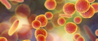
Pneumonia is an acute infectious inflammation with the presence of foci of infiltration in the lung tissue and intra-alveolar exudation. It is the most common among inflammatory lung diseases. The incidence rate increases with age. In elderly and senile people, community-acquired pneumonia occurs in 22-51%, a pattern emerges - the older the patient, the higher the risk of pneumonia and its complications. Among patients, men predominate by 10%.
Causes of the disease
A focus of inflammation can form in any area of the lung. Unlike the left-sided form, lower lobe pneumonia on the right is more common because the bronchus is located at a slight angle, so the air movement is somewhat worse. Therefore, stagnant processes and poor mucus discharge more often provoke the onset of disease on the right side.
Swelling of the lung tissue and filling it with exudate in a small area or segment is called lobar pneumonia. In the case when several such segments are affected, pneumonia will be called “lobar pneumonia”.
Note. Diagnosing chronic forms in this case is not entirely correct, since left-sided lower lobe pneumonia always occurs acutely and the diagnosis “chronic” is erroneous.
The medical history of a person with such a disease should contain information about the medical history, how the disease began, and the causes of infection.
Among the latter may be:
- aspiration from the stomach or pharynx;
- infectious infection of the respiratory tract;
- aerosol route of infection, when the infection occurs through the use of air conditioners and inhalers; the pathogen Legionella is often the cause of the disease;
- when pathogenic microflora enters the lungs through the bloodstream, for example, with vascular thrombosis in the pelvis, sepsis can develop.
What causes inflammation of the pulmonary parenchyma in children?
Inflammation of the pulmonary parenchyma in children on the right is caused in most cases by viruses. As a result, doctors observe the peak of disease epidemics in the autumn-winter period.
With strong immunity against the background of influenza, inflammation of the pulmonary parenchyma on the right is rarely detected in children.
When the body's protective abilities are insufficient, bacteria penetrate into the damaged bronchial epithelium, causing the pathological process.
Features of viral pneumonia in a child:
- Acute process;
- Fever;
- Dry cough in the initial stages of the disease;
- Extensive damage to lung tissue.
The above-described specificity of pathological changes in the right lung in children led to the fact that the World Health Organization (WHO) created a unique medical field - pediatric pulmonology. She is engaged in the isolation of specific pathogens and the development of principles for the treatment of childhood pneumonia.
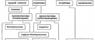
In Russia, the ICD 10 Classification is used purely empirically. Domestic doctors rely on the established clinical classification of the disease. It involves identifying x-ray morphological forms of the disease: focal, segmental, lobar, total.
In a young child, pneumonia quickly flows from one form to another. Without proper therapy, a lobar lesion develops from focal inflammation within a few days. These facts are presented so that readers understand that the disease cannot be neglected, since its consequences are dangerous not only to health, but also threaten the life of the child.
Etiology and pathogenesis
Left-sided lower lobe pneumonia
Pneumonia develops when defense mechanisms are weakened, most often in very young and fairly elderly people as a result of hypothermia or infection. Often the disease is an aggravation of a long-term cold or advanced bronchitis that has not been treated.
The presence of chronic diseases can act as a risk factor and determine susceptibility to illness. Most often, pneumonia is recorded in people with diabetes, heart and kidney failure, and circulatory pathologies.
Secondary pneumonia can occur against the background of measles, scarlet fever, meningitis and other diseases. People from a lower social class, alcoholics, and smokers are more prone to the disease.
Left-sided lobar pneumonia is an infectious disease; the table shows the main groups of pathogenic microflora that cause it.
Table. Infectious agents - pathogens of pneumonia:
| Infectious group | Most common pathogens | Photo of the group representative |
| Bacteria | Among the gram-positive ones are pneumo-, staphylo-, and streptococci. Gram-negative - intestinal, Haemophilus influenzae and Frildender's bacillus, pseudomonas, enterobacteria and others. |
|
| Viruses | Influenza and parainfluenza, herpes and other respiratory viruses. |
|
| Mycoses | With very weak immunity or immunodeficiency, pathogenic fungi can develop in the lungs. |
|
| Atypical pathogens | Mycoplasma, Legionella and Chlamydia. |
|
When a microbe enters the lungs, a change in cellular immunity occurs in the area of the inflammatory process. At the beginning, the lesion is in one place, but as it progresses it affects large areas of the left lung.
At the same time, microcirculation processes in the alveoli change due to platelet aggregation and pathophysiological changes in cellular receptors that respond to the development of the inflammatory process. In bronchopulmonary tissues, cellular nutrition and gas exchange are disrupted.
This occurs due to the oxidation of the cytoplasmic elements of the plasmalemma (mainly cell membrane lipids), which causes abnormal functioning.
Classification
There are several options for ranking pneumonia into different types.
Most often we have to talk about:
- Non-hospital form, i.e. when a person fell ill with left-sided lower lobe pneumonia without being in a hospital facility. Depending on the severity of the disease, the severity of the condition, age characteristics, the presence of concomitant complications or chronic diseases, four non-hospital forms are distinguished. People falling under the first two categories can be treated on an outpatient basis, the third requires mandatory hospitalization, and the fourth requires the patient to be in intensive care or intensive care unit.
- Hospital-acquired pneumonia is diagnosed when the patient became infected in the hospital, but provided that the inflammation formed after a two-day (or longer) stay in the hospital. Based on this, the hospital form has two types: early up to five days and late, if the patient stayed in a hospital for a longer period.
How is right upper lobe pneumonia manifested and treated?
As the name of the disease suggests, right upper lobe pneumonia develops in the upper part of the right lung. This disease is difficult. The victim is overcome by shortness of breath, fever with a possible transition to a state of delirium.
Pneumonia affects the right lung much more often than the left. This is due to the anatomical structure of the body: the right bronchus is shorter and wider, so it is easier for infections to spread through it.
Pneumonia is another name for pneumonia. The disease was very dangerous until the discovery of penicillin. At this time, the disease is well treated, especially if diagnosed at the beginning of its development. But about 5 percent of those infected still die to this day. Therefore, pneumonia must be taken seriously.
How does upper lobe pneumonia manifest?
Diseases of an infectious-inflammatory nature are difficult to distinguish from each other, so the sick person does not always understand whether he just has a cold or whether a more serious illness is already beginning to develop.
With right-sided pneumonia, the inflammatory process develops in the right lung. Accordingly, the left side affects the left.
Pneumococcus and Klebsiella bacteria are to blame for the development of the disease. The human body into which they enter, for example, by airborne droplets, does not immediately react to their presence. Pathogens linger for some time on the mucous membranes, for example, the nose or larynx. The disease has not yet begun to overcome a person, but his immune system understands that foreign organisms have appeared, and it is time to prepare to fight them.
If for some reason the immune system weakens, bacteria move from the mucous membranes to the lungs. Here they actively reproduce. The cause of decreased immunity may be hypothermia, a cold, or repeated contact with a sick person.
Upper lobe pneumonia differs from others in the severe course of the disease. The patient's health deteriorates very sharply. He feels a prolonged fever. Possible delirium. The whole body is poisoned, the health of other systems is disrupted.
Elderly people and those who have problems with the immune system are most susceptible to this disease.
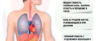
Symptoms of the disease resemble fever:
- the patient is constantly shivering;
- he experiences muscle aches;
- he is plagued by severe headaches.
Upper lobe pneumonia appears suddenly. If in the evening the patient experienced some discomfort, it was so insignificant that he did not in any way connect it with a possible illness. And in the morning there are difficulties with breathing: it becomes shallow. A person is even afraid to take a deep breath, as it causes pain. A painful cough begins, dry and exhausting.
The resulting temperature does not go down, and if it is possible to do this, it is only for a short time. The following symptoms gradually appear:
- digestive problems, nausea;
- the whites of the eyes turn yellow due to the destruction of red blood cells in the blood;
- rashes appear on the lips;
- at rest, shortness of breath does not stop.
Sometimes a condition similar to that of meningitis occurs. Sometimes the patient is haunted by hallucinations.
Diagnosis and treatment of right-sided pneumonia
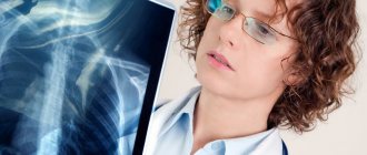
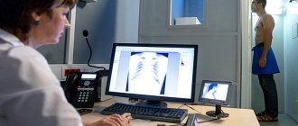
The presence of pneumonia is determined using radiography.
At the appointment, the doctor examines the patient and interviews him. The specialist must listen to wheezing in the lungs, because this method remains the best in identifying pathology to this day. Even with modern technology, an experienced doctor will be able to correctly hear and understand the nature of the noise in the respiratory organs.
An x-ray shows how much of the lung is affected by inflammation. This is a very objective method. It is also good because in the absence of an experienced doctor it will always help to make a correct diagnosis.
Other diagnostic methods are laboratory blood tests and bacterial culture of sputum. Changes in leukocytes, ESR, etc. are determined from the blood. And sputum shows the type of pathogen. But this analysis is ready only a few days after the biomaterial is collected. Doctors do not wait for results, but prescribe treatment immediately using a standard regimen. And after receiving data from the laboratory, additional drugs are prescribed.
Right-sided pneumonia of the upper lobe is fraught with dangerous complications if left untreated. It happens that pathology leads to disability of the patient and even death.
Treatment of the disease is carried out using antibacterial therapy. Penicillins, ampicillins and much more are used depending on the type of pathogen. The patient takes them strictly as prescribed by the doctor. The entire treatment process is controlled by radiographs and laboratory tests.
It happens that the patient is admitted to the hospital in serious condition. In this case, treatment begins with normalizing the patient's condition. This may include ventilation of the lungs using artificial means, adjusting the balance of water and salts, restoring blood pressure and other measures.
The doctor may prescribe antipyretic, antiallergic, anti-inflammatory, and painkillers.
Without fail, the patient takes medications that help strengthen or correct immunity.
All medications can be supplemented with therapeutic massage, physiotherapeutic procedures, and physical therapy.
Pneumonia is a serious disease, so all treatment procedures are prescribed only by a doctor. Often the patient needs appropriate care and special procedures. Therefore, upper lobe pneumonia is most often treated in a hospital setting.
Symptoms
Coughing with phlegm at a high temperature is a sign of pneumonia
Left-sided lower lobe pneumonia in people of different ages has different symptoms. The signs are similar in that, regardless of the person’s age, the body temperature is usually high, a productive cough with difficult to clear sputum, which may be accompanied by blood, a severe cough may be accompanied by pain, especially with a very deep breath.
Signs in children
In children, symptoms indicating pneumonia are:
- rhinitis;
- productive cough;
- febrile temperature;
- increased breathing rate;
- loss of appetite;
- headache.
Note. Children under one year of age may have pneumonia without obvious symptoms. This poses a particular danger, since as the disease progresses, the consequences are fraught with serious complications.
Symptoms in adults
When suffering from left-sided lower lobe pneumonia in adults, the following symptoms occur:
- febrile fever for more than three days;
- chills;
- deep, intense cough with sputum;
- shortness of breath at rest and during physical work;
- weakness;
- cardiopalmus;
- headache.
Often, other signs are added to the indicated symptoms, which often happens in the presence of another pathology. These symptoms may indicate a wide range of diseases, so to clarify the diagnosis, you should definitely consult a doctor (general practitioner, pulmonologist).
The specialist will conduct an examination, prescribe special tests and appropriate treatment. The differential diagnosis should exclude bronchitis, bronchial asthma, and tuberculosis.
Note. Bronchopulmonary syndrome with left-sided lower lobe pneumonia has the following characteristics: pain when inhaling from the lower left side of the chest increases with coughing or strong inhalation, the cough can be dry, which is characteristic of the onset of the disease, or with viscous thick sputum, sometimes with blood, shortness of breath is present, The intercostal and thoracic muscles are involved in the respiratory act.
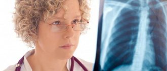
Diagnostics
Pneumonia on CT image
In most cases, an experienced pulmonologist makes the correct diagnosis at the stage of conversation and physical examination, but the patient should be sent for an X-ray examination to confirm and evaluate those processes that are visible only on the negative.
Diagnostic instructions:
- Physical examination . With left-sided lower lobe pneumonia, the patient has pallor of the skin, swelling of the wings of the nose when breathing, and the participation of the intercostal muscles in the act of breathing is noticeable. During auscultation (listening to the lungs), sonorous fine-bubble rales and a dull percussion sound, hard bronchial breathing are heard, which will be localized in the lower part of the left lung, the nature of the sound depends on the stage of the disease.
- A blood test helps determine the nature of the pathogen. With pneumonia, there may be an increased number of leukocytes in the peripheral blood (leukocytosis> 10-12•10⁹/l with a shift in the leukocyte formula to the left), which indicates a bacterial infection; if the norm of lymphocytes is exceeded, they speak of a viral infection. An increase in ESR is evidence of an inflammatory process.
- Sputum examination allows you to find out the type of pathogen and exclude tuberculosis.
- X-ray . As a rule, a frontal examination of the chest is prescribed, but often an X-ray examination is carried out in two planes - direct and lateral. This helps to determine the size of the infectious focus of inflammation and its location. Inflammation is displayed on the image in the form of uneven and unclear darkening of a delineated spotty nature. X-rays are taken twice - at the beginning and after treatment (for control).
- Computed tomography (CT) is the most accurate method, which also uses X-rays, but the level of radiation is several times higher. CT allows you to accurately determine the type of infiltration of the lung tissue and accurately determine the stage of the disease. The cost of this research is significant. Computed tomography is prescribed by a doctor according to indications.
- Spirometry . A spirograph is a device that analyzes the volume and speed of inhalation and exhalation. Based on the data obtained, the presence and intensity of obstruction and respiratory rate can be determined.
- Bronchoscopy is performed under anesthesia to determine the diagnosis in difficult situations. The method allows you to perform a tissue biopsy and distinguish cancer or tuberculosis from pneumonia.
- For atypical pneumonia, ELISA is prescribed - an enzyme-linked immunosorbent test (ELISA), and PCR is used to identify the type of infection.
X-ray is the most reliable way to diagnose
Community-acquired pneumonia: diagnosis, treatment. Prevention of community-acquired pneumonia
Community-acquired pneumonia is one of the most common infectious diseases of the respiratory tract. Most often, this disease is the cause of death from various infections. This occurs as a result of a decrease in people’s immunity and the rapid adaptation of pathogens to antibiotics.
What is community-acquired pneumonia?
This is an infectious disease of the lower respiratory tract. Community-acquired pneumonia in children and adults develops in most cases as a complication of a viral infection. The name of pneumonia characterizes the conditions under which it occurs. A person falls ill at home, without any contact with a medical facility.
Pneumonia in an adult
Adults most often suffer from pneumonia as a result of bacteria entering the body, which are the causative agents of the disease. Community-acquired pneumonia in adults does not depend on geographical areas and socio-economic relations.
Throughout life, a person's respiratory tract and lungs are constantly exposed to pathogens: viruses and parasites. On the way to the lungs, bacteria encounter protective barriers, which are represented by the upper respiratory tract and oropharynx. If these barriers are overcome by pathogenic organisms - bacteria, viruses and fungi, an infection begins to develop.
What is pneumonia like?
This disease is conventionally divided into three types:
- Mild pneumonia is the largest group. She is treated on an outpatient basis, at home.
- The disease is of moderate severity. Such pneumonia is treated in hospital. The peculiarity of this group is that most patients have chronic diseases.
- Severe form of pneumonia. She is being treated only in the hospital, in the intensive care unit.
Community-acquired pneumonia occurs:
- Focal. A small area of the lungs is inflamed.
- Segmental. Damage to one or several parts of the organ is typical.
- Share. Some part of the organ is damaged.
- Total. The entire lung is affected.
Community-acquired pneumonia can be unilateral and bilateral, right-sided and left-sided.
Symptoms
- Body temperature rises.
- Chills and weakness appear.
- Performance and appetite decrease.
- Sweating appears, especially at night.
- Head, joints and muscles hurt.
- Consciousness becomes confused and orientation is disturbed if the disease is severe.
- Pain in the chest area.
- Herpes may appear.
- Abdominal pain, diarrhea and vomiting.
- Shortness of breath that occurs during physical activity. This does not happen when a person is at rest.
Causes
Community-acquired pneumonia develops when microbes enter a weakened human body and cause inflammation. The causes of the disease are as follows:
- Hypothermia of the body.
- Viral infections.
- Concomitant diseases: diabetes, heart, lungs and others.
- Weakened immunity.
- Excessive consumption of alcoholic beverages.
- Prolonged stay on bed rest.
- Postponed surgeries.
- Elderly age.
Pathogens
- Pneumococci (most often the cause of the disease).
- Staphylococci.
- Atypical pathogens: mycoplasmas and chlamydia.
- Klebsiella.
- Viruses.
- Pneumocystis.
- Escherichia coli.
- Haemophilus influenzae.
Diagnostics
During the examination, it is very important to identify and evaluate the clinical symptoms of the disease, such as fever, chest pain, cough with sputum. Therefore, if a person has community-acquired pneumonia, a medical history must be created for each patient. In it, the doctor writes down all the patient’s complaints and prescriptions. To confirm the diagnosis, a radiation examination is performed: chest x-ray. Clinical manifestations of community-acquired pneumonia are:
- Cough with the release of mucopurulent sputum, which contains streaks of blood.
- Chest pain when breathing and coughing.
- Fever and shortness of breath.
- Trembling voice.
- Wheezing.
Sometimes the symptoms differ from those typical for a given disease, which makes it difficult to make a correct diagnosis and determine a treatment method.
Radiation examination
The patient is prescribed an x-ray if he has community-acquired pneumonia. Diagnosis using the radiation method involves examining the organs of the chest cavity in the anterior part. The picture is taken in frontal and lateral projection. The patient undergoes an X-ray examination as soon as he sees a doctor, and then half a month after treatment with antibacterial agents has begun. But this procedure can be performed earlier if complications arise during treatment or the clinical picture of the disease changes significantly.
The main sign of community-acquired pneumonia during an X-ray examination is compaction of the lung tissue; darkening is detected in the image. If there are no signs of compaction, then there is no pneumonia.
Lower lobe right-sided pneumonia
Many patients go to the hospital when they are bothered by symptoms such as shortness of breath, cough accompanied by mucous sputum, fever up to 39 degrees, pain with a tingling sensation on the right side under the rib. After listening to the patient’s complaints, the doctor examines him, listens and probes where necessary. If there is a suspicion that the patient has community-acquired right-sided pneumonia, which, as a rule, is much more common (which is why we pay special attention to it), he is prescribed a full examination:
- Laboratory tests: general, clinical and biochemical blood tests, urine and sputum tests.
- Instrumental studies, which include chest x-ray, fibrobronchoscopy and electrocardiogram. The shape of the darkening on the x-ray image allows you to clarify the diagnosis, and fiberoscopy helps to identify the involvement of the bronchi and trachea in the process of inflammation.
If the results of all tests confirm that the patient has right-sided community-acquired pneumonia, the medical history is supplemented. Before starting therapy, the results of studies for all indicators are recorded in the patient’s chart. This is necessary in order to make adjustments as necessary during treatment.
Laboratory and instrumental studies may show inflammation of the lower right lobe of the lung. This is a different story of the disease. Community-acquired lower lobe pneumonia - this will be the diagnosis. When it is accurately established, the doctor prescribes treatment individual for each patient.
How to treat community-acquired pneumonia?
Patients with this diagnosis can be treated both in a hospital and at home. If a patient has community-acquired pneumonia, a medical history is required, regardless of the place of treatment. Patients undergoing outpatient treatment are divided into two groups. The first category includes people under 60 years of age who do not have concomitant diseases. The second category includes people over 60 or people with concomitant diseases (of any age). When a person has community-acquired pneumonia, treatment is carried out with antibacterial drugs.
For patients of the first group the following are prescribed:
- "Amoxicillin" dosage of 0.5-1 g or "Amoxicillin/clavulanate" - 0.625 g at a time. Taken 3 times a day.
- An alternative to these drugs may be: Clarithromycin or Roxithromycin in dosages of 0.5 g and 0.15 g, respectively. Take twice a day. Azithromycin may be prescribed, which is taken once a day in an amount of 0.5 g.
- If there is a suspicion that the disease is caused by an atypical pathogen, the doctor may prescribe Levofloxacin or Moxifloxacin 0.5 g and 0.4 g, respectively. Both drugs are taken once a day.
If patients of the second group have community-acquired pneumonia, treatment is carried out using the following drugs:
- Amoxicillin/clavulanate is prescribed three times a day, 0.625 g or twice a day, 1 g; Cefuroxime should be taken in the amount of 0.5 g at a time, twice a day.
- Alternative drugs may be prescribed: Levofloxacin or Moxifloxacin, 0.5 g and 0.4 g, respectively, once a day orally. Ceftriaxone is prescribed 1-2 g intramuscularly, also once a day.
Treatment of the disease in children
Community-acquired pneumonia in children with an uncomplicated form of the disease, depending on age, is treated with the following drugs:
- Children under 6 months are prescribed: “Josamycin” twice a day for a week at the rate of 20 mg per kilogram of body weight. Maybe Azithromycin - the daily dose should not exceed 5 mg per kilogram of body weight, the duration of treatment is 5 days.
- Children under 5 years of age are prescribed Amoxicillin 25 mg/kg orally twice a day, treatment duration is 5 days. They may prescribe Amoxicillin/clavulanate at a dose of 40-50 mg per kilogram of body weight or Cefuroxin Axetil at a dosage of 20-40 mg/kg, respectively. Both drugs are taken twice a day, the duration of treatment is 5 days.
- Children over 5 years of age are prescribed Amoxicillin at a dosage of 25 mg/kg in the morning and evening. If there is a suspicion of atypical pneumonia, prescribe Josamycin orally, increasing the dosage to 40 mg/kg per day for a week or Azithromycin according to the scheme: 1 day - 10 mg/kg, then 5 mg/kg for 5 days. If there is no positive result in treatment, you can replace Amoxicillin at the rate of 50 mg/kg once a day.
Preventive measures to prevent the disease
Prevention of community-acquired pneumonia is carried out using pneumococcal and influenza vaccines. If necessary, they are administered simultaneously, only in different hands. A 23-valent unconjugate vaccine is used for this purpose. It is introduced:
- People who are over 50 years old.
- Persons living in nursing homes.
- Adults and children with chronic diseases of the lungs, heart and blood vessels or who are under constant medical supervision.
- Children and adolescents (from six months to adulthood) who have been taking aspirin for a long time.
- Pregnant women in the 2nd and 3rd trimesters.
- Doctors, nurses and other staff of hospitals and outpatient clinics.
- Employees of patient care departments.
- Family members of those people who are at risk.
- Medical workers caring for patients at home.
Prevention of community-acquired pneumonia is:
- A healthy lifestyle that includes physical exercise, regular long walks in the fresh air, and active recreation.
- A balanced healthy diet with a standardized content of proteins, vitamins and microelements.
- Annual vaccination of children and adults against influenza, which is done before the onset of the cold season. Very often the flu causes complications. A person gets sick with pneumonia, which has a complicated course.
- Life without hypothermia and drafts.
- Daily cleaning and ventilation of the premises.
- Frequent hand washing and rinsing of nasal passages.
- Limit contact with ARVI patients.
- During the period of massive spread of infection, take honey and garlic. They are excellent immunostimulating agents.
- If you or your child gets sick with the flu, do not self-medicate, but call a doctor.
syl.ru
Treatment
Pneumonia cannot go away on its own and requires complex treatment. You should not treat it yourself because in this case you can get quite serious complications.
Treatment in mild cases is carried out at home, and in difficult situations hospitalization and daily medical supervision are required. Most often, children and old people, as well as those with chronic diseases in the later stages, are admitted to the hospital.
Etiotropic treatment
Regardless of the type of pneumonia, the patient is required to be prescribed antibiotics. Since bacteria have acquired resistance to some antibacterial drugs of different generations, it is possible to prescribe several drugs at the same time.
The duration of treatment depends on the degree of the disease and the body’s response to the proposed therapy. As a rule, the average duration of treatment is 10-14 days.
Antibiotic use ends three days after the fever disappears. Antiviral drugs are prescribed if pneumonia is caused by a viral infection.
The note. For all types of pneumonia, it is recommended to prescribe vitamin-mineral complexes and immunomodulators.
Symptomatic treatment
Lazolvan is an effective mucolytic
To relieve symptoms accompanying the disease, the following are indicated:
- expectorants (ACC or others) and mucolytics (lasolvan or others) to enhance the secretion, dilute and facilitate the discharge of sputum;
- antipyretics (paracetamol or others) are indicated at temperatures above 38 degrees;
- analgesics for severe pain during coughing.
If there are concomitant diseases, the patient is prescribed additional drug therapy according to the established diagnosis. It is important to adhere to the treatment regimen and dosage prescribed by your doctor, although the recommended doses in the package insert may differ.
You should not use medications that suppress cough, as this will make it difficult to clear sputum and delay recovery. You should drink more than two liters of fluid per day, which helps thin the mucus and remove harmful substances from the body, especially with a significant medication load.
Additional treatment
Breathing exercises
When treating left-sided lower lobe pneumonia, the best results can be achieved with an integrated approach. It is recommended to use folk remedies simultaneously with drug therapy.
Good fortified decoctions or teas with rose hips, raspberries, lingonberries, mint and other herbs. At the pharmacy or at the market you can buy a breast mixture with coltsfoot, marshmallow, thyme and oregano, which has expectorant properties and soothes coughs.
You can make honey compresses on the chest, and if you are not allergic, do inhalations with eucalyptus, propolis, calendula or chamomile.
In addition, auxiliary treatment methods are recommended that will speed up the healing process:
- exercise therapy;
- breathing exercises (from the second week of treatment);
- physiotherapy;
- breathing in rarefied air or visiting salt caves;
- acupressure (must be performed exclusively by a medical massage therapist).
These types of therapy can be used after recovery, which will have a preventive effect.
The note. In the recent past, cupping and mustard plasters were used to treat pneumonia; currently, these types are not used due to discomfort and low effectiveness.
Hardening helps avoid respiratory diseases and pneumonia
Symptoms and treatment of left-sided lower lobe pneumonia
Left-sided lower lobe pneumonia refers to focal diseases, the occurrence of which is explained by the presence of a virus, fungus or bacteria that affects the lower respiratory tract. Left-sided pneumonia is much less common than right-sided pneumonia. This is explained by the peculiarity of the structure of the bronchi. This disease can be transmitted by airborne droplets or be a consequence of a previous illness, surgery, or decreased immunity while taking certain medications. Often, chronic respiratory diseases can cause pneumonia.
Symptoms of the disease
At the initial stage of its development, left-sided lower lobe pneumonia does not differ from other types of pneumonia, since the symptoms of the disease are similar. This pathological process may initially be completely asymptomatic, which is very dangerous, especially for newborns or small children.
Characteristic signs of pneumonia, which is localized in the lower lobes of the left lung, are:
- elevated body temperature (more than 38 degrees);
- chills;
- headache;
- dizziness;
- pain in the left lung or left chest (where the pathological process occurs);
- increasing chest pain when inhaling;
- increased sweating;
- nausea;
- vomit;
- cough, sometimes with sputum that may be streaked with blood.
It is impossible to diagnose left-sided lower lobe pneumonia based on symptoms alone. To find out the cause of the inflammatory process, in addition to a general examination, which includes listening to the patient’s chest for the presence of wheezing and uncharacteristic bronchial breathing, the doctor additionally prescribes a blood and sputum test (if it is separated). The most common and effective way to detect pneumonia is radiography.
If sputum does not come out on its own during a cough, a sample is taken for analysis using bronchoscopy. An analysis such as sputum examination will reveal the true cause of the disease, since quite often completely other diseases can be hidden under the guise of pneumonia.