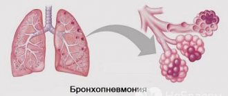
Lobar pneumonia is an acute infectious disease in which one or more lobes of the lung are affected, fibrinous effusion appears in the alveoli, and fibrinous deposits appear on the pleura. Croupous pneumonia affects mainly adults. The disease is characterized by a pronounced clinical picture and symptoms of intoxication. Patients with lobar pneumonia are admitted to the therapy clinic.
To examine patients at the Yusupov Hospital, doctors use modern equipment from leading European, American and Japanese companies. Pulmonologists use European treatment protocols and take an individual approach to the choice of treatment method for each patient. Medicines are administered through the digestive tract, intramuscularly, intravenously and by inhalation. Thanks to complex treatment, the length of stay of patients in hospital is reduced.
Causes and stages of lobar pneumonia
The causative agent of lobar pneumonia is pneumococcus types I-IV. Sometimes lobar pneumonia is caused by Friedlander's diplobacillus. In most cases, acute pneumonia begins in complete health and in the absence of contact with patients with pneumonia. This suggests that the development of lobar pneumonia occurs under the influence of microorganisms located in the upper respiratory tract. An important factor provoking the development of pneumonia is decreased immunity.
There are 4 stages of development of lobar pneumonia. The hot flash stage is characterized by severe hyperemia of the lung tissue and congestion in the capillaries. Its duration is from 12 hours to 3 days. In the stage of red hepatization, blood cells exit through the walls of capillaries and small veins due to a violation of their tone and permeability, and effusion appears in the alveoli. The exudate is rich in fibrin, which coagulates and prevents air from entering the alveoli. The lungs become dense, similar to the liver. The duration of the second stage is from 1 to 3 days.
In the gray hepatization stage, the release of red blood cells through the vascular wall stops. In addition to fibrin, the effusion contains alveolar epithelium and leukocytes. Due to the abundant content of leukocytes, the affected lung has a characteristic gray-greenish tint. The duration of this stage varies from 2 to 6 days. During the resolution stage, fibrin dissolves and liquefies under the influence of proteolytic enzymes. This stage is the longest.
If treatment is prescribed in a timely manner and it is effective, the pathological process may lose its characteristic cyclicity and break off in the early stages of development. If the resorption of exudate is impaired, complications of lobar pneumonia develop. Overgrowth of connective tissue in the lesion may occur - carnification and cirrhosis of the lung. Sometimes purulent melting occurs, and pneumonia is complicated by an abscess or gangrene of the lung.
With lobar pneumonia there are phenomena of dry pleurisy with fibrinous layers and the development of adhesions. In the case of lymphogenous generalization by microorganisms, purulent mediastinitis and pericarditis occur. With the hematogenous spread of bacteria, metastatic ulcers appear in the brain, purulent meningitis, peritonitis, acute ulcerative or polypous-ulcerative endocarditis, and purulent arthritis develop.
Etiology of the disease
In most cases, the main cause of lobar pneumonia is a bacterium called Friendler's bacillus.
However, this form of pneumonia can also be caused by typical pathogens (streptococcus, pneumococcus, Klebsiella, staphylococcus and E. coli) against the background of reduced immunity.
Many people confuse lobar pneumonia with focal pneumonia. In fact, these are two different forms of pneumonia. The main reason for the development of the disease is the pathogenic activity of various microorganisms.
The main pathogens of the disease penetrate the lung tissue in several ways:
- Airborne. This is the most common route of penetration of microorganisms in lobar, focal and other types of pneumonia.
- Hematogenous.
- Lymphogenic.
Symptoms of lobar pneumonia
Lobar pneumonia has an acute onset. In patients in full health, body temperature rises to 39 ° C, chills and chest pain appear. In the initial stage of the disease, the cough is dry, then it becomes productive, with the release of “rusty” sputum. There is severe shortness of breath, the chest on the affected side lags behind when breathing.
In the initial phase of inflammation, percussion reveals a dull tympanic sound over the lesion. During auscultation, harsh breathing with prolonged exhalation, mild crepitus, and wet and dry rales in a limited area are heard. In the compaction phase of lobar pneumonia, the following symptoms appear:
- a sharp increase in vocal tremors, bronchophony during palpation of the chest;
- with percussion – dull sound;
- vesicular breathing is not audible, crepitus disappears, and pleural friction noise is often heard.
In the resolution phase, vocal tremors gradually normalize, bronchophony disappears, and abundant, sonorous crepitus appears over a long period of time. Loud fine-bubble rales are heard, bronchial breathing gradually gives way to harsh and then vesicular breathing.
When examining the cardiovascular system, a rapid pulse is determined. In the case of severe lobar pneumonia, it is poorly filled, arrhythmic, blood pressure is reduced, and the heart sounds are muffled.
Diagnosis of lobar pneumonia
Pulmonologists at the Yusupov Hospital draw up an individual examination plan for a patient with lobar pneumonia, which includes:
- collection of blood, urine, sputum samples;
- biochemical blood test (total protein, electrophoresis of serum proteins, bilirubin, fibrinogen);
- bacteriological culture of sputum for sensitivity of flora to antibiotics;
- Electrocardiography.
The leading method for diagnosing lobar pneumonia is an X-ray examination of the chest organs. X-ray of the lungs is performed in two projections. The hot flash stage is characterized by an intensification and enrichment of the pulmonary pattern due to hyperemia. Transparency is normal or slightly reduced. The root of the lung on the affected side is somewhat expanded, its shadow is uniform. When the process is localized in the lower lobe, a decrease in excursion of the corresponding dome of the diaphragm is recorded.
During the hepatization stage, radiologists determine an intense decrease in the transparency of the lung tissue, corresponding to the affected area. The size of the affected area of the lungs is normal or slightly enlarged. There is a slight increase in shadow intensity towards the periphery. In the medial sections of the darkening, “stripes of enlightenment” are visible. The root of the lung on the affected side is expanded, its shadow is uniform. The adjacent pleura is compacted.
The resolution stage is characterized by a decrease in the intensity of the shadow of the affected area. The shadow is fragmented, it decreases in size, the root of the lung is expanded.
Patients with lobar pneumonia undergo a study of the function of external respiration, and according to indications, a pleural puncture is performed. Multislice computed tomography is performed in the following situations:
- in the presence of obvious clinical signs of pneumonia and no changes on the radiograph;
- if, during examination of a patient with suspected lobar pneumonia, atypical changes are revealed (obstructive atelectasis, abscess or pulmonary infarction);
- recurrent pneumonia, in which infiltrative changes occur in the same lobe (segment);
- prolonged pneumonia, pneumonia, in which infiltrative changes in the lung tissue do not resolve within 4 weeks.
In the absence of a productive cough, doctors at the Yusupov Hospital perform fiberoptic bronchoscopy, transtracheal aspiration, and transthoracic biopsy. If the patient has pleural effusion and there are conditions for safe pleural puncture, pleural fluid is examined.
Diagnosis
For pneumonia, diagnosis is carried out in a hospital. Measures taken at home will not be enough in most cases. The signs that appear with pleuropneumonia differ in specificity. Therefore, the examination does not take much time. To identify an accurate diagnosis, the following methods are used:
- percussion;
- auscultation;
- bronchoscopy;
- lung tissue biopsy;
- laboratory research;
- X-ray.
Using the latter method, the affected area, interstitial changes, and the nature of darkening are determined. With pneumonia, the vascular pattern increases, the density of functional tissue increases, and the roots of the bronchial tree expand. The clinical examination scheme includes CBC, OAM, biochemical blood test, sputum microscopy, culture, and serological tests.
Blood analysis
Treatment of lobar pneumonia
Patients with lobar pneumonia are admitted to a therapy clinic. The wards have a central ventilation system. Each room is equipped with air conditioning to ensure comfortable temperature conditions. In severe cases of pneumonia, patients are treated in the intensive care unit, which is equipped with modern cardiac monitors that allow continuous monitoring of the functioning of the respiratory and cardiovascular systems and determination of the oxygen content in the blood. If necessary, patients undergo artificial ventilation using stationary and portable ventilators.
For lobar pneumonia, 2 antibiotics are prescribed simultaneously (benzylpenicillin, ampicillin, amoxicillin clavulanate, cefuroxime, cefotaxime, ceftriaxone). One antibiotic is administered intravenously, the second intramuscularly. Doctors at the Yusupov Hospital provide comprehensive treatment for lobar pneumonia, including:
- immunoreplacement therapy (intravenous administration of fresh frozen or native plasma, immunoglobulin).
- correction of microcirculatory disorders (heparin, rheopolyglucin);
- correction of dysproteinemia (albumin, retabolil);
- detoxification therapy (saline solutions, 5% glucose solution);
- oxygen therapy (oxygen is supplied through a mask or catheters);
- corticosteroid therapy (prednisolone and other glucocorticoids).
In cases of severe intoxication, doctors at the Yusupov Hospital perform plasmapheresis in patients with lobar pneumonia. Antioxidant therapy consists of ingesting ascorbic acid and rutin. Eufillin, Atrovent, Berodual have a bronchodilator effect. Expectorant drugs (lazolvan, acetylcysteine) improve the drainage function of the bronchi. Expectorants and bronchodilators during intensive care are administered through a nebulizer.
Prevention
Prevention of the disease includes strengthening the body, hardening it, and quitting smoking. You need to lead an active lifestyle, if possible, play sports, run in the morning. After 65 years of age, mandatory vaccination with pneumococcal vaccine.
To prevent lobar pneumonia in children, it is necessary to strengthen children's immunity. You need to take your baby for walks more, monitor his diet, and include fresh vegetables and fruits in his diet. It is also necessary to exclude hypothermia of the child’s body. The child needs to be provided with adequate sleep and rest at least 8 hours a day. If the baby gets sick, it is necessary to carry out timely therapy.
Thus, this is a rather dangerous pathology that can lead to various complications. Lobar pneumonia is especially difficult in children, whose immunity is still quite weak. Therefore, it is necessary to treat it in time, and to prevent it, it is recommended to carry out special preventive measures.
Physiotherapeutic methods of treating lobar pneumonia
To treat patients with lobar pneumonia, pulmonologists at the Yusupov Hospital widely use physiotherapeutic procedures. Ultrasonic aerosol inhalation is used to introduce mucolytic antibiotics and heparin into the respiratory tract. Decimeter wave treatment is used almost immediately after the fever has passed.
Pulsed UHF therapy is performed for patients with reduced immunity. Antibacterial drugs are introduced into the respiratory system using magnetophoresis. Massage, physical therapy and breathing exercises improve the drainage function of the bronchi.
If you suspect lobar pneumonia, call the Yusupov Hospital, where patients are hospitalized 24 hours a day, 7 days a week. Doctors, without waiting for the results of a bacterial examination of sputum, begin antibacterial therapy. Pulmonologists use individual treatment regimens for pneumonia with effective, safe medications.
Author
Dmitry Nikolaevich Staroverov
Head of the department of anesthesiology and resuscitation with intensive care and intensive care wards - anesthesiologist-resuscitator
Forecast
With timely diagnosis and treatment of lobar pneumonia, the prognosis for recovery is favorable. Otherwise, it worsens significantly, especially in people with immunodeficiency.
The use of antibiotics reduces the risk of complications and death. Such cases are possible in elderly patients suffering from heart failure, as well as with complications of the meningeal type. Late therapy, as well as incorrectly selected dosages of medications, can lead to a protracted process and transition of the disease to the chronic stage.
The insidiousness of lobar pneumonia lies in the difficulty of diagnosis at an early stage of its development. The main task is to relieve inflammatory symptoms as quickly as possible, prevent oxygen starvation of the brain and suppress bacterial activity.
Our specialists
Alexander Vyacheslavovich Averyanov
Doctor of Medical Sciences, Professor, Doctor of the Highest Qualification Category
Make an appointment
Svetlana Anatolyevna Zhabina
General practitioner, pulmonologist, candidate of medical sciences, first category doctor
Make an appointment
Vladimir Vladimirovich Kvasovka
Deputy General Director for Medical Affairs, general practitioner, gastroenterologist, candidate of medical sciences
Make an appointment