Pneumococcal infection is very common. According to statistics, over one and a half million people around the world die from it every year. Moreover, more than 50% of the victims are children of primary preschool age and newborn babies. More often, diseases provoked by pneumococci are diagnosed when the immune system is weakened. They are treatable.
Total information
Pneumococcal pneumonia is one of the most common forms of bacterial pneumonia - in their structure, it occurs in every third person diagnosed with bacterial pneumonia.
Young children and the elderly are most often affected.
Males get sick somewhat more often than females - this is explained by the fact that men smoke more often than women, and smoking is considered one of the significant factors contributing to the occurrence of the disease described. In addition, men more often work in workplaces with harmful conditions for the respiratory system - this is also a contributing factor.
note
For every woman with bacterial pneumonia, there are three men with the same diagnosis.
Causes and risk factors of the disease
Pneumococcal inflammation is caused by gram-positive pneumococcal bacteria. Their sources are carriers or infected people. Carriage is typical for a quarter of the adult population and half of children attending educational institutions.
In addition, a high percentage of carriage is observed among medical workers, large manufacturing enterprises, and people living in camps or barracks. Most often, pathology caused by pneumococci affects children under five years of age and elderly people. In older people, this disease can be fatal. At risk are patients with chronic diseases of the liver, heart, lungs, immunodeficiency and after undergoing transplant surgery.
Infection occurs by contact or airborne droplets. In cold weather, the probability of the latter increases several times. Factors that worsen prognosis for a diagnosis of pneumococcal pneumonia include:
- extensive lung damage;
- smoking;
- alcoholism;
- work in hazardous production;
- cardiovascular pathologies with stagnation in the pulmonary circulation;
- chronic diseases of the respiratory system;
- chronic fatigue;
- weak immunity;
- children under 2 years of age.
Causes of pneumococcal pneumonia
The immediate cause of the development of this pathology is pneumococcus. The pathogen has a special coating (capsule) that protects it from the effects of negative factors (in particular, antibacterial agents). The characteristics of the capsule determine the persistence of the pathogen, which means its ability to cause more or less pronounced pathology. About 90 varieties of the causative agent of pneumonia have been described, about a quarter of which, due to the characteristics of the capsule, are capable of causing a particularly severe form of the described disease, since they are unhindered in the body.
note
The severity of symptoms and the severity of the pathology depend not only on pneumococcus, but also on secondary microflora, which can join, since the body is already weakened in the fight against pneumococcus. There are several gradations of such pathogens.
Depending on the ability to cause a pathological process, a secondary infection is:
- pathogenic;
- opportunistic.
The first are infectious agents that cause a pathological process in the lung tissue in any case.
The second is microflora, which lives peacefully in the body of a healthy person and does not cause him harm, but becomes aggressive in suitable conditions.
By specificity, a secondary infection that can join with pneumococcal infection is:
- nonspecific;
- specific.
In the first case, it is a pathogen that can provoke not only pneumonia, but also other infectious pathologies in other organs and tissues.
In the second case, it is an infectious agent, the presence of which determines the occurrence of a certain infectious disease, although it can also cause nonspecific pathologies (including pneumonia). A classic example is Mycobacterium tuberculosis, without which pulmonary tuberculosis does not occur.
With pneumococcal pneumonia, staphylococci, streptococci, bacteroides, intestinal, pseudomonas and hemophilus influenzae are often associated.
A number of contributing factors have also been identified, against the background of which the incidence of pneumococcal pneumonia increases. This:
- pathologies of the immune system;
- general weakening of the body, which usually occurs after operations, injuries, critical conditions, and so on;
- damage to the respiratory system of an inflammatory, degenerative-dystrophic and other nature;
- hypothermia;
- irrational antibacterial therapy;
- smoking;
- infection by the influenza virus.
Important
Half of the cases of pneumococcal pneumonia occur during influenza pandemics.
The role of Streptococcus pneumoniae in the occurrence of lung diseases
Streptococcus pneumoniae (pneumococcus) is a bacterium that causes the infectious disease pneumonia. Streptococcal pneumonia is very acute and severe. Also, streptococcal pneumococcus can cause various purulent-inflammatory diseases (for example, lung abscess), meningitis and other complications in humans.
Characteristics of the pathogen, epidemiology
Microorganisms are rounded small cells that are arranged in twos (diplococci); when they enter the human body, they can form a protective capsule around themselves, which makes them more resistant to the body’s immune system. A sick person spreads streptococcus pneumoniae (infection). The pathogen enters the body through the upper respiratory tract.
Children and weakened people suffer the disease very acutely. The incidence increases in the autumn-spring period, which is associated with environmental humidity (this makes it easier for bacteria to enter the respiratory tract, plus favorable conditions for life).
What happens at the site of introduction of streptococcus into the lung tissue or Pathogenesis
Streptococcus pneumoniae enters the body through the upper respiratory tract from a sick person or a bacteria carrier (when there are no clinical manifestations). The body's defenses (macrophages, immunoglobulins, tissue covering the upper respiratory tract) stand in the way of infection. Frail people or young children are more likely to develop streptococcal pneumonia. Having passed all the “barriers” on the way to the lung tissue, the microorganism reaches the lungs.
Streptococcal pneumococcus has special receptors that allow it to detect and attach to the lung tissue, subsequently damaging all its structures. At the site of pathogen penetration, necrosis of the lung tissue occurs—destruction.
Development of pathology
Pneumococcus enters the pulmonary parenchyma in the following ways:
- when inhaled - respiratory;
- with blood flow - hematogenous;
- with lymph flow - lymphogenous.
The main route is the respiratory route - the pneumococcus actually penetrates into the lungs. By hematogenous and lymphogenous routes, infection enters the lung tissue from chronic infectious foci, which at that time had already arisen in the body, already existing in the body:
There are four phases in the development of the described disease:
- microbial edema - with exudation into the alveoli of the lungs;
- red liver - with the appearance of fibrinogen and red blood cells in the effusion;
- gray hepatitis - with a large number of leukocytes in the pulmonary effusion;
- resolution phase – with involution (reverse development) of these disorders.
How does lung tissue change during pneumococcal pneumonia? On her part, violations such as:
- hyperemia (redness);
- swelling of tissues;
- increase in local temperature.
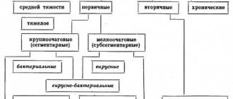
Depending on the various characteristics, the following types of pneumococcal pneumonia are distinguished:
- by localization - one- and two-sided;
- by type of lesion - focal, diffuse;
- by severity – mild, moderate, severe;
- according to the presence of complications - uncomplicated, complicated.
Symptoms of pneumococcal pneumonia
The clinical picture of the disease being described is a combination of local and general symptoms.
Local symptoms of pneumococcal pneumonia are:
- pain;
- cough;
- dyspnea.
The pain characteristics will be as follows:
- by location - in the chest;
- by distribution – irradiation is not observed;
- by nature – aching, pressing;
- by severity – medium severity;
- by occurrence - they appear almost immediately, but at first they are unexpressed, then as the pathology progresses they intensify.
The cough with this disease is usually mild, at first dry, then with the release of a small amount of viscous mucous whitish sputum.
note
With pneumococcal pneumonia, shortness of breath does not appear immediately, but as the pathology progresses. At the same time, it is difficult for the patient to inhale and exhale.
Pneumococcus is capable of poisoning the body with its own toxins, such as:
- products of their vital activity;
- exotoxins - toxic substances that pneumococcus secretes into tissues in order to adapt to them;
- endotoxins are actually the “filling” of the microbial body.
When such toxins affect the body, signs appear that indicate poisoning of the entire body. These are symptoms such as: general weakness;
- hyperthermia - an increase in body temperature to 37.8-39.0 degrees Celsius;
- chills. If it is combined with hyperthermia, then the disease is called fever;
- fatigue, malaise, weakness;
- increased fatigue when performing any work. To complete it, the patient must take more frequent and longer breaks than under normal circumstances;
- headache;
- sleep disturbance;
- loss of appetite.
In addition, specific signs of a general condition disorder may be observed, such as:
- myalgia – muscle pain;
- arthralgia – joint pain.
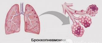
Against the background of pneumococcal infection, two scenarios of pneumonia can develop:
- lobar (lobar) pneumonia;
- focal inflammation (lobular pneumonia, bronchopneumonia).
Signs of lobar pneumonia develop quickly. The picture is polymorphic - consists of local and general symptoms - such as hyperthermia, chills, weakness, lack of appetite, headache, cough, chest pain.
Focal pneumonia mainly occurs against the background of influenza and manifests itself with the same symptoms, but less pronounced. Some of them are able to “hold on” for a long time – sometimes up to 3-4 weeks.
Symptoms and treatment
The most common signs of pneumonia in children are sudden high fever, chills, runny nose, pallor, rapid pulse, cough, shortness of breath and stabbing chest pain. In older patients, these complaints are less common; pneumonia is more likely to develop after an upper respiratory tract infection.
Worsening symptoms of the underlying illness may indicate pneumonia.
Meningitis is often expressed in infants by high fever, vomiting and seizures, and in older children by headaches. Inflammation of the middle ear causes fever and severe ear pain in children.
Otitis in children
Although otitis media causes severe ear pain and fever, clinical symptoms can vary greatly. Therefore, the disease is not always easy to diagnose. Approximately 38% of otitis media are caused by str pneumoniae. These are mainly pathogens such as Haemophilus influenzae, Moraxella catarrhalis, Streptococcus pyogenes, Staphylococcus aureus, Pseudomonas and Enterobacteriaceae.
Otitis media is treated with nasal drops, antipyretics and painkillers, as well as local heat. But drops in the ears do not make sense. In general, healing occurs quickly. Of course, complications can develop - meningitis, brain abscess. In this regard, the doctor must decide whether antibiotic therapy is required in this case.
Because an individual's course is unpredictable, many infectious disease specialists advise parents to seek medical attention for all children under 2 years of age, as well as for children with severe underlying illness or influenza.
Pneumococcal infection in adults
Pulmonary inflammation is the most common disease caused by str pneumoniae in people over 50 years of age. The illness usually begins suddenly and with a high fever, often also with chills, cough and painful breathing.
In old age, these typical symptoms are less common, making diagnosis difficult. In older people, pneumonia often manifests itself with only a mild cough, increased breathing rate, fatigue, weakness, and weight loss. Sometimes the temperature does not rise or remains only slightly above normal. The lungs are affected more quickly than in young people. Older people are especially susceptible to complications.
As the most common complication, approximately 2% of patients develop pleural emplyema (purulent pleurisy). This is a purulent accumulation in the pleural cavity.
Pneumococcus and antibiotics
Pneumococcal infection is usually treated with penicillin. This weapon, however, is becoming increasingly blunted as more strains of the pathogen become resistant. The reason lies primarily in the widespread, often inappropriate use of antibiotics, which leads to the fact that pathogens become accustomed to antibiotics due to natural selection.
The rate of penicillin-resistant S pneumoniae ranges around 5%. This is relatively low, of course, but the trend is growing. In some European countries in 2009, more than 30% of pathogens were resistant to penicillin.
Despite antibiotics and intensive treatment, approximately 10% of all sick children who develop dangerous complications (pneumonia, meningitis) still die. The rest suffer from paralysis, deafness, epilepsy or other serious illnesses.
In addition, severe pneumococcal infections, such as inflammation of the meninges, are very acute. This means that the disease moves forward, under certain circumstances, much faster than the antibiotic can act. Half of deaths occur within 48 hours of infection.
Pathogens can be detected in the blood or urine using special tests. To confirm meningitis, a blood test is required and a lumbar puncture is also necessary.
Pneumococcal infection is treated with antibacterial medications, primarily penicillin. For severe infections, antibiotics are given intravenously.
Diagnosis of pneumococcal pneumonia
Diagnosis of pneumococcal pneumonia is carried out on the basis of the patient’s complaints, anamnesis (history) of pathology, and the results of additional research methods (physical, instrumental, laboratory).
When studying the anamnesis, first of all, the following details are clarified:
- whether this disease was preceded by influenza;
- how long does the pathology last?
- whether treatment was carried out and whether it was effective.
By performing a physical examination, the following pathological changes can be identified:
- on general examination, the patient’s general condition is between satisfactory and moderate. The skin and visible mucous membranes are pale, with cyanosis;
- upon local examination, the patient breathes quickly and shallowly. With focal pneumonia, which can drag on for several weeks, hypoxia occurs and increases, which is why symptoms such as “drumsticks” and “watch glasses” appear. In the first case, there is a thickening of the terminal phalanges of the fingers, in the second - a characteristic convexity of their nail plates;
- upon palpation (palpation) - a weakening of vocal tremor is determined (small vibration of the chest wall when the patient pronounces sounds and words, which is detected by placing the palms on the chest of the subject);
- with percussion (tapping) - in the case of gradual progression of the pathological process, the percussion sound is dulled, this is especially clearly noted in the case of a protracted pathological process in the case of the development of focal pneumonia;
- during auscultation (listening with a phonendoscope), it is determined that vesicular breathing is hard and weakened, and multiple small-caliber moist rales are also heard.
The following instrumental methods are used in the diagnosis of pneumococcal pneumonia:
- X-ray and fluoroscopy of the chest organs - a characteristic enhancement of the pulmonary pattern is revealed on X-ray images, the same abnormalities are visible on fluoroscopy, but also when carried out on the screen of an X-ray unit, the excursion (movement) of the lungs can be assessed;
- computer methods – thanks to computer sections, it is possible to assess the condition of the lung tissue at different depths. For this purpose, computed tomography (CT and multislice computed tomography (MSCT)) is performed - the latter is an improved CT method, thanks to which diagnosticians receive more information about the condition of the lung parenchyma, and more accurately;
- respiratory tests - the patient makes a series of inhalations and exhalations, as well as breath holding, while the volume of air is measured, based on the results of the examination, a conclusion is drawn about the severity of respiratory failure, which occurs with the progression of the described disease;
- radioisotope research - is based on the ability of radioisotope elements that have entered organs and tissues to create a color image during a tomographic examination. Radioisotopes are administered to the patient along with a pharmacological drug intravenously and then tomographic diagnostics are carried out; based on the created color image, conclusions are drawn about the condition of the lung tissue and the presence of foci of inflammation in it;
- biopsy - by puncturing the chest wall with a long thin needle, fragments of lung tissue are collected, their color, consistency, structure are assessed, and then sent to the laboratory for examination under a microscope.
Laboratory research methods that are used to study the described disease in a particular person are as follows:
general blood test - an increase in the level of leukocytes (leukocytosis) is detected with a shift in the leukocyte formula to the left, an increase in ESR (erythrocyte sedimentation rate);- bacterioscopic examination - sputum is examined under a microscope, pneumococcus is detected and identified in it and, if present, a secondary infection;
- bacteriological examination - they inoculate sputum on special nutrient media, wait for the growth of colonies, and identify pneumococcus and secondary infectious pathogens based on their characteristics (color, branching, structure, etc.). The method is also valuable in that it is used to determine the sensitivity of microflora to antibiotics - for this, sputum is inoculated on media with antibiotics;
- biochemical blood test - an increased amount of C-reactive protein is detected, which signals the development of inflammatory pathology;
- histological examination - the tissue structure of the biopsy is determined under a microscope, foci of inflammation with a large number of leukocytes are detected;
- cytological examination - the cellular structure of the biopsy is determined under a microscope.
Methods for detecting streptococcus
In pneumonia, sputum and blood are of important diagnostic value. The microorganisms themselves are detected in the sputum, and antibodies (certain proteins that are produced by the immune system in response to the disease) are detected in the blood.
What methods can you use to detect an infection?
- microscopy (pathological material is stained with a special paint and examined under a microscope);
- antigen in the blood (antigen is a trace of pneumococcus in the blood);
- bacteriological method (growing bacteria on nutrient media);
- serological (several sera are used for comparison);
- biological (infection of laboratory animals).
The first two methods are used to quickly diagnose the condition, but do not always provide accurate results, while the latter is most often used for experimental purposes.
Differential diagnosis
Differential (distinctive) diagnosis of pneumococcal pneumonia is carried out with the same diseases as differential diagnosis of other types of pneumonia. These are diseases and pathological conditions such as:
- viral pneumonia – inflammation of the lungs caused by viruses;
- interstitial pneumonia - inflammatory damage to the connective (supporting) tissue of the lungs;
- pneumosclerosis - germination of the pulmonary parenchyma with connective tissue;
- pneumofibrosis - significant germination of the pulmonary parenchyma with scar connective tissue, in which it is possible to replace the parenchyma with it;
- pulmonary tuberculosis – its infectious and inflammatory lesion by Mycobacterium tuberculosis (Koch bacillus);
- amyloidosis of the lungs - the formation and accumulation in their tissues of amyloid (a pathological protein-polysaccharide complex), which is not normally produced in any tissue of the human body;
- hemosiderosis of the lungs - the formation and accumulation of hemosiderin, which is a natural pigment;
- tracheitis - an inflammatory process in the mucous membrane lining the inside of the trachea;
- bronchitis - inflammatory damage to the bronchial mucosa;
- bronchiolitis is an inflammation of the mucous membrane of the bronchioles (the smallest bronchi).
Clinical picture
The average duration of the incubation period after infection is from one to three days.
Symptoms are similar to other types of pneumonia:
- heat;
- fever;
- signs of intoxication: chills, weakness, headache, nausea, loss of appetite, etc.;
- chest pain, especially if the pleura is involved.
Much will depend on the type of pathogenesis. With lobar pneumonia, the symptoms develop rapidly and very clearly, so suspicion of pneumonia arises almost immediately.
Characteristic is the appearance of shortness of breath and cough (at first non-productive, then with sputum, often rusty in color). The condition of lobar pneumonia is assessed as severe (tachycardia, respiratory failure, complications such as abscess, pleurisy, etc.), so the patient is urgently hospitalized for inpatient treatment.
Focal pneumonia develops somewhat differently; the symptoms are not so obvious. As a rule, the development of inflammation is preceded by ARVI. The person is weakening.
The symptoms are similar to those that appear with the croupous form, however, the temperature can be up to 38 degrees or around this mark. The patient's condition in this case is assessed as average; complications are not excluded, but are not often recorded.
Note. In the case of bronchopneumonia, treatment may take up to a month or even more, since in this case it is more difficult to quickly eliminate infiltrative changes in the lungs.
Complications
Complications that most often occur with pneumococcal pneumonia are:
pneumosclerosis;- pneumofibrosis;
- pulmonary hypertension - an increase in blood pressure in the vessels that make up the pulmonary circulation;
- cor pulmonale – a compensatory increase in the size of the right atrium and ventricle, which causes pulmonary hypertension;
- malignant neoplasia. In most cases, this is lung cancer - a tumor of bronchial epithelial cells;
- respiratory failure - a violation of varying degrees of severity of the respiratory function of the lungs, against the background of which the supply of oxygen to the alveoli and the release of carbon dioxide from them deteriorates;
- multiple organ failure - dysfunction of a number of organs and tissues, which appears as a result of hypoxia (oxygen starvation) resulting from respiratory failure;
- sepsis – generalized spread of an infectious pathogen through the blood and/or lymph throughout the body with penetration into organs and tissues, their infection and the emergence of infectious foci;
- gangrene of the lung - its suppuration followed by necrosis;
- lung abscess - a limited abscess in the form of a closed cavity with purulent contents;
- pleurisy is an inflammatory lesion of the pleura (a connective tissue film, one layer of which covers the lungs, and the other lines the inside of the chest wall);
- Pleural empyema is a purulent-inflammatory lesion that has no definite boundaries.
Treatment of pneumococcal pneumonia
Depending on the severity of the pathology, the patient is treated on an outpatient basis or hospitalized.
The main goals of therapy are:
- elimination of the infectious agent;
- relief of inflammatory changes in lung tissues;
- restoration of respiratory function of the lungs.
Treatment of the pathology is conservative, it can be general (systemic) and local.
The main general purposes are:
- antibacterial drugs - they are prescribed taking into account the sensitivity of pneumococcus to them. You should also take into account the sensitivity of a secondary infection that could join;
- mucolytics - they are prescribed to thin sputum and facilitate its passage;
- bronchodilators - these drugs are necessary to improve patency along the bronchial tree;
- infusion therapy - it is carried out for detoxification purposes. The patient is given intravenous drips of various solutions - saline and protein, electrolytes, glucose, fresh frozen plasma, blood serum and others;
- antihistamines - necessary to eliminate sensitization, since the body is sensitive to pneumococcal toxins;
- vitamins – in the form of pharmaceutical complexes and injection preparations.
As a local treatment, the following are carried out:
- chest massage;
- her vibration massage;
- breathing exercise;
- physiotherapeutic methods - UHF, microwave, inhalation.
note
After stopping the inflammatory process, it is advisable to refer the patient to sanatorium treatment.
Prevention
The main methods of preventing pneumococcal pneumonia are:
- prevention, timely diagnosis and adequate treatment of diseases that contribute to the occurrence of pneumococcal pneumonia - immune, respiratory, hormonal and others;
- identification and elimination of chronic foci of infection;
- to give up smoking;
- rational antibacterial therapy - the use of antibacterial agents only as prescribed by a doctor and under his supervision;
- regular preventive examinations with a pulmonologist and therapist, even if the patient does not present any complaints, performing additional examination methods (in particular, fluorography and radiography).
Prevention of Pneumococcal infection in children:
Prevention of pneumococcal infection is carried out with the polyvalent polysaccharide vaccine “PNEUMO 23”, which is produced in France by Sanofi Pasteur. It is a mixture of purified capsular polysaccharides of the 23 most common serotypes of pneumococcus.
It is administered to children from 2 years of age who are at risk for pneumococcal infection: children with asgothenia, immunodeficiency, chronic kidney disease, sickle cell anemia, chronic heart disease. The vaccine is administered once subcutaneously or intramuscularly, the dose is 0.5 ml. This vaccine rarely causes adverse reactions and is highly immunogenic. After vaccination, antibodies in the blood remain for up to 5 years. A contraindication to the administration of pneumococcal vaccine is hypersensitivity to the vaccine components.
Children with an immunodeficiency state in case of contact with a patient with pneumococcal infection can be administered gamma globulin 0.2 ml/kg intramuscularly.
Forecast
The prognosis for pneumococcal pneumonia varies.
Pneumococcus is an opportunistic infectious agent; it lives in the human nasopharynx; out of a hundred people in the population, it is detected in every tenth. This means that when favorable conditions are created for the pathogen, the risk of the occurrence and progression of pathology is quite high. If it has already occurred, then the prognosis depends on the duration and severity of its course, the extent of lung damage, the development of complications, the body’s reaction to the pathology and its complications.
The prognosis for focal pneumonia is more favorable than for the diffuse form of this disease. Typically, difficulties in treatment arise when complications develop.
note
The frequency of deaths is rarer than with other types of pneumonia - they usually occur in advanced conditions.
The prognosis worsens in cases such as:
- the patient’s late visit to the clinic for help;
- self-medication using folk remedies;
- treatment by charlatans;
- late diagnosis of pneumococcal pneumonia;
- inadequate treatment;
- resistance (insensitivity) of the pathogen to antibacterial drugs;
- accession and propagation of secondary infection;
- children and old age;
- concomitant somatic diseases of the respiratory and other systems;
- the occurrence of complications and their inadequate treatment.
Kovtonyuk Oksana Vladimirovna, medical observer, surgeon, consultant doctor
just today
( 40 votes, average: 4.28 out of 5)
Pulmonary atelectasis: causes, symptoms, treatment
Pneumoconiosis: classification, stages, symptoms, treatment and prevention