COPD (chronic obstructive pulmonary disease) is a bronchopulmonary progressive disease in which there is a restriction of the air flow that circulates through the respiratory tract. As a result, all systems of the human body suffer from a lack of oxygen and excess carbon dioxide.
The disease develops due to damaging factors that have a long-term effect on the respiratory system: chronic inflammation of the lower respiratory tract, constant inhalation of air with harmful components (soot, chemicals). Smoking ranks first among the unfavorable factors causing the development of this serious disease.
How does the respiratory system work?
The respiratory system consists of the airways and lungs. The supply of oxygen to the body's cells occurs through its transfer to the alveoli of the lungs and then into the blood.
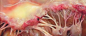
Alveoli are the structural part of the lungs. They are thin-walled bubbles with a diameter of 0.2 mm, braided by a network of capillaries. The lungs contain about 500 million alveoli. Through capillaries, oxygen enters the blood and is distributed throughout all systems of the body. And also through capillaries, the waste product - carbon dioxide - is delivered from the body to the alveoli.
The lower respiratory tract is the trachea and bronchi. When you inhale, oxygen is transported through them into the lungs with air, and when you exhale, carbon dioxide is released.
The bronchi are a paired organ that branches from the trachea to the right and left lungs. They are an extensive network of tubes that fill the entire volume of the lungs. The thin tips of the bronchi end in many alveoli.
In addition to the transport function, the bronchi have another important one – protective. The air passing through them is warmed, disinfected and filtered.
The protective function works due to the structure of the epithelium of the inner walls of the bronchi and the bactericidal mucous secretion that is produced in the bronchi.
COPD - what kind of disease is it?
For various reasons, the body's protective barrier does not always work. The mucous membranes are damaged, and then an immune response occurs in the tissues - inflammation. This can happen due to allergens, inhalation of harmful substances, pathogenic bacteria and viruses.
With inflammation of the bronchi (bronchitis), the tissues swell, turn red, the amount of mucus increases sharply, and it becomes viscous. If the diameter of the bronchi narrows, their ability to conduct air correspondingly deteriorates. This phenomenon is called obstruction. With obstruction, the patient experiences difficulty breathing, shortness of breath, and possible spasm. If inflammation affects the alveoli, pneumonia occurs.
Obstruction is characteristic of bronchial asthma, a complicated form of acute and chronic bronchitis (obstructive bronchitis).
But what is COPD and how does it differ from these diseases? The fact is that obstruction can be reversible. In asthma, the cause of obstruction is bronchospasm, when the bronchial muscles contract, leading to a narrowing of the lumen of the bronchi. The use of antispasmodics and bronchodilators relieves the attack.
In acute bronchitis, after recovery, the tissue heals and restores its functions.
Chronic obstructive disease is characterized by irreversible processes in the tissues of the bronchi and alveoli. In this case, changes occur at the organic level, and the obstruction is difficult to eliminate with drugs. As a result, the respiratory system can no longer cope with its tasks, and the person experiences chronic respiratory failure.
The body's tissues chronically lack oxygen, and carbon dioxide is not eliminated properly. In addition, the altered mucous membrane cannot cope with microbes and viruses, and the patient begins to suffer from infectious diseases. Chronic obstruction tends to progress.
The disease affects the bronchi and lungs, and includes chronic obstructive bronchitis and pulmonary emphysema. Most patients have signs of both diseases.
Diagnostics
A diagnosis of COPD should be considered in any person who has a cough, excess sputum production, and/or shortness of breath.
It is necessary to take into account the risk factors for developing the disease in each patient. If any of these symptoms are present, it is necessary to conduct a respiratory function test. These signs are not diagnostically significant individually, but the presence of several of them increases the likelihood of the disease. Chronic cough and excess sputum production often long precede ventilation disorders leading to the development of shortness of breath.
Key symptoms for diagnosing COPD
Chronic cough: bothers the patient constantly or periodically; observed more often during the day, less often at night. Cough is one of the leading symptoms of the disease; its disappearance in COPD may indicate a decrease in the cough reflex, which should be considered as an unfavorable sign.
Chronic sputum production: at the beginning of the disease the amount of sputum is small. The sputum is mucous in nature and is released mainly in the morning. However, as the disease worsens, its quantity may increase, it becomes more viscous, and the color of the sputum changes.
Shortness of breath: progressive (increases over time), persistent (daily). Intensifies with stress and during respiratory infectious diseases.
History of risk factors:
– smoking and tobacco smoke;
– industrial dust and chemicals;
– smoke from home heating appliances and fumes from cooking.
During a clinical examination, an extended expiratory phase in the respiratory cycle is determined; above the lungs - upon percussion - a pulmonary sound with a box-like tint; upon auscultation of the lungs - weakened vesicular breathing, scattered dry rales.
The diagnosis is confirmed by examining respiratory function.
Determination of forced vital capacity (FVC), forced expiratory volume in the first second (FEV1) and calculation of the FEV1/FVC index.
X-ray of the chest organs.
Exclusion of diseases manifesting similar clinical symptoms.
Symptom assessment
The rate of progression and severity of COPD symptoms depends on the intensity of exposure to etiological factors and their combined effect. In typical cases, the disease makes itself felt over the age of 40 years.
Cough is the earliest symptom, appearing by 40–50 years of age. By this time, during the cold seasons, episodes of respiratory infection begin to occur, which at the beginning are not associated with one disease. Subsequently, the cough takes on a daily character, rarely worsening at night. The cough is usually unproductive; can be paroxysmal in nature and provoked by inhalation of tobacco smoke, changes in weather, inhalation of dry cold air and a number of other environmental factors.
Sputum is released in small quantities, usually in the morning and is mucous in nature. Exacerbations of an infectious nature are manifested by a worsening of all signs of the disease, the appearance of purulent sputum and an increase in its quantity, and sometimes a delay in its release. The sputum has a viscous consistency, often containing “lumps” of secretion. As the disease worsens, the sputum becomes greenish in color and an unpleasant odor may appear.
Bronchopulmonary infection, although common, is not the only cause of exacerbation. Along with this, an exacerbation of the disease may develop due to the increased effect of exogenous damaging factors or inadequate physical activity. In these cases, signs of damage to the respiratory system are less pronounced. As the disease progresses, the intervals between exacerbations become shorter.
As the disease progresses, shortness of breath can vary from a feeling of lack of air during habitual physical activity to severe manifestations at rest.
Shortness of breath felt during physical activity occurs on average 10 years after the onset of cough. Dyspnea is the reason most patients consult a doctor and is the main cause of disability and anxiety associated with the disease. As pulmonary function declines, shortness of breath becomes more severe. With emphysema, the onset of the disease is possible with shortness of breath. This occurs in situations where a person comes into contact with finely dispersed (less than 5 microns) pollutants at work, as well as with hereditary deficiency of α1-antitrypsin, leading to the early development of panlobular emphysema.
The Medical Research Council Dyspnea Scale (MRC) is used to quantify the severity of dyspnea.
Patients with COPD visit doctors, as a rule, with complaints of cough, sputum production and shortness of breath increasing in intensity.
Anamnesis
The peculiarity of the development of COPD is that the duration of development of the disease can span several decades. The insidiousness of this disease is that it does not give clinical manifestations for a long time and is asymptomatic.
When studying the anamnesis, it is desirable to establish the frequency, duration and characteristics of the main symptoms of exacerbations and evaluate the effectiveness of previous treatment measures. COPD is a primarily chronic disease.
Thus, a detailed clinical picture of the disease due to tobacco smoking occurs 20–40 years from the time when a person began to smoke regularly. The American Thoracic Society standards emphasize that the onset of the first clinical symptoms in patients with COPD is usually preceded by smoking at least 20 cigarettes per day for 20 years or more.
In this regard, when talking with a patient, it is necessary to indicate the length of smoking, the number of cigarettes the patient smokes per day (smoking intensity).
If the patient smokes or has smoked, then it is also necessary to study the smoking history and calculate the smoking index (SI) ≪pack/years≫ using the formula:
IR > 10 packs/year is a significant risk factor for the development of COPD.
There is another calculation formula for the smoking index. The number of cigarettes smoked per day is multiplied by the number of months in a year during which a person smokes. If the result exceeds 120, then the patient must be considered a heavy smoker. In the presence of cough and sputum, these symptoms should be assessed as manifestations of bronchitis of a smoker (≪smoker).
COPD occurs for a long time without clear clinical signs; at least sick people do not have active complaints of coughing and sputum production. In these situations, the doctor, when talking with such patients, must use special questionnaires, the answers to the proposed questions of which help the patient to more clearly characterize the state of his health, and the doctor to obtain clearer information.
When collecting anamnesis, it is recommended to carefully analyze episodes of cough, its duration, nature, time of occurrence and pay attention to increased sputum production. The appearance of blood in the sputum deserves special attention, which gives reason to suspect another cause of cough: lung cancer, tuberculosis and bronchiectasis.
When questioning the patient, it is necessary to pay attention and evaluate the following parameters:
1) individual risk factors for each individual patient; the disease can significantly increase in its manifestations when several risk factors are combined in the same person: smoking, contact with industrial pollutants, infectious diseases of the respiratory tract, other negative environmental factors, genetic predisposition;
2) the dynamics of the development of symptoms;
3) frequency of exacerbations and hospitalizations for COPD;
4) medications taken, regularity of taking them;
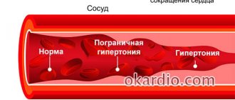
5) the presence of concomitant diseases: heart disease (CHD, hypertension), pathology of the gastrointestinal tract, etc.
Assessment of objective status
The results of an objective examination of COPD patients depend on the severity of bronchial obstruction and emphysema, the presence of complications such as respiratory failure and chronic cor pulmonale. However, examination results do not always reflect the severity of the disease. Even signs such as incoordination of respiratory movements and central cyanosis do not always characterize the degree of airway obstruction, and the absence of clinical symptoms does not exclude the presence of COPD in patients.
The examination of the patient should be carried out carefully, attention should be paid to the appearance of the sick person, his behavior, the reaction of the respiratory system to a conversation, and movement around the office.
When starting to examine the chest organs, it is necessary to pay attention to the number of respiratory movements in one minute and compare this indicator with shortness of breath. Tachypnea and dyspnea may not correlate in their manifestations.
When examining the chest, you should pay attention to its shape and participation in the act of breathing. The chest becomes barrel-shaped (especially in the emphysematous type of COPD), and the excursion of the lower edge of the lungs is limited. Recession of the intercostal spaces is one of the diagnostic signs of increased work of breathing.
When percussing the chest, a boxy percussion sound is noted; the lower borders of the lungs are lowered by an average of one rib, which is associated with the development of emphysema.
The auscultatory picture of the lungs may be characterized by a predominance of emphysema or bronchial obstruction. Thus, in patients with pulmonary emphysema, breathing is weakened vesicularly. On the contrary, in patients with severe bronchial obstruction, the main auscultatory symptom is dry, predominantly wheezing, aggravated by forced exhalation, simulating coughing, and in the supine position.
With irreversible bronchial obstruction, persistent signs of respiratory failure predominate, pulmonary hypertension increases, and chronic cor pulmonale is formed. It is difficult to identify signs of compensated cor pulmonale during physical examination. Heart sounds are difficult to hear. As the disease progresses, such patients develop diffuse cyanosis and worsen shortness of breath. First, transient and then permanent hypoxia and hypercapnia are observed, and blood viscosity often increases, which is caused by secondary polycythemia. A decompensated pulmonary heart is formed, the liver increases in size, and peripheral edema appears.
Diagnosis of COPD
In severe and irreversible bronchial obstruction, two clinical forms of the disease are distinguished: emphysematous and bronchitis, the main differences of which are given in Table. 2.
Identification of two forms of COPD has prognostic significance. Thus, in the emphysematous form, the development of the cor pulmonale occurs in later stages compared to the bronchitis form. In practice, patients with a mixed form of the disease are more common.
Summarizing the above, it should be emphasized that physical examination data are insufficient to establish a diagnosis of COPD. They provide guidelines for the further direction of diagnostic research using instrumental and laboratory methods.
Pulmonary function test
The study of respiratory function is the most important step in the diagnosis of COPD. This study is necessary to make a diagnosis, determine the severity of the disease, select individual therapy, assess the effectiveness of its implementation, clarify the prognosis of the course of the disease and conduct an examination of work ability.
To identify patients in the early stages of COPD development, all patients with chronic cough and sputum production, a history of risk factors, even in the absence of shortness of breath should undergo spirometry.
In patients with obstructive respiratory diseases, when making a functional diagnosis, it is necessary to measure the forced vital capacity of the lungs - FVC, forced expiratory volume in the first second - FEV1, and determine the ratio of these parameters (FEV1 / FVC).
The most sensitive parameter in diagnosing airflow limitation is the FEV1/FVC ratio. This parameter is decisive at all stages of COPD and at all degrees of severity of the disease. A decrease in FEV1/FVC of less than 70% during the period of remission of the disease indicates obstructive disorders, regardless of the severity of COPD. Determining the percentage of FEV1/FVC is most convenient in clinical practice, since there are no universally applicable absolute values for FEV1 and FVC. A decrease in FEV1/FVC less than 70% is an early sign of airflow limitation, even if FEV1 remains >80% of the normal value. Obstruction is considered chronic if it occurs at least 3 times within one year, despite therapy.
The FEV1 value in the post-bronchodilator test reflects the stage and severity of the disease.
Determination of peak expiratory flow volume (PEF) is the simplest and fastest method for assessing the state of bronchial patency, but has low sensitivity. PEF values may remain within normal limits for a long time in patients with COPD. This method is also characterized by low specificity for COPD, since a decrease in PEF values can also occur in other respiratory diseases.
At the same time, peak flowmetry can be used as a screening method to identify a group at risk for developing COPD and to establish the negative impact of various pollutants.
In COPD, determination of PEF is a necessary method of monitoring during an exacerbation of the disease and especially at the stage of rehabilitation of patients.
Bronchodilation test
The test is performed during the initial examination without an exacerbation of the disease. When performing a bronchodilation test, the change in FEV1 value is assessed:
• to determine the maximum achieved FEV1 indicators and establish the stage and severity of COPD;
• to exclude BA (positive test);
• to assess the effectiveness of therapy, make decisions on treatment tactics and the volume of therapy;
• to determine the prognosis of the course of the disease.
Choice of prescribed drug and dose.
As bronchodilator agents in the form of metered-dose inhalers when conducting tests in adults, it is recommended to prescribe the following, taking into account the severity of COPD:
• short-acting β2-agonists (salbutamol 2 - 4 doses = 200-400 mcg) with measurement of the bronchodilation response after 15-30 minutes;
• anticholinergic drugs – ipratropium bromide (4 doses = 80 mcg) with measurement of the bronchodilation response after 30–45 minutes.
For inhalation of the above-mentioned drugs using a nebulizer, the following dosages are recommended:
• inhalation of a salbutamol solution at a dose of 2.5–5 mg through a nebulizer, followed by a study (after 15–30 minutes) of the bronchodilation response;
• inhalation of an IB solution at a dose of 500 mcg through a nebulizer followed by a study (after 30–45 minutes) of the bronchodilator response;
• inhalation of a berodual solution at a dose of 2 ml (1 mg of fenoterol and 500 mcg of ipratropium bromide) through a nebulizer, followed by a study (after 30–45 minutes) of the bronchodilation response.
To avoid distortion of the results and to correctly perform the bronchodilator test, it is necessary to cancel the therapy in accordance with the pharmacokinetic properties of the drug taken (short-acting β2-agonists - 6 hours before the start of the test, long-acting β2-agonists - 12 hours before, long-acting theophyllines - 24 hours before the start of the test). hours).
Method for calculating bronchodilation response
The reversibility of obstruction is a variable value and can be different in the same patient during periods of exacerbation and remission of the disease.
The choice of reversibility index used depends on the clinical situation and the specific reason for which reversibility is being examined. The use of a reversibility indicator, which is less dependent on the initial parameters, allows for a more correct comparative analysis of data from different researchers.
The simplest way is to measure the bronchodilation response by the absolute increase in FEV1 in ml [FEV1 abs. (ml) = FEV1 dilate. (ml) – FEV1 ref. (ml)]. A very common method for measuring reversibility is the ratio of the absolute increase in FEV1, expressed as a percentage, to the initial one [ΔFEV1% ref.]:
A reliable bronchodilator response in its value should exceed spontaneous variability, as well as the response to bronchodilators in healthy individuals. Therefore, an increase in FEV1≥15% of predicted and ≥200 ml is recognized as a marker of a positive bronchodilation response; when such an increase is obtained, bronchial obstruction is considered reversible.
When assessing a bronchodilator test, it is important to take into account adverse reactions from the cardiovascular system: tachycardia, arrhythmia, increased blood pressure, as well as the appearance of symptoms such as agitation or tremor.
FEV1 monitoring
An important method to confirm the diagnosis of COPD is monitoring FEV1 - an annual spirometric determination of this indicator. In adulthood, there is normally an annual fall in FEV1 of 30 ml per year. Large epidemiological studies conducted in different countries have established that patients with COPD are characterized by a drop in FEV1 of more than 50 ml per year.
Laboratory and instrumental diagnostics
Sputum analysis
Sputum examination is a mandatory diagnostic procedure. Cytological examination of sputum provides information about the nature of the inflammatory process and its severity, and also allows one to identify atypical cells. Given the advanced age of most patients with COPD, there should always be oncological suspicion. If the doctor doubts the diagnosis, it is recommended to conduct several (3–5) cytological studies in a row. The method of studying induced sputum is also used, i.e. collected after inhalation of hypertonic sodium chloride solution. This method of obtaining sputum and its after-
The next study is more informative for identifying atypical cells.
In patients with COPD, sputum is usually mucous in nature; its main cellular elements are macrophages. As the disease worsens, the sputum becomes purulent and its viscosity increases. An increase in the amount of sputum, its high viscosity and yellow or green color are signs of an exacerbation of the infectious inflammatory process.
To tentatively identify the group affiliation of the pathogen, an assessment of the results of Gram staining of smears is used. Cultural microbiological examination of sputum should be carried out in case of uncontrolled progression of the infectious process to select rational antibiotic therapy.
Blood analysis
A clinical blood test is a mandatory method of examining a patient. With exacerbation of the disease, as a rule, neutrophilic leukocytosis with a band shift and an increase in ESR are observed. With a stable course of COPD, no significant changes in the content of peripheral blood leukocytes are observed. With the development of hypoxemia in patients with COPD, polycythaemic syndrome is formed, which is characterized by an increase in the number of red blood cells, a high level of hemoglobin, low ESR, an increase in hematocrit (in women > 47%, in men > 52%) and increased blood viscosity. These changes in blood tests are typical for patients with severe bronchitis-type COPD.
X-ray methods
Chest X-ray is a mandatory test when diagnosing COPD. During the initial X-ray examination, other diseases accompanied by clinical symptoms similar to COPD are excluded,
in particular, lung cancer and tuberculosis. X-ray of the chest organs is performed in postero-anterior and lateral projections. If a diagnosis of COPD is established during an exacerbation of the disease, an X-ray examination can exclude pneumonia, spontaneous pneumothorax as a result of rupture of bullae and other complications, including pleural effusion.
In mild COPD, significant radiographic changes are usually not detected.
In the bronchitis variant of COPD, X-ray data provide important diagnostic information about the condition of the bronchial tree: increased density of the bronchial walls, deformation of the bronchi.
X-ray diagnostics are especially informative for identifying and assessing emphysema. In the posteroanterior projection, flattening and low position of the diaphragm are recorded, and in the lateral projection, a significant increase in the retrosternal space (Sokolov’s sign) is recorded. The angle formed by the lines of the diaphragm and the anterior chest during pulmonary emphysema is 90° or more (normally it is acute). The emphysematous variant of COPD is characterized by a depletion of the vascular pattern of the lungs.
The development of cor pulmonale, as a rule, is manifested by hypertrophy of the right ventricle, and the enlarged shadow of the heart extends mainly in the anterior direction, which is determined in the retrosternal space; the vessels of the roots of the lungs are noticeably emphasized. A correlation has been established between the pressure in the pulmonary artery and the diameter of its descending part (X-ray methods are not decisive in the diagnosis of cor pulmonale).
CT scan
A more in-depth method of x-ray diagnostics is computed tomography. This method is optional; it is resorted to when it is necessary to clarify the nature of emphysema and determine treatment tactics. Computed tomography, especially high resolution, can significantly increase the level of diagnosis of various forms of emphysema. Centriacinar emphysema is most often localized in the apical parts of the lungs, and distal acinar emphysema is most often determined subpleurally and subseptally. Panacinar emphysema is found mainly in the posterior-basal parts of the lungs (the cause of its occurrence is associated with occupational hazards); this form of emphysema has a poor prognosis.
In the bronchitis variant of COPD, computed tomography allows one to examine the anatomical characteristics of bronchial lesions and determine the extent of these lesions in the proximal or distal part of the bronchus. With the help of computed tomography, bronchiectasis is better diagnosed and its localization is clearly established.
Electrocardiography
ECG data in most cases allows us to exclude the cardiac origin of respiratory symptoms. An ECG also allows, in a number of patients, to identify signs of overload and/or hypertrophy of the right heart with the development of a complication such as cor pulmonale; In addition, it is possible to register cardiac arrhythmias.
Blood Gas Study
Blood gas measurements are performed in patients with increasing sensations of shortness of breath, a decrease in FEV1 < 50% of predicted, or with clinical signs of respiratory failure or right heart failure.
Respiratory failure is defined when PaO2 < 8.0 kPa (< 60 mm Hg), regardless of the increase in PaCO2 at sea level when breathing air. It is preferable to take samples for analysis by arterial puncture. Finger and ear oximetry is reliable for determining blood saturation SaO2 and can be the means of choice for examining patients by clinicians. The study is carried out using a pulse oximeter, which
makes it possible to determine blood oxygen saturation and identify patients with hypoxemia.
Cytological examination of sputum, clinical blood test, chest x-ray, analysis of ventilation and gas exchange function of the lungs, ECG are among the necessary diagnostic program for examining patients with moderate to severe COPD.
Additional research methods
Exercise test (step test)
In the initial stages of the disease, disturbances in the diffusion capacity and gas composition of the blood at rest may be absent and appear only during physical activity (step test, see Appendix). To objectify and document the degree of decrease in exercise tolerance, it is recommended to conduct an exercise test. Exercise testing is used when the severity of shortness of breath does not correspond to a decrease in FEV1. It is also used to select patients for rehabilitation programs.
When conducting a walking test, the patient is given the task of walking as far as possible in 6 minutes, after which the distance traveled is recorded. It is recommended to monitor SaO2 during the study. A patient with a decrease in FEV1 to 1.0 l (≈ 40% of the proper values of this parameter) usually walks about 400 m during this time.
Echocardiography
Allows you to identify and evaluate signs of pulmonary hypertension, dysfunction of the right and left hearts, and determine the severity of pulmonary hypertension.
Bronchological examination
It is performed when making a differential diagnosis of COPD with other diseases (including cancer, tuberculosis) that manifest similar respiratory symptoms, as well as to assess the condition of the bronchial mucosa.
The study includes:
• examination of the bronchial mucosa;
• taking bronchial contents for subsequent studies.
The following methods can also be included:
• bronchoalveolar lavage with determination of cellular composition in order to clarify the nature of inflammation;
• biopsy of the bronchial mucosa.
The quality of life
Quality of life is an integral indicator that determines the patient’s adaptation to the presence of the disease and the ability to perform the patient’s usual functions related to his socio-economic situation at work and at home. To determine the quality of life, special questionnaires are used. The most famous is the St. George's Hospital questionnaire.
When formulating the diagnosis of COPD, the severity of the disease is indicated: mild (stage I), moderate (stage II), severe (stage III) and extremely severe (stage IV), exacerbation or remission of the disease, exacerbation of purulent bronchitis (if any) ; presence of complications (cor pulmonale, respiratory failure, circulatory failure), indicate risk factors, smoking index. It is recommended to indicate the clinical form of COPD (emphysematous, bronchitis) in severe cases of the disease.
The nomenclature of COPD in accordance with the International Classification of Diseases, Injuries and Causes of Death, X revision (1992) is presented in the appendix.
How does COPD develop?
The disease develops gradually. Its mechanism is triggered by unfavorable external influences that provoke an inflammatory process in the organs of the respiratory system. Another important factor is genetic predisposition, which causes an imbalance in the destruction and synthesis of healthy tissue.
During inflammation, pulmonary proteases are released - enzymes that break down the molecules of the mucous tissue of the bronchi and lungs. Normally, this process is balanced by the work of antiproteases, which are “responsible” for the restoration of molecules. One of the proteases is alpha-1-antitrypsin. In some cases, people with COPD have a deficiency of alpha-1 antitrypsin. This means that the activity of antiproteases is reduced, and the process of tissue destruction prevails over the restoration process.
If the healthy structure of the bronchial mucosa does not have time to recover, it is replaced by fibrous connective tissue, like scar tissue. If the harmful effects do not stop, this rough tissue gradually grows, thickening the walls of the bronchi and narrowing their diameter. Also, during the inflammatory process, hypersecretion of mucus occurs, which clogs the passage of the bronchi. After some time, the process becomes irreversible and begins to progress even if the harmful factors are eliminated.
If the bronchi thicken when damaged, the walls of the alveoli, on the contrary, are destroyed. As a result, a pathological expansion of the air spaces occurs, the parenchyma becomes more weakly attached to the respiratory tract, which leads to difficulty in the release of air during exhalation. Destroyed alveoli can form bullae (air cysts) with a diameter of more than 1 cm. Such pathologies lead to loss of elastic strength of the lungs and their hyperairiness.
What factors lead to the development of the disease?
Smoking
According to statistics, about 75% of COPD cases are caused by heavy smoking. Smoking 20 cigarettes a day for more than 20 years or longer is highly likely to lead to a clinically established diagnosis. If you regularly smoke passively for about 20 hours a week, your risk of getting sick doubles.
Adverse Occupational Exposures
Work in conditions of increased gas, dust and chemical contamination - that is, when inhaling substances that damage the mucous membrane of the respiratory tract, for example, in asbestos, gypsum factories, mines.
The risk of disease increases if work or living conditions involve inhalation of tars resulting from the combustion of wood, coal and other biofuels, with insufficient ventilation of the room.
Severe bronchial asthma
If asthma continues for a long time with severe attacks, bronchial obstruction gradually ceases to be reversible and becomes chronic. This is especially likely if the diagnosis was established in childhood.
Chronic infections
Recurrent infectious diseases of the lower respiratory tract can lead to chronicity and the development of obstruction. In turn, people with COPD often suffer from respiratory tract infections. Chronic obstruction leads to stagnation of mucus in the lower respiratory tract, which promotes the proliferation of bacteria. This in turn increases inflammation and further damage to bronchopulmonary tissue. This means it leads to faster progression of the disease.
Symptoms of COPD
Cough is the earliest sign of the disease. At first it is episodic, then becomes daily. A smoker's cough can be considered an early symptom of developing obstructive disease.
Sputum separation. Relatively early symptom. During the period of remission there may be no sputum.
Dyspnea. Appears approximately 10 years after the onset of the disease. At a later stage, shortness of breath appears even with minor physical exertion, up to severe respiratory failure.
Breath. Weak, whistling, shortened on exhalation. Breathing through clenched teeth.
Weight loss.
Barrel chest.
With the development of respiratory failure, the patient's skin acquires a bluish tint, swelling and puffiness of the face occur. The ends of the phalanges of the fingers thicken.
In advanced forms of the disease, a person may develop signs of carbon dioxide poisoning (usually at night).
Breathing exercises for COPD
Special breathing exercises make their “mite” in the treatment of COPD:
- Starting position: lie down on your back. As we exhale, we pull our legs towards ourselves, bend them at the knees, and grab them with our hands. We exhale the air to the end, inhale with the diaphragm, return to the starting position.
- Fill a jar with water and insert a cocktail straw. We take in as much air as possible when inhaling, and slowly exhale it into the tube. We perform the exercise for at least 10 minutes.
- We count to three, exhaling more air (pulling in the stomach). At “four” we relax the abdominal muscles and inhale through the diaphragm. Then we sharply contract the abdominal muscles and clear our throat.
Diagnostics
The disease develops long before the onset of functional disorders, which can be recorded instrumentally. Therefore, unfortunately, early diagnosis of COPD is almost impossible.
When diagnosing, the following methods are used:
- X-ray and CT (computed tomography) of the chest;
- study of the function of the respiratory system using spirometry and other tests;
- sputum cultures for microflora;
- determining the level of alpha-1-antitrypsin;
- ECG and echocardiography to exclude cardiac causes of shortness of breath and identify cardiac complications.
When diagnosing, it is important to differentiate COPD from diseases with a similar clinical picture - asthma, heart failure.
Treatment
The strategic goal in the treatment of stable obstructive disease is to prevent exacerbations and increase the functionality of the respiratory system.
For this purpose, a set of measures is used:
- Quitting smoking. Is crucial in therapy.
- Inhaled bronchodilators are drugs that dilate the bronchi.
- Inhaled corticosteroids – reduce inflammation and relieve symptoms.
- Phosphodiesterase-4 inhibitors have anti-inflammatory and bronchodilatory effects.
- Oxygen therapy is treatment with oxygen.
- Normalization of nutrition.
- Physiotherapy.
- In some cases, surgery is indicated.
Patients with COPD are advised to get vaccinated against influenza to avoid complications and sudden progression of the disease.
Treatment of COPD by severity
At the beginning of therapy, non-drug improvement of patients is carried out. This includes reducing exposure to harmful factors in the air you breathe, becoming aware of potential risks and ways to improve the quality of the air you breathe.
Important! Regardless of the stage of COPD, the patient should quit smoking.
Treatment of chronic obstructive pulmonary disease involves:
- reduction in the severity of clinical symptoms;
- improving the patient’s quality of life;
- prevention of progression of bronchial obstruction;
- preventing the development of complications.
Therapy is carried out in two main forms: basic and symptomatic.
Basic is a long-term form of treatment and involves the use of drugs that dilate the bronchi - bronchodilators.
Symptomatic therapy is carried out during exacerbations. It is aimed at combating infectious complications, ensuring the liquefaction and removal of sputum from the bronchi.
Important! Pneumococcal vaccinations are recommended once every 5 years and influenza vaccinations every season. This helps prevent the development of complications and aggravation of COPD.
Medicines used in treatment:
- bronchodilators;
- combinations of glucocorticoids and beta2 agonists;
- glucocorticosteroids in inhalers;
- phosphodiesterase-4 inhibitor – Roflumilast;
- Methylxanthine Theophylline.
Main methods of therapy:
- If there is severe shortness of breath, then short-acting bronchodilators are used: Terbutaline, Berrotec, Salbutamol, Fenoterol, Ventolin.
These medications can be used up to four times a day. Limitations to their use are heart defects, tachyarrhythmias, glaucoma, diabetes, myocarditis, thyrotoxicosis, aortic stenosis. Important! It is necessary to carry out inhalations correctly; for the first time it is better to do this in the presence of a doctor who will point out mistakes. The medicine is injected while inhaling, this will prevent it from settling in the throat and ensure distribution in the bronchi. After inhalation, you should hold your breath for 10 seconds while inhaling. - If the patient has a wet cough, then drugs are prescribed to help thin it - mucolytics. The best remedies are drugs based on acetylcysteine: ACC, Fluimucil in the form of water-soluble powder and effervescent tablets. There is acetylcysteine in the form of a 20% solution for inhalation using a nebulizer (a special device that converts the liquid form of the drug into an aerosol). Acetylcysteine inhalations are more effective than powders and tablets taken orally, since the substance immediately appears in the bronchi.
In the treatment of moderate COPD, medications that help remove sputum and bronchi dilators are effective. And for bronchitis COPD - anti-inflammatory drugs. At the same time, non-drug therapy methods and medications are used, which are combined depending on the patient’s condition. Sanatorium-resort treatment gives an excellent effect.
Principles of therapy:
- Medicines that slow down bronchial obstruction are used regularly or periodically.
- To relieve exacerbation of the disease, inhaled glucocorticoids are used. They can be used in conjunction with andrenomimetics, which are designed for long-term action.
- As a complement to drug treatment, physical therapy is used, which increases patients' resistance to physical activity, reduces fatigue and shortness of breath.
COPD differs from other ailments in that as it progresses, the volume of therapeutic procedures increases, but none of the drugs used affects the decrease in bronchial patency.
Third degree
Treatment of patients with the third stage of COPD severity:
- Continuous anti-inflammatory therapy is carried out.
- Large and medium doses of glucocorticosteroids are prescribed: Becotide, Pulmicort, Beclazone, Benacort, Flixotide in the form of aerosols for inhalation through a nebulizer.
- Combination medications may be used, including a long-acting bronchodilator and a glucocorticosteroid. For example, Symbicort, Seretide, which are the most effective modern therapeutic drugs intended for the treatment of stage 3 COPD.
Important! If your doctor has prescribed an inhaled corticosteroid, you should definitely ask how to use it correctly. Improper inhalation negates the effectiveness of the drug and increases the likelihood of side effects. After each inhalation you need to rinse your mouth.
Fourth degree
Treatment of patients with extremely severe COPD:
- In addition to bronchodilators and glucocorticosteroids, oxygen therapy (inhalation of oxygen-enriched air from a portable can) is prescribed.
- Surgical treatment is carried out only if the age and health of the patient allows (there are no diseases of other organs and systems).
- In the most severe cases, artificial ventilation is performed.
- If COPD is accompanied by an infection, then doctors supplement the therapy with antibiotics. Fluoroquinols, cephalosporins, and penicillin derivatives are used depending on the patient’s condition and existing concomitant ailments.
We suggest you read: How to charge your e-shock without charging
Treatment of COPD requires significant joint efforts between doctors and patients. Long-term changes in the lungs cannot be eliminated overnight through standard therapy. Due to chronic changes in the respiratory system, the bronchi become damaged - they become overgrown with connective tissue and narrow, which is irreversible.
How to treat COPD? Doctors say that this type of chronic pulmonary pathology cannot be completely cured. The development of the disease is stopped by timely prescribed therapy. In most cases, it helps improve the condition. Few achieve complete restoration of normal functioning of the respiratory system (lung transplantation is indicated for severe COPD). After confirmation of the medical report, lung disease is eliminated with medications in combination with folk remedies.
Drugs
The main “doctors” in the case of respiratory pathology are considered to be bronchodilator drugs for COPD. For a complex process, other medications are also prescribed. An approximate course of treatment looks like this:
- Beta2 agonists. Long-acting drugs – “Formoterol”, “Salmeterol”; short - salbutamol, terbutaline.
- Methylxanthines: Aminophylline, Theophylline.
- Bronchodilators: tiotropium bromide, oxitropium bromide.
- Glucocorticosteroids. Systemic: Methylprednisolone. Inhalation: Fluticasone, Budesonide.
- Patients with severe and extremely severe COPD are prescribed inhaled medications with bronchodilators and glucocorticosteroids.
Treatment of COPD with folk remedies is recommended in combination with medications. Otherwise, there will be no positive results from traditional medicine recipes. Several effective grandmother’s recipes for combating COPD:
- Take 200 g of linden blossom, the same amount of chamomile and 100 g of flax seeds. We dry the herbs, chop them, and infuse them. For one glass of boiling water put 1 tbsp. l. collection Take 1 time per day for 2-3 months.
- Grind 100 g of sage and 200 g of nettle into powder. Pour boiled water over the mixture of herbs and leave for an hour. We drink half a glass twice a day for 2 months.
- Collection for removing sputum from the body during obstructive inflammation. We will need 300 g of flaxseeds, 100 g of anise berries, chamomile, marshmallow, licorice root. Pour boiling water over the mixture and leave for 30 minutes. Strain and drink half a glass every day.
Complications
The course of COPD is characterized by the following complications:
- Respiratory failure.
- Recurrent respiratory tract infections.
- Pulmonary hypertension (increased pressure in the pulmonary circulation).
- Failure of the right ventricle of the heart (cor pulmonale).
- Pneumothorax (accumulation of air in the pleural cavity).
- Lungs' cancer.
- Cardiac ischemia.
- Anxiety and depressive disorders.
- Weight loss.
The complicated course of the disease ends in death in 50% of cases within 10 years after diagnosis. The prognosis largely depends on whether the patient managed to quit smoking and how long he had as a smoker before.
Concomitant diseases
With COPD, the patient has a high chance of suffering not only from the obstruction itself, but also from the diseases that accompany it. Among them:
- Cardiovascular diseases, from coronary heart disease to heart failure. They occur in almost half of the cases and are explained very simply: with a lack of oxygen in the body, the cardiovascular system experiences great stress: the heart moves faster, blood flows faster through the veins, and the lumen of blood vessels narrows . After some time, the patient begins to notice chest pain, a racing pulse, headaches and increased shortness of breath. A third of patients whose COPD is accompanied by cardiovascular diseases die from them.
- Osteoporosis. Occurs in a third of cases. Not fatal, but very unpleasant and also caused by a lack of oxygen. Its main symptom is brittle bones. As a result, the patient's spine is bent, his posture deteriorates, his back and limbs hurt, night cramps in the legs and general weakness are observed. Endurance and finger mobility decrease. Any fracture takes a very long time to heal and can be fatal. Often there are problems with the gastrointestinal tract - constipation and diarrhea, which are caused by the pressure of the curved spine on the internal organs.
- Depression. Occurs in almost half of patients. Often its dangers remain underestimated, and the patient meanwhile suffers from low tone, lack of energy and motivation, suicidal thoughts, increased anxiety, feelings of loneliness and learning problems. Everything is seen in a gloomy light, the mood constantly remains depressed. The reason is both the lack of oxygen and the impact that COPD has on the patient’s entire life. Depression is not fatal, but it is difficult to treat and significantly reduces the enjoyment that the patient could get from life.
- Infections. They occur in seventy percent of patients and cause death in a third of cases. This is explained by the fact that lungs affected by COPD are very vulnerable to any pathogen, and it is difficult to relieve inflammation in them. Moreover, any increase in sputum production means a decrease in air flow and the risk of developing respiratory failure.
- Sleep apnea syndrome. With apnea, the patient stops breathing at night for more than ten seconds. As a result, he suffers from constant oxygen starvation and may even die from respiratory failure.
- Cancer. It occurs frequently and causes death in one case out of five. It is explained, like infections, by the vulnerability of the lungs.
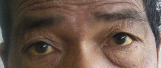
In men, COPD is often accompanied by impotence, and in older people it causes cataracts.