Increased pressure in the pulmonary artery is a serious pathology. Drugs for the treatment of pulmonary hypertension include several groups of medications - endothelin receptor antagonists, inhibitors of enzymes that prevent vascular relaxation, prostacyclins, calcium channel blockers, diuretics and a number of other drugs. All of them should be used only on the recommendation of a cardiologist and after a thorough, multifaceted examination. These medications are taken for life, and improvement occurs only after a few months.
Causes
The causes of idiopathic hypertension are not known. Presuming factors include complicated heredity, microthromboembolism (sedimentation of blood clots) in the vascular bed of the lung tissue, dysfunction of the endothelium (internal lining) of the pulmonary capillaries.
The causes of secondary pulmonary arterial hypertension are diseases such as:
- Cardiac and vascular diseases:
- post-infarction cardiosclerosis,
- postmyocardial cardiosclerosis – scar replacement of normal heart muscle after its inflammation,
- congenital and acquired heart defects,
- tumors of cardiac tissue,
- arterial hypertension,
- cardiomyopathy,
- pulmonary embolism,
- vasculitis – lesions of the vascular wall of an inflammatory, allergic or toxic nature.
- Diseases of the bronchopulmonary system:
- chronic obstructive disease, characterized by the presence of chronic obstructive bronchitis and emphysema, more often occurring in long-term smokers,
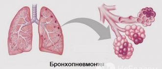
- chronic bronchitis, bronchiolitis, pneumonia with frequent and prolonged course,
- severe course of bronchial asthma.
- Other diseases:
- lesions of the vascular wall caused by systemic connective tissue diseases - systemic lupus erythematosus, scleroderma, etc.
- portal hypertension syndrome with increased pressure in the liver vessels in people with liver cirrhosis,
- HIV infection,
- congenital metabolic disorders - pathology of the thyroid gland, glycogenosis (a disease of glycogen metabolism in the body), hemoglobinopathies, etc.
- A special place is occupied by pulmonary hypertension of newborns, which can also be primary or secondary. The first option develops in a newborn child without signs of lung disease. The second option occurs in a baby with cardiac or pulmonary pathology, as well as with asphyxia (suffocation) and hypoxia (oxygen starvation) during childbirth. The causes of persistent pulmonary hypertension in newborns include:
- respiratory distress syndrome (acute respiratory disorders),
- aspiration (inhalation) of meconium during childbirth,
- pneumonia and sepsis in the neonatal period,
- premature (intrauterine) closure of the open foramen ovale and ductus arteriosus, designed to “switch off” blood flow through the lungs of the fetus, and normally closing at the time of the newborn’s first cry or in the first months of life,
- congenital diaphragmatic hernia, in which organs located in the abdominal cavity compress the left lung, due to which the blood flow of the right lung is increased.
The acute form of hypertension occurs in the shortest possible time and is caused by massive pulmonary embolism, status asthmaticus, and pulmonary edema.
Indications for the use of antihypertensive drugs
The pathology clinic is nonspecific. Therefore, a drug regimen is prescribed only after a comprehensive laboratory and instrumental examination of the patient.
With pulmonary hypertension, the following clinical symptoms occur, for which the following medications are indicated:
- Dyspnea. It is constant and intensifies with physical and psycho-emotional stress. This symptom occurs even with mild hypertension in the respiratory system.
- Increased systolic and diastolic blood pressure. Changes in the pulmonary system provoke pathological processes in the cardiovascular organs.
- Painful sensations in the chest similar to angina pectoris.
- Syncope. They are short-term loss of consciousness due to changes in pressure.
- Asthenic syndrome. It includes constant fatigue, weakness, ringing in the ears and flashing spots before the eyes.
- Hemoptysis. Cough discharge often contains streaks of blood.
Symptoms of the disease
Due to the fact that pulmonary hypertension develops slowly in most cases, clinical manifestations in the early stages may be absent for a long time.
As hypertension progresses, subcompensation of the disease occurs, and the first signs of the disease appear. These include shortness of breath, first due to exertion, and then at rest. In addition to shortness of breath, the patient notes a bluish discoloration of the skin of the face and extremities (acrocyanosis), fatigue, general weakness, and decreased exercise tolerance. Dizziness and fainting are possible due to decreased oxygen supply to the brain. All these symptoms are a manifestation of incipient respiratory failure.
Due to the fact that the described symptoms occur in many heart diseases, it is necessary to consult a doctor as soon as possible and clarify the cause of the appearance of such symptoms.
Due to the constantly high pressure in the capillaries of the lungs, the right ventricle, whose function is to push blood into the pulmonary artery, cannot cope with the increased load, and right ventricular failure is formed. Its signs are swelling first of the lower extremities, and then of the entire body, enlargement of the abdomen, discomfort and pain in the right half of the abdomen, caused by stagnation of fluid in the liver and its enlargement up to cardiac cirrhosis of the liver.
At the stage of decompensation, the patient experiences diffuse cyanosis throughout the body, severe edema, shortness of breath at rest and in the supine position. Dystrophic changes develop in all organs and tissues.
Depending on the anatomical and functional disorders that occur during the disease, as well as on the level of physical activity tolerance, degrees of hypertension are distinguished:
- 1st degree, or transient (labile) – characterized by an unstable increase in pressure in the pulmonary artery of more than 30 mm Hg, occurring after physical exertion. As a rule, symptoms bother the patient under significant loads or do not occur at all.
- Stage 2, or stable hypertension, is characterized by a constant increase in pressure in the pulmonary artery above 25 mm Hg at rest and above 30 mm Hg during exercise. Symptoms occur with previously well-tolerated exercise (walking, climbing stairs, etc.).
- 3rd degree, or irreversible - right ventricular failure develops with all its clinical manifestations, which in the absence of treatment quickly progresses to the terminal stage and death.
How to prevent and forecasts?
Disguising a disease as another is a problem even for medical experts. Patients are often treated for incorrect diagnoses, so in the future it is necessary to develop the diagnosis of the disease in order to avoid deaths. It is worth increasing patient survival. Doctors are not losing hope of finding out why young and middle-aged people are at risk.
It is necessary to create regional centers and allow highly qualified specialists from different countries to study the disease for the sake of timely assistance to patients with pulmonary hypertension and the discovery of preventive agents. This pathology ranks third in incidence rate after myocardial infarction and stroke. These numbers encourage doctors to take a closer look at this issue, because a disease that causes so many victims needs to be treated.
Speaking of prognoses, they depend on the severity and form of the disease. The most dangerous primary CTEPH. Survival rate in the first year of the disease is 68%, and after 5 years - 30%. With stable high pressure in the pulmonary vessels and a positive response to treatment, the outcome is favorable. At the decompensated stage (the final stage of disease progression), survival is no more than 5 years.
Chronic thromboembolic pulmonary hypertension can be prevented by leading a healthy lifestyle, giving up bad habits, stress and significant physical activity. For persons with pathologies that provoke increased thrombus formation, it is important to carry out thrombolytic therapy. With sufficient and timely treatment, the prognosis for life and work is favorable, but it is impossible to completely cure the pathology without surgical intervention.
- Prevention of respiratory diseases
- Pneumonia in children - causes, symptoms, diagnosis and treatment
Diagnostics
Due to the fact that pulmonary hypertension can progress to severe right ventricular failure quite quickly (within 2-8 years from the time of diagnosis), it is necessary to be wary of suspecting hypertension in persons with existing heart and lung diseases, as well as with systemic diseases.
To do this, this category of patients must regularly visit their doctor with the following examination methods performed annually:
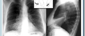
- Chest X-ray allows you to determine the enlargement of the borders of the heart with hypertrophy of the right ventricle and the strengthening of the pulmonary pattern due to the vascular component,
- In addition to signs of the main pathology (myocardial ischemia, post-infarction cardiosclerosis, etc.), the ECG allows us to identify signs of right ventricular hypertrophy and overload of the right chambers of the heart,
- Echocardiography helps not only to diagnose myocardial diseases, but also to indirectly measure the pressure in the pulmonary artery, on the basis of which the issue of conducting an invasive diagnostic method is decided - catheterization of the pulmonary artery with a more accurate measurement of pressure in it.
pulmonary hypertension on x-ray
To clarify the diagnosis if hypertension is suspected due to thromboembolism of small branches of the pulmonary artery, methods such as:
- Radionuclide scanning of the lungs, based on the ability of atoms labeled with radioisotopes to penetrate the blood, as a result of which the radiation from these atoms is captured by an appropriate screen and a picture is obtained that recreates the blood flow in the lungs,
- Pulmonary angiography - injection of a contrast agent into the pulmonary artery followed by x-rays,
- Computed tomography of the lungs.
For timely diagnosis of the disease, the first three examination methods, carried out regularly, at least once a year, and more often if indicated, are sufficient.
In newborns, hypertension can also be detected by cardiac ultrasound and radiography.
List of medications for hypertension - names, descriptions of drugs
Here is a list of medications for hypertension, their pharmaceutical and "commercial" names.
This page will make it easier to find information about the drugs you are interested in. To quickly find the medicine you need, use the "find on page" search in your Internet browser. For detailed information about medications (indications for use, dosage, side effects, compatibility with other medications), see individual articles linked here.
Diuretic medications for hypertension: detailed list
Pulmonary artery hypertension develops against the background of other diseases, which may have completely different causes. Hypertension develops due to the proliferation of the inner layer of the pulmonary vessels. In this case, their lumen narrows and disruptions in the blood supply to the lungs occur.
This disease is quite rare, but, sadly, most cases are fatal.
The main diseases that lead to the development of this pathology include:
- Chronical bronchitis;
- fibrosis of lung tissue;
- congenital heart defects;
- bronchiectasis;
- hypertension, cardiomyopathy, tachycardia, ischemia;
- thrombosis of blood vessels in the lungs;
- alveolar hypoxia;
- increased red blood cell count;
- vascular spasms.
There are also a number of factors that contribute to the occurrence of pulmonary hypertension:
- thyroid diseases;
- intoxication of the body with toxic substances;
- long-term use of antidepressants or appetite suppressants;
- use of drugs taken intranasally (inhaled through the nose);
- HIV infections;
- oncological diseases of the circulatory system;
- cirrhosis of the liver;
- genetic predisposition.
Pulmonary hypertension can form as a result of prolonged compression of the blood vessels of the lungs. This condition can occur with chest injuries, excess weight and tumors.
Treatment of pulmonary hypertension
Treatment for hypertension should be started promptly as soon as the diagnosis is confirmed, even if only minor symptoms are present. Treatment should primarily be aimed at eliminating the underlying disease. Of the drugs that have proven themselves well in clinical trials and are first-line drugs in the treatment of pulmonary hypertension, the following are indicated:
- Calcium channel antagonists – nifedipine, amlodipine, diltiazem. It is preferable to prescribe retard forms, which have a longer lasting effect.
- Drugs called prostanoids have a powerful relaxing effect on the vascular wall. Drugs such as epoprostenol and treprostinil are used as injections, and to achieve an effect they must be used for a long time, which is not very convenient and not cheap for the patient. Therefore, in recent years, iloprost has been developed, which is used by inhalation in the form of an aerosol and has proven clinical effectiveness.
- Endothelin receptor antagonists prevent remodeling and changes in the elasticity of the vascular wall of the pulmonary arteries. One of the most studied drugs is bosentan in tablet form.
In addition to those listed, medications are required for the treatment of underlying cardiac (diuretics, antihypertensive drugs, nitrates for myocardial ischemia, etc.) or bronchopulmonary disease (timely antibiotic therapy, inhaled drugs for the treatment of bronchial asthma, etc.).
In newborns, for the treatment of pulmonary hypertension in the first days of life, oxygen therapy is used through a mask or with the help of artificial ventilation, as well as the supply of a mixture of oxygen with nitric oxide, which has a relaxing effect on the blood vessels of the lungs and, therefore, reduces the load on the child’s heart. In recent years, significant success has been achieved in the use of extracorporeal membrane oxygenation of a child's blood, in which the blood taken using a device is saturated with oxygen through a special circuit and then returned to the bloodstream.
In addition to medication, surgical treatment is prescribed according to indications, for example, for heart defects, pulmonary embolism, significant occlusion of the coronary arteries causing myocardial infarction, etc.
Buy online
Epidemiology. Pathogenesis
One of the most important areas of scientific work by E.I. Chazov studied thrombosis and embolism, in particular pulmonary embolism (PE). In the monograph “Essays on Emergency Cardiology” [1] E.I. Chazov accurately describes the picture of this disease, using vivid clinical examples, and identifies the difficulties of differential diagnosis of pulmonary embolism.
In the book “Thrombosis and embolism in the clinic of internal diseases” [2] E.I. Chazov identifies thrombosis and pulmonary embolism as one of the pressing problems of the therapeutic clinic.
The work provides epidemiological data on pulmonary embolism, describes its clinical picture and diagnosis, and mentions possible mechanisms of disease development.
The monograph also raises the question of the connection between pulmonary embolism and chronic thromboembolic pulmonary hypertension (CTEPH): “Recently, many authors have been intensively studying the problems of the occurrence of cor pulmonale and hypertension of the pulmonary circulation. There are reports that such conditions can develop as a chronic process as a result of repeated embolisms or thromboses in the small branches of the pulmonary artery. There is even an assumption that this process has a significant impact on the occurrence and course of Aertz disease, one of the characteristic symptoms of which is hypertension of the pulmonary circulation... We believe that thromboembolism of small branches of the pulmonary artery is of known importance for the development of primary pulmonary hypertension, but this issue requires further research."
Now many of the assumptions and judgments expressed more than 40 years ago by E.I. Chazov, seem simplified. But the foundation that was laid in these works served as the basis for further research in this direction.
Work on CTEPH is actively continuing at the Research Institute of Clinical Cardiology named after. A.L. Myasnikov Federal State Budgetary Institution National Medical Research Center of Cardiology, Ministry of Health of the Russian Federation.
The following is a modern understanding of CTEPH, its epidemiology, pathogenesis, diagnosis and basic approaches to treatment.
CTEPH is a precapillary form of pulmonary hypertension (PH), which, according to the modern classification, along with other pulmonary artery obstructions (tumors, vascular anomalies, emboli), belongs to group IV PH [3].
There are currently no accurate epidemiological data on the prevalence of CTEPH both in our country and in the world. Among all forms of PH, CTEPH occurs in 1.5–3% of cases [3]. The uniqueness of CTEPH is that it is a potentially curable form of PH, since it has a good functional and hemodynamic effect with timely and successful surgical treatment.
It is generally accepted that most often CTEPH is a late complication of acute PE. According to an international study, 75% of patients with CTEPH had a history of at least one episode of registered PE [4]. In 56% of cases, the cause of acute pulmonary embolism was deep vein thrombosis of the lower extremities [5]. In 50% of cases after acute pulmonary embolism, complete lysis of thrombotic masses does not occur against the background of adequate anticoagulant therapy; this is confirmed by lung scintigraphy data demonstrating the presence of persistent perfusion defects [6]. However, only 0.1–9.1% of patients develop CTEPH within the first two years after an episode of acute PE [7–10].
Obstruction of the branches of the pulmonary artery by thrombotic masses and secondary changes in the microvasculature of the lungs lead to an increase in pulmonary vascular resistance (PVR) and pulmonary artery pressure (PAP), which in turn contributes to the development of severe dysfunction of the right heart with the formation of isolated right ventricular heart failure in the initial stages , and in advanced cases of the disease - biventricular heart failure, which determines an extremely unfavorable prognosis in this category of patients [11].
Most studies overestimate the incidence of CTEPH after an episode of acute PE. One reason for this overestimation is that many patients already had CTEPH at the time of their first episode of PE. Also, in most studies, CTEPH was diagnosed several months after an episode of acute PE, which is not typical for this disease, since, as a rule, CTEPH develops after the so-called light interval - a period of asymptomatic disease, which most often lasts several years. Thus, in a study by L. Guérin et al. [12], which included 146 patients with a diagnosis of acute PE; during the observation period (26 months), CTEPH was suspected in 8 due to the appearance of typical symptoms and echocardiographic signs. After right heart catheterization (RHC), the diagnosis was confirmed in 7 (4.8%) of 8 patients. However, during the recorded first episode of acute pulmonary embolism, only 2 patients had pulmonary artery systolic pressure (PASP) below 50 mmHg, in the remaining 5 cases PASP was from 62 to 102 mmHg, which is not typical for the first episode TELA. This is due to the fact that the unadapted right ventricle, due to its anatomical and physiological characteristics, cannot cope with an acute increase in pulmonary pressure above 50 mm Hg. Therefore, according to the authors, it is likely that CTEPH was present in these patients before. Thus, the frequency of CTEPH after an acute episode of PE in the study by L. Guerin et al. was no more than 1.5%.
According to the European CTEPH Registry, on average it takes 1 year from the first signs of the disease to diagnosis. This is primarily due to the non-specificity of the first signs of the disease: shortness of breath developing in the patient, decreased tolerance to physical activity can be manifestations of a large number of diseases, and the lack of alertness of first-line doctors leads to a delay in making the correct diagnosis.
Today, there are a considerable number of theories trying to explain the reason for the incomplete lysis of thrombotic masses, but none of them can fully reveal the pathogenesis of this process [13, 14].
It is known that with the development of certain clinical conditions, an increase in platelet activity is observed, which in turn predisposes to pathological thrombus formation and the formation of CTEPH. When comparing 433 patients with CTEPH and 254 patients with other forms of PH, such as the presence of intracardiac shunts, infected pacemakers, splenectomy, previous thrombosis of the veins of the lower extremities (especially recurrent course), non-0 (I) blood type, the presence of lupus anticoagulant/antiphospholipid antibodies , thyroid hormone replacement therapy and a history of malignant neoplasms, as well as autoimmune diseases and blood diseases of a congenital and acquired nature, have been identified as factors that increase the risk of developing CTEPH [15–17]. Also, based on the results of several studies, it has been suggested that during embolism with large thromboemboli and recurrent pulmonary embolism, the physiological fibrinolytic system is depleted, its activity is not enough to completely lyse the clots [10, 18]. This kind of inferiority of the physiological fibrinolytic system raises suspicion regarding the presence of an innate tendency to hypercoagulable states due to congenital pathology of the hemostatic system. However, it is known that such classical thrombophilias as protein C and S deficiency and antithrombin III deficiency, as well as factor V and II mutations, are no more common in patients with CTEPH than in the healthy population [17]. Only elevated levels of antiphospholipid antibodies and lupus anticoagulant were reported in patients with CTEPH more often than in patients with idiopathic PAH. In other studies, 41% of patients with CTEPH had elevated levels of coagulation factor VIII, von Willebrand factor, an adhesion glycoprotein that stabilizes and activates factor VIII [19]. Also, with CTEPH, there appear to be abnormalities in the structure of fibrinogen molecules [20-22], accompanied by modification of the structure of fibrin and the clot itself, which manifests itself as resistance to physiological thrombolysis and, thus, slows down the process of resolution of thrombotic masses. In a study comparing fibrinogen in CTEPH patients with a healthy control group, fibrinogen in CTEPH patients was shown to be more resistant to plasmin-mediated lysis. Researchers have suggested that this is due to altered fibrin and/or fibrinogen structure. In addition, an altered structure of fibrinogen was also found in the vascular wall of the pulmonary artery, which, according to the authors, may influence the transition of acute PE to CETEPH [22]. Another study examined the role of a thrombin-activated fibrinolysis inhibitor. The level of this indicator was significantly increased in patients with CTEPH than in patients with pulmonary arterial hypertension and the control group, and correlated with the resistance of the thrombus to lysis, which demonstrates the significant role of this indicator in the pathogenesis of the disease [23].
Along with the above, another key mechanism in the resolution of thrombotic masses is neoangiogenesis. In clot samples obtained after thromboendarterectomy (TEE), it was found that angiogenesis in patients with CTEPH was defective, which was also proven in experimental models [24, 25].
Endothelial dysfunction and remodeling of vessels in the pulmonary circulation in CTEPH
Mechanical occlusion of the pulmonary artery by thrombotic masses is not the only mechanism for the formation of PH in CTEPH. All patients with CTEPH have varying degrees of severe microvasculopathy, which is a kind of diffuse spasm of the distal pulmonary bed with an artery diameter of about 0.1-0.5 mm due to the release of vasoconstrictor and procoagulant substances from platelets and endothelium [3]. This pathology was first described by K. Moser and C. Bloor when studying histological samples of lung tissue from patients with L.G. Researchers have observed intimal thickening and pulmonary vascular remodeling in the form of eccentric intimal fibrosis, intimal fibromuscular proliferation, and plexiform lesions [26, 27]. The same lesion was previously seen in patients with idiopathic PAH, which provided the basis for the use of drugs approved for the treatment of idiopathic PAH in CTEPH.
In the literature, such changes are explained as a result of redistribution of pulmonary blood flow between pulmonary arteries unaffected by thrombi. Under the influence of unusually high pressure and tension, endothelial dysfunction and remodeling of the vessels of the pulmonary circulation occur, which leads to the progression of the disease, a gradual increase in PAP and PVR. However, this microvasculopathy is observed not only in unaffected areas of the lung, but also in the distal segments of occluded arteries, which cannot be explained by simple redistribution of blood flow. In a study by R. Dorfmüller et al. [28] found that in patients with PH there are anastomoses between the systemic and pulmonary circulations (via the bronchial arteries). The researchers suggested that this type of compensation is formed to maintain perfusion in the ischemic area (the blood supply zone of the thrombosed pulmonary artery), and as mentioned above, the effect of high systemic arterial pressure on the pulmonary vascular bed leads to endothelial dysfunction and vascular remodeling of the pulmonary circulation. Continued vascular remodeling of the pulmonary circulation may ultimately lead to in situ
[29].
Most often, severe diffuse distal pulmonary disease is suspected when the degree of mechanical obstruction of the pulmonary arteries does not correlate with PVR [30]. Such patients have a higher risk of perioperative and postoperative complications when performing TEE from the branches of the pulmonary artery and a greater likelihood of persistent residual PH [31-33].
It is known that the development of severe microvasculopathy limits the effectiveness of not only surgical treatment, but also endovascular treatment using transluminal balloon angioplasty of the pulmonary arteries (TPA).
Thus, to be able to control the course of the disease, it is necessary to identify and treat CTEPH before the development of irreversible remodeling of the distal pulmonary bed.
Diagnosis of CTEPH
To determine treatment tactics, first of all, correct, timely diagnosis is necessary [3] (Fig. 1).
Rice. 1. Diagnostic algorithm for suspected CTEPH.
The diagnosis of CETEPH is established by identifying the following indicators obtained at least 3 months after the start of optimal anticoagulant therapy if:
1. The average pressure in the pulmonary artery is 25 mm Hg. and higher with a pulmonary artery wedge pressure (PAWP) of 15 mm Hg. and below.
2. Pulmonary vascular resistance more than 2 units. Wood (160 dyn. s/cm5).
3. At least one segmental perfusion defect according to ventilation-perfusion lung scintigraphy or obstruction of the branches of the pulmonary artery according to multispiral computed tomography angiography or invasive pulmonary angiography, which is the “gold standard” for visualizing the pulmonary arterial bed [3].
Characteristic of CTEPH are lesions of the pulmonary arteries in the form of chronic occlusions (saccular and wedge-shaped lesions), ring-shaped stenoses, intravascular structures in the form of membranes and bridges. Depending on the pathological changes in the pulmonary vascular bed, the lesion can be divided into the proximal type - a technically resectable form of CTEPH, and the distal type - lesions at the segmental level and microvascular lesions, which are considered inoperable [3].
In addition to the predominant localization of thrombotic masses in the pulmonary vascular bed when determining the operability of a patient, indicators of central hemodynamics are extremely important. It is known that with PVR more than 1200 dynes. s/cm5, bilateral TEE surgery is accompanied by a higher incidence of complications and has a higher risk of mortality [30].
An assessment of the patient's operability should be carried out in an expert center by a multidisciplinary commission with the obligatory participation of a cardiologist, a cardiovascular surgeon with experience in performing TEE operations, and a specialist in x-ray endovascular diagnosis and treatment [3].
Molecular mechanisms of CTEPH development. Points of influence of pathogenetic therapy
When studying the molecular mechanisms of the development of PH in CTEPH, it was revealed that deficiency of nitric oxide (NO), prostacyclin, as well as increased production of endothelin-1 are the main links in the pathogenesis of the disease [31]. According to research results, the NO pathway seems to be one of the most important today [34, 35]. Endogenous NO is formed from L-arginine using calcium-dependent NO synthase, which oxidizes L-arginine to L-citrulline, NO and water [36]. In a healthy body, NO plays a key role in maintaining the ventilation-perfusion ratio in the lungs by decreasing NO levels in areas of low ventilation, leading to vasoconstriction and increasing NO levels in areas of increased ventilation, leading to vasodilation. Ultimately, redistribution of blood flow to well-ventilated areas of the lung ensures effective oxygenation. It is known that NO regulates the dilation of pulmonary vessels by activating soluble guanylate cyclase in arterial smooth muscle cells and increasing the level of cGMP, leading to a decrease in vascular tone. In addition, cGMP has antiproliferative and antifibrosing effects [37].
Despite the successes achieved in the treatment of PH, the use of prostacyclin analogues, endothelin receptor antagonists, phosphodiesterase inhibitors (IFES) type 5 and their combinations did not allow to fully control the course of the disease, and the use of inhaled NO and its donors was ineffective for long-term therapy due to rapid destruction and development of tolerance. This is why the need for a selective, NO level-independent vasodilator was so obvious.
IFES type 5 sildenafil increases the effects of endogenous NO by increasing cGMP levels, however, the majority of patients with PH do not respond to treatment with sildenafil due to the extremely low level of endogenous NO, which at this level cannot affect cGMP sufficiently [38] .
Direct NO-independent stimulation of sGC was first demonstrated in 1994 when F. Ko et al. reported the cGMP-stimulating properties of benzylindazole YC-1. Subsequently, two more effective classes of cGMP stimulators were developed: guanylate cyclase stimulators or heme-dependent activators (BAY 41−2272, BAY 41−8543, BAY 63−2521, riociguat) with a pyrazolopyridinylpyrimidine core and guanylate cyclase stimulators or heme-independent activators (BAY 58 −2667, cinaciguat, HMR-1766, atataciguat) [39, 40]. Each of these classes of drugs has demonstrated positive effects in experimental models of PH, but various pharmacokinetic effects, such as inhibition and induction of cytochrome P450 isoenzymes, prevented BAY 41−2272 and BAY 41−8543 from undergoing preclinical testing. Currently, there is proven effective drug therapy for patients with inoperable CTEPH. The drug of choice, riociguat, increases cGMP biosynthesis through direct stimulation and by sensitizing the enzyme under conditions of low endogenous NO concentrations [41]. The presence of dual action in sGC stimulation made riociguat more effective compared to PDE5. Other classes of drugs, such as PDE-5 inhibitors, endothelin receptor antagonists, and prostanoids, are referred to in current guidelines as class IIb [33]. In table 1 indicated
Table 1. Studies examining the effectiveness of pathogenetic therapy for CTEPH Note. FC - functional class; CI—cardiac index; CO - cardiac output. a list of studies examining the effectiveness of pathogenetic therapy for CTEPH [42].
Modern methods of treating CTEPH
From the early 60s of the twentieth century to the present day, the “gold standard” for the treatment of CTEPH remains the operation of bilateral TEE, which, if successfully performed, in addition to normalizing hemodynamic parameters and improving functional status, allows for a 5-year survival rate of 74-89% [ 42, 43]. According to J. Lewczuk et al., the 2-year survival rate of patients with CTEPH with mPAP more than 30 mmHg in the absence of surgical treatment is 12% [44].
Considering the technical features of the operation, the mortality rate in a center where this type of intervention is performed should not exceed 5% [43, 45]. Unfortunately, not only insufficient equipment and professionalism of the center’s specialists are limitations for surgical treatment. According to the international registry, only 63% of patients with CTEPH are considered operable [46], the remaining 37%, according to the decision of the interdisciplinary commission, have an inoperable form, most often these include patients with a distal type of pulmonary vascular lesion, as well as with severe concomitant pathology [47, 48].
The high mortality rate of patients with CTEPH considered inoperable and the rapid development of endovascular treatment methods have led to the emergence of a new alternative method for the treatment of CTEPH - TPA. In 1988, Voorburg et al. for the first time reported effective endovascular intervention on the pulmonary arteries in a 30-year-old patient with CTEPH. After staged angioplasty of the pulmonary arteries, a decrease in mPAP was noted from 46 to 35 mmHg. Subsequently, this method of treatment was returned only in 2001 by Feinstein et al. The researchers demonstrated good results in improving hemodynamic parameters and functional status in 18 patients with CTEPH. However, the number of life-threatening complications, such as reperfusion pulmonary edema, which developed in 11 of 18 patients, and pulmonary artery perforation, which led to death, did not allow the method to be introduced into daily clinical practice at that time. Ten years later, Japanese scientists revived this method of treatment, improving the technique of performing the procedure, developing a whole algorithm for preparing patients before endovascular treatment [49-51].
Since 2020, the European Society of Cardiology and the European Respiratory Society have introduced TPA for the distal type of lesion into the structure of the treatment algorithm for CTEPH with class IIb recommendations. Today, a large number of endovascular interventions on the pulmonary arteries are performed annually throughout the world. A list of the largest studies from 2001 to 2020 is presented in table. 2 [52].
Table 2. List of the largest studies on TLA
According to the literature, the number of perioperative and postoperative complications during TLA is on average 10–52% [53]. This relatively large number of complications is explained by the structural features of the vascular wall of the pulmonary artery. The thinner and more distensible pulmonary artery can become perforated if the catheter or balloon is not carefully manipulated, leading to reperfusion edema.
In an analysis of all results published to date, the 30-day mortality rate in patients undergoing TLA ranged from 0 to 14.3% [52]. Long-term results from six studies showed that 2-year survival rates for patients after TLA ranged from 89 to 100%. According to the same review, the 2-year mortality of patients treated with TLA and those on medical therapy was 1.3% versus 13.2%, respectively, but there was no significant difference in the 2-year mortality of patients treated with TLA and TEE ( 2.1% versus 4.8%, respectively) [54, 55].
When studying all the complications that arose during TLA, it was found that hemorrhages into the lung parenchyma occurred in 17.8%, hemoptysis in 14.0%, and perforation of the pulmonary artery in 2.9%. In 5.5% of cases, patients required invasive ventilation [56–64].
According to the results of a multicenter retrospective analysis of TPA data in 7 medical institutions in Japan (308 patients, 1408 sessions, on average, 4 interventions were performed per patient), hemodynamic parameters significantly improved in 249 patients [65]. According to the CPOS data, the achieved results were maintained without negative dynamics in the long-term period (before TLA, sPAP was 43.2±11.0 mmHg, after the last TLA procedure - 24.3±6.4 mmHg, in the long-term period - 22.5±5.4 mmHg). Most reports of TPA that included more than 10 patients indicate that, on average, mPAP values decrease by 10 to 20 mmHg. from the initial level after 4-5 TLA procedures. It should be noted that all patients received at least one pathogenetic drug and oxygen therapy [65].
The number of the most common complication, reperfusion pulmonary edema, was reduced after the development of the PEPSI (Pulmonary Edema Predictive Scoring Index), the calculation of which allows one to assess the risk of reperfusion edema depending on the number of open vessels and the degree of improvement in blood flow through them [67]. The PEPSI index is the product of the initial level of PVR in Wood units (dyne s/cm5/80) and the degree of expected increase in blood flow after angioplasty of each segmental artery. To ensure maximum safety during each intervention, the PEPSI index value should not exceed 35. The degree of increase in blood flow (∆PFG) is similar to the TIMI assessment in the coronary arteries.
According to publications of foreign researchers, the TLA procedure is performed more often through the femoral vein. In Russia, at the Federal State Budgetary Institution National Medical Research Center of Cardiology, after the development of N.M. Danilov et al. [66] of access through the superficial veins of the forearm, only this access is used for diagnostic CPOS and TLA. An important advantage of the new access is the possibility of performing an intervention in post-thrombophlebitic syndrome, an installed vena cava filter and a clipped inferior vena cava.
Over the past 5 years, there has been an evolution in the strategy and technique of performing TLA. For example, earlier Japanese authors recommended that when performing the procedure for the first ballooning, use a balloon corresponding to the size of the pulmonary artery, which often caused tearing of the intima of the pulmonary artery with subsequent complications in the form of hemoptysis. It has now been shown that blood flow is sufficiently restored after the use of balloons of smaller diameter compared to the proper diameter of the artery [68]. Additionally, it has been shown that the diameter of the balloon vessel increases further over time and eventually the pressure gradient on both sides of the affected area equalizes [69].
It is believed that to increase the effectiveness and safety of TLA, it is necessary to select the optimal substrate (artery) on which the intervention will be performed. For this purpose, an angiographic classification of types of pulmonary artery lesions was developed (Fig. 2)
Rice. 2. Angiographic classification according to the type of lesions of the pulmonary arteries. Type A - ring-shaped lesion; type B - network type lesion; type C - subtotal lesion; type D - total occlusion; type E—severe distal lesion. [64].
When studying the types and nature of lesions of the pulmonary arteries according to invasive angiopulmonography and MSCT angiopulmonography, it turned out that the frequency of complications and the success rate of the intervention directly depend on the type of lesion of the selected target vessel. Efficacy was higher and complication rates were lower for lesion types A and B (ring and network lesions). Angioplasty of totally occluded vessels had the least success, and the greatest number of complications developed in the treatment of subtotal lesions [64].
As experience accumulates, the areas of application of TLA expand. The method began to be used in patients with residual PH, as well as in patients with combined lesions of the pulmonary vessels (proximal and distal types of pulmonary artery lesions). A hybrid approach to the treatment of inoperable CTEPH is confidently entering practice, in which surgical treatment is preceded by a series of TLA until the improvement in hemodynamic parameters necessary for surgical intervention. Additional indications for TLA may be the ineffectiveness or impossibility of prescribing pathogenetic drug therapy. Contraindications to TLA are a list of conditions and concomitant diseases that limit any endovascular intervention using contrast agents (severe renal failure, allergy to a contrast agent, anemia, acute inflammatory process, etc.). It should be emphasized that severe PH, high PVR, hypoxemia and reduced cardiac output are not contraindications, but rather make TPA the treatment of choice [70].
There are few data comparing different treatment methods for CTEPH: drug therapy, TLA, TEE. Despite initially poorer hemodynamic outcomes in patients undergoing TEE or TLA, the study found that patients had significantly better survival at 5 years than patients receiving medical therapy alone (98% versus 64%, respectively) [68].
In table 3 presented
Table 3. Major studies that compared 3 treatments for CTEPH. Major studies that compared 3 treatments for CTEPH [52].
In addition to improvements in hemodynamics and functional status, there is now accumulating evidence indicating that TPA promotes reverse remodeling of the right heart [71].
Thus, the fight against the consequences of vascular thrombosis in the pulmonary circulation continues, new treatment methods are emerging and old ones are being improved. The direction of efforts to combat CTEPH, a clearer understanding of the mechanisms of development of this disease, laid down by Evgeny Ivanovich Chazov, allows us to seriously hope for success in the battle against this terrible disease.
The authors declare no conflict of interest.
Information about authors
Chazova Irina Evgenevna
— Deputy General Director, Director of the Institute of Clinical Cardiology named after. A.L. Myasnikova Federal State Budgetary Institution "National Medical Research Center of Cardiology" of the Ministry of Health of Russia, Academician of the Russian Academy of Sciences, Professor, Doctor of Medical Sciences.
Karabasheva Madina Borisovna
— postgraduate student of the department of hypertension; e-mail (corresponding author)
Danilov Nikolay Mikhailovich
- Doctor of Medical Sciences, Art. Researcher, Department of Hypertension
Matchin Yuri Georgievich
— Doctor of Medical Sciences, head of the laboratory of x-ray endovascular diagnostic methods and treatment on an outpatient basis
Possible consequences of pulmonary hypertension
The risk of complications increases with the development of right ventricular heart failure, but even in the early stages of hypertension remains very significant. Complications include cardiac arrhythmias, acute cardiopulmonary failure, massive, acutely developed pulmonary embolism, which in most cases ends in lightning death. Each of them can be fatal.
Prevention of the development of complications is a timely visit to the doctor, regular examination and constant use of medications prescribed by the doctor.
Epidemiology
According to various prospective studies, the incidence of CTEPH varies from 0.4 to 14.7% among survivors of acute PE. M. Zhang et al. (2018) in their systematic review and meta-analysis of 15 cohort studies conclude: CTEPH is not a rare complication of acute PE, and the authors note the particular importance of careful identification and application of a comprehensive systematic approach to screening, especially in patients at risk [4]. In the “classic” work assessing the epidemiology of CTEPH (V. Pengo et al., 2004), the cumulative incidence of symptomatic CTEPH secondary to acute PE was 1.0% at 6 months, 3.1% at 1 year, and 3.8% of cases in the second year in a prospective long-term study. The authors also conclude that CTEPH is a relatively common and serious complication of pulmonary embolism, and also advocate the need for systematic measures for the early detection and prevention of CTEPH [5].
Forecast
The prognosis in the absence of treatment is unfavorable, since most patients die in the first year from the appearance of severe clinical symptoms and diagnosis. With timely diagnosis and use of medications, the prognosis is favorable, and the five-year survival rate is about 95%.
Children who have had pulmonary hypertension in the neonatal period and received adequate treatment have a favorable prognosis - the survival rate of children is more than 75%. Most young patients do not lag behind their peers in development. Neurological complications can develop in children who have undergone prolonged mechanical ventilation.
Risk group
Pulmonary hypertension due to chronic thromboembolism occurs in people suffering from the following pathologies:
- intestinal inflammation;
- heart defects;
- thyroid insufficiency;
- malignant neoplasms;
- thrombophilia;
- obesity;
- pregnancy and childbirth;
- varicose veins;
- smoking;
- inactive lifestyle;
- hormonal disbalance;
- traumatic injury to the chest organs;
- condition after removal of the spleen;
- esophageal vein thrombosis;
- previous osteomyelitis.
Pathology most often occurs at a young age, as well as in people who often suffer from infectious diseases.