Pneumothorax is an independent disease of the outer tissue of the lungs - the pleura.
The name of the disease comes from the Greek pneuma - air and thōrax - chest. That is, it is logically clear that this refers to the accumulation of air in the chest (it was determined that it was in the pleural cavity). It is worth clarifying that there is a small amount of air in the pleural cavity, but the pressure in the cavity is negative to ensure sufficient expansion of the lungs when inhaling. But when suddenly “extra” air appears there, breathing problems are immediately created with possible accompanying complications.
A small pneumothorax may end on its own without consequences. However, there are also severe cases (for example, in the case of chest injury), which can only be resolved through surgery.
Pneumothorax - what is it?
For reference. Pneumothorax is the accumulation of any gases in the pleural cavity.
Most often, atmospheric air acts as such a gas. In order to understand how this pathology occurs, you need to know what the pleural cavity is.
The pleura is a thin serous membrane that covers the outside of the lung tissue, separating it from other organs. It moisturizes deeper tissues, protects them from damage and foreign agents.
The pleura consists of two layers: visceral and parietal. The first fits very tightly to the lungs, limiting each of their lobes from each other. The second one lines the chest from the inside.
A slit-like space is formed between them - the pleural cavity. The latter contains several milliliters of fluid, which ensures the sliding of the pleura. If the fluid becomes low, friction occurs between the sheets, which is accompanied by acute pain.
For reference. If there is a lot of fluid, hydrothorax occurs.
There is normally negative pressure in the pleural cavity. It performs a suction function, which helps the lungs expand as you inhale and collapse as you exhale. The absence of negative pressure changes the biomechanism of breathing.
A minimal amount of air in the pleural cavity does not affect lung function. The fact is that the pleura is a self-regulating tissue, it can absorb liquid and produce it again, it can suck in air if there is not enough of it.
Important. Large amounts of gas cannot be utilized by the pleura.
With pneumothorax, the pressure changes from negative to positive. At first, it becomes difficult for the lung to expand, since the space in the chest is already occupied by air. Then there is so much air that it compresses the lung, causing atelectasis.
For reference. Pneumothorax is most often unilateral, since the pleura of the right lung does not communicate with the pleura of the left. Thus, only one lung suffers, and the second, relatively speaking, works for two.
Sometimes the pathological process affects both halves of the chest. Then the function of external respiration is sharply disrupted.
Attention. In addition to hydrothorax and pneumothorax, there is a combined option. When the upper half of the pleural cavity contains air, and the lower half contains liquid. This condition is called hydropneumothorax.
It occurs, for example, with external injury with damage to blood vessels. Then blood acts as the liquid.
Symptoms
Manifestations of lung collapse can vary depending on the volume of the free cavity, the type of disease and the rate at which air fills the voids. First of all, when the lung collapses, severe pain occurs in the sternum, which intensifies with inspiration, and especially with coughing. The onset of the spontaneous type is acute in most cases. The patient experiences shortness of breath and difficulty breathing, which is more pronounced the larger the collapsed part of the lung. If a significant segment collapses, other signs of pneumothorax may appear:
- tachycardia;
- cyanosis;
- hypotension;
- feeling of heaviness in the chest;
- general weakness, fatigue with little physical activity.
The severity of the manifestations depends on the type of pathological condition; symptoms with a tense appearance increase as the lung is compressed. Among the manifestations that characterize spontaneous pneumothorax, psycho-emotional ones can be noted: the fear and panic that a person experiences when he realizes that it has suddenly become difficult for him to breathe.
Causes of the disease
The penetration of air into the pleural cavity is preceded by its depressurization. This happens when the layers of the pleura are damaged. Statistically, mechanical damage is the most common option.
Mechanical reasons
Such damage includes injuries and wounds.
For reference. The most common cause of the pathology is a car accident. When braking sharply, the driver's body moves forward by inertia, so he can hit his chest on the steering wheel. In this case, rib fractures occur, fragments of which easily damage the pleura and lungs. Air from the alveoli enters the pleural fissure. There are no visible chest injuries.
Another common cause is an open wound. It occurs when struck with a knife or a bullet hole appears. The wounding object passes through all tissues, damaging the pleura, and pneumothorax occurs. In this case, the beginning of the wound channel is clearly visible.
For reference. Separately, pneumothorax of an iatrogenic nature can be distinguished. It can be random. For example, when placing a central venous catheter or during puncture.
In the first case, the doctor damages the pleura unintentionally. In the second, the pleura should be damaged, but if the manipulation technique is followed, an insignificant amount of air enters the chest cavity. If the lung is accidentally hit, pneumothorax occurs.
Attention. Sometimes pneumothorax is created artificially to treat certain diseases.
Less commonly, a number of pathological conditions that occur in the chest cavity lead to pneumothorax.
Consequence of pathology of the lungs and mediastinum
- Emphysematous changes in the alveoli. This is a pathology in which lung tissue loses its elasticity. All over her
Throughout or in some areas, foci of hyperairiness appear. Here the alveolar tissue contains collagen fibers that can stretch but cannot contract; air enters this area and remains there. Such areas easily rupture and air enters the pleural cavity. - Cavities in the lung. This is the name for abscesses filled with pus and cysts filled with air and other contents. If they are located on the surface of the lung, rupture of their wall leads to the release of contents into the pleural cavity. This is how pneumothorax and hydropneumothorax occur.
- Persistent bronchial asthma and COPD. These diseases lead to remodeling of the bronchial tree, and later of the lung tissue. What they all have in common is a frequent, persistent cough. During coughing, the pressure in the lungs and bronchi increases greatly and the affected areas of the lungs can rupture. The air from them will enter the pleural cavity.
- Fibrous-cavernous tuberculosis or metatuberculous foci. When they are located on the surface of the lung tissue, the same mechanism for the formation of pneumothorax is possible as with abscesses and other cavities.
- Ruptures of the hollow organs of the mediastinum. These organs include the esophagus, trachea, main bronchi, and sometimes the stomach if there is a diaphragmatic hernia. Such conditions occur very rarely and often lead to the death of the patient.
- Oncological diseases. Tumors that are located between the pleural cavity and hollow organs, including the airways, digestive tract, and lungs, can also cause pneumothorax. The fact is that tumors grow uncontrollably into the walls of organs and then disintegrate, opening the way for air flow.
Etiology
Experts in the field of pulmonology understand spontaneous pneumothorax as a lung disease of an idiopathic or spontaneous nature. This means that the disease is not associated with lung injury, diagnostic procedures or medical intervention.
Primary spontaneous pneumothorax most often affects people who:
- have a subpleurally localized emphysematous bulla - it is detected by thoracotomy and acts as a causative factor in 75% of situations;
- have a certain body type. Clinicians have found that in the vast majority of cases, pathology occurs in thin and tall people. Nevertheless, this does not mean at all that the disease does not develop in persons of a different constitutional structure;
- For many years they have been addicted to bad habits, namely smoking cigarettes. Smoking increases the likelihood of developing pneumothorax in a healthy person up to 20 times;
- belong to the male sex - in men the disease is detected several times more often than in women;
- constitute the working age category.
Secondary spontaneous pneumothorax has a pathological basis, which is why the range of etiological factors is represented by the following diseases:
- cystic fibrosis and COPD;
- bronchial asthma and Pneumocystis pneumonia;
- tuberculosis and interstitial diseases affecting the lungs;
- Beck's sarcoidosis and abscess pneumonia;
- Wegener's granulomatosis and lymphangioleiomyomatosis;
- rheumatoid arthritis and Marfan syndrome;
- Bechterew's disease and polymyositis;
- dermatomyositis and scleroderma;
- bronchiectasis and pulmonary syphilis;
- oncological tumors of the lungs.
The most rare causes of spontaneous pneumothorax are:
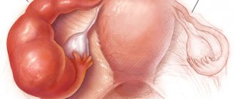
- thoracic endometriosis - forms in young females in the first few days of menstruation. The disease most often affects females aged 30 to 40 years;
- rupture of an abscess into the pleural cavity.
Even newborn babies can get pneumothorax, but this happens extremely rarely, only in 2% of cases. This often happens due to:
- disturbances in the process of postpartum expansion of the lung;
- rupture of lung tissue, which can occur during artificial ventilation of the lungs;
- congenital anomalies of the development of lung tissue - in this case, cysts or bullae are found in babies;
- respiratory distress syndrome.
Mechanism of development of spontaneous pneumothorax
Pneumothorax - classification
During diagnosis, it is important to determine what type of pneumothorax is. The classification is quite complex; it consists of several parameters that make it possible to accurately describe the pathological process, its location and cause. Pneumothorax is classified as follows.
By etiology
There are three etiological varieties:
- Spontaneous - primary or secondary pneumothorax, developing for no apparent reason in the absence of trauma or damage;
- Artificial – this includes iatrogenic damage that occurs during therapeutic and diagnostic procedures;
- Traumatic – occurs with any wounds or injuries to the chest.
By severity
The severity of the pathology depends on the volume of trapped air:
- Local pneumothorax. The air compresses individual areas of the lung; it is not able to resolve on its own, but it does not occupy the entire volume of the pleural cavity.
- Extensive pneumothorax. Air occupies the entire pleural cavity, there is so much of it that it completely compresses the lung.
By distribution
There are two possible options:
- Unilateral. In this case, it is either left-handed or right-handed. The pleural cavity of one lung does not communicate with the pleural cavity of the other, therefore the pathological process can affect only one half of the chest. This type of pneumothorax is more common.
- Bilateral. This is a rare phenomenon that occurs when the pleura is damaged on both the left and right sides. A patient with such a pathology requires immediate medical attention.
According to complexity
Pneumothorax can be complicated or uncomplicated. In the first case, in addition to the main disease, other pathological processes that are pathogenetically related to it develop, for example, bleeding. In the second case, pneumothorax is the only pathology.
According to the method of communication with the external environment
In case of pneumothorax, the pleural space must communicate with the external environment at some period of time.
In one case, the message is constant and air enters the pleural fissure with each inhalation, and leaves it with each exhalation. This type of pathology is called open.
In another case, air enters once in large quantities and the hole closes. This type of pneumothorax is called closed.
Attention. The third option is the most dangerous, it is called valve. The opening through which air enters is covered by pleural tissue, which functions as a valve. On inhalation, the valve moves and air freely penetrates into the pleural cavity; on exhalation, the tissue returns to its place and the hole closes. With each inhalation, the volume of gas between the layers of the pleura increases.
Laboratory research:
1. When analyzing arterial blood gases, hypoxemia (PaO2 < 80 mm Hg) is observed in 75% of patients with pneumothorax. 2. The presence of underlying lung disease and the size of pneumothorax are closely related to changes in arterial blood gas composition. The main cause of hypoxemia is the collapse and decreased ventilation of the affected lung with preserved pulmonary perfusion (shunt effect). Hypercapnia develops rarely, only in patients with severe underlying lung diseases (COPD, cystic fibrosis); respiratory alkalosis is quite often present. 3. With VSP PaO2 < 55 mm Hg. Art. and PaCO2 > 50 mm Hg. Art. observed in 15% of patients. 4. ECG changes: deviation of the electrical axis of the heart to the right, decrease in voltage, flattening and inversion of T waves in leads V1-V3. 5. X-ray of the chest organs. To confirm the diagnosis, a chest x-ray is necessary (the optimal projection is anteroposterior, with the patient in an upright position). The radiographic sign of pneumothorax is the visualization of a thin line of visceral pleura (less than 1 mm) separated from the chest. Often with pneumothorax, a shift of the mediastinal shadow to the opposite side is detected. Since the mediastinum is not a fixed structure, even a small pneumothorax can lead to displacement of the heart, trachea and other elements of the mediastinum, therefore a contralateral shift of the mediastinum is not a sign of tension pneumothorax. Approximately 10–20% of pneumothoraxes are accompanied by the appearance of a small pleural effusion (within the sinus), and if the pneumothorax does not expand, the amount of fluid may increase. In the absence of signs of pneumothorax, according to the radiograph in the anteroposterior projection, but in the presence of clinical data in favor of pneumothorax, radiographs in the lateral position or lateral position on the side (decubitus lateralis) are indicated, which allows confirming the diagnosis in an additional 14% of cases. An X-ray sign of pneumothorax in a patient in a horizontal position (more often with artificial ventilation of the lungs: mechanical ventilation) is the sign of a deep sulcus (deep sulcus sigh) - a deepening of the costophrenic angle, which is especially noticeable when compared with the opposite side. 6. Computed tomography. CT is a more reliable method compared to radiography for the diagnosis of small pneumothoraxes, for the differential diagnosis of large emphysematous bullae and pneumothorax. CT is indicated to determine the cause of VSP (bullous emphysema, cysts, interstitial lung diseases, etc.).
Determination of the size of pneumothorax.
The size of pneumothorax is one of the most important parameters that determine the choice of treatment tactics for patients with PSP. A simple approach to determining the volume of pneumothorax is proposed.
Pneumothoraxes are divided depending on the distance between the apex of the lung and the dome of the chest: small pneumothorax with a distance of less than 3 cm, large - more than 3 cm; and also depending on the distance between the lung and the chest wall: less than 2 cm and more than 2 cm, respectively.
Clinic
It is important to recognize pneumothorax as soon as possible and begin providing first aid.
When examining a patient with pneumothorax, the physician observes the following:
- Position in bed. The patient lies on the sore side and tries to compress the sore half of the chest as much as possible. This reduces the excursion of the lungs, the movement of the pleura, and, as a result, pain. If there is an open wound, the patient presses it with his hand.
- Changing the configuration of the chest. With severe pneumothorax, the affected side increases in size, the intercostal spaces are smoothed out, and the distance between the ribs increases. If pneumothorax bilateral chest becomes barrel-shaped.
- Skin color. The patient is pale and covered in sweat. The fingertips and nasolabial triangle may turn bluish.
Treatment
For patients diagnosed with pneumothorax, initial treatment occurs in a hospital setting. A pleural puncture is performed to remove air from the pleural cavity and restore the normal position of the lung. In open collapse, sutures are first placed in the open area to stop air from entering. In case of valvular pneumothorax, a puncture is performed, thus turning it into an open one.
Further treatment of all types is carried out approximately the same. Use aspiration with a thick needle. In order not to provoke a sharp expansion of the lung in case of complete pneumothorax and in case of damage to lung tissue, a drainage system is placed in the pleural cavity.
One of the main aspects of pneumothorax treatment is analgesia. This is necessary for the patient both during the development of pathology and during the period of restoration of lung volume. To prevent re-development of the disease, artificial provocation of the formation of scars and adhesions using sclerosing drugs can be used.
During the recovery period, the patient is advised to strictly follow medical recommendations. It is necessary to stop smoking, not subject the body to significant stress, and adhere to a balanced diet. Also, in case of a tuberculosis process that has provoked the development of pneumothorax, breathing exercises and physical therapy under the guidance of an instructor are indicated.
Pneumothorax - symptoms
Pneumothorax can be suspected not only by the appearance of the patient, but also by his complaints. The disease manifests itself as follows:
way:
- Chest pain. It always occurs when a hole appears in the pleura and then when air passes through it.
- Dyspnea. The patient breathes shallowly to reduce chest excursion, and the respiratory rate increases.
- Cough. A dry, obsessive cough occurs when there is a hole in the visceral pleura.
- State of shock. Painful or traumatic shock may occur, accompanied by tachycardia and hypotension.
In addition to the listed general symptoms, there are individual manifestations characteristic of each type of pneumothorax. They are able to indicate its origin. There are five types of pneumothorax.
Spontaneous pneumothorax
Spontaneous pneumothorax is called a pneumothorax that is not preceded by damage.
It is divided into two types based on origin: primary and secondary. The first occurs if there were no pathological processes in the chest before its appearance.
For reference. Primary is a rare type of pathology that occurs due to genetic changes in the connective tissue of the lungs. For example, with a lack of alpha1-antitrypsin.
Secondary occurs against the background of an existing pathology. In this case, pneumothorax is preceded by symptoms of the underlying disease. Pneumothorax itself is its complication.
Mechanical
It is divided into two types: open and closed.
The first occurs in case of injury from a piercing, cutting or firearm. In this case, the diagnosis is simplified: a deep bleeding wound is visible, from which air comes out as you exhale.
A closed wound is associated with injury to the cartilaginous or bony part of the ribs. The main symptom is sharp pain at the fracture site, pathological mobility of broken ribs, and a crunching sound when palpated.
Iatrogenic
It is a consequence of accidental or intentional damage to the pleura by a doctor.
Attention. Accidental pneumothorax is quite easy to suspect: the patient feels a sharp pain during manipulation with a sharp object in the chest area.
Intentional pneumothorax is created for therapeutic purposes if it is necessary to ensure functional rest of the lung. This method treats tuberculosis that is resistant to standard therapy.
Menstrual
The reasons for this phenomenon are not completely clear. It is classified as spontaneous recurrent pneumothorax, developing within three days after the onset of menstruation.
There are two main mechanisms.
The first is associated with pulmonary endometriosis - the growth of tissue of the uterine mucosa on the surface of the lung. During menstruation, this tissue behaves in the same way as in the uterus - it sloughs off, creating an opening in the lung.
The second option is more dubious. During menstruation, air enters the uterus through the vagina, and from there it moves into the abdominal cavity. In women with fenestrations—small holes in the diaphragm—this air can enter the pleural space.
For reference. Recognizing menstrual pneumothorax is not so easy. To do this, the symptoms of pneumothorax must be associated with a specific phase of the menstrual cycle. Since these issues are dealt with by doctors of different specialties, the diagnosis may not be made immediately.
Pneumothorax in infants
Before birth, the baby’s lungs do not function, so any pathology in them remains unrecognized.
If the fetus has cysts, bullae, tissue deficiencies, or connective tissue pathology in the lungs, it may have a pneumothorax at birth.
The fact is that with the first inhalation, the lungs expand for the first time, and the pressure in them increases significantly. Places where the tissue does not correspond to normal are torn, and the child experiences an accumulation of air.
Attention. The main symptom is an increase in respiratory failure after the first breath, intense crying. Newborns develop complications more often and faster.
Kinds
Based on the nature of the communication with the upper respiratory tract, three variants of lung collapse are distinguished.
- When air enters the pleural cavity during inhalation and escapes through a fistula in the visceral pleura during exhalation, an open pneumothorax develops.
- If air enters in the same way, but cannot escape back because the hole is blocked, a closed pneumothorax occurs. This is the easiest option of all possible.
- Valvular develops when a valve is formed in the area of damage to the visceral pleura, preventing the escape of air.
With closed collapse, air is absorbed within a fairly short time, and tension pneumothorax is characterized by a progressive accumulation of air in the pleural cavity.
Depending on the volume of gas accumulated in the pleural cavity, spontaneous pneumothorax can be limited or total. Most often, partial collapse of the lung is observed. According to the nature of its spread, lung collapse can be unilateral or bilateral. The most severe case, entailing complete cessation of respiratory function and rapid death in the absence of immediate help, is total bilateral collapse of the lungs.
Artificial pneumothorax is provoked specifically during an operation called thoracoplasty. In severe cases of disseminated tuberculosis, such surgical intervention is sometimes necessary.
Complications
Possible complications of the disease include the following pathological processes:
- Subcutaneous hyperairiness. In this case, air penetrates not only into the pleural cavity, but also under the skin. In this case, the skin rises slightly, and when pressure is applied to it, crepitus occurs.
- Mediastinal hyperairiness. In this case, air enters the mediastinum through a lesion in the parietal pleura. This compresses the mediastinal organs, including the heart and pericardium.
- Inflammation of the pleura. It occurs when, in addition to air, pathogenic microorganisms penetrate into the pleural fissure.
- Bleeding. Blood can flow outward or into the natural spaces of the chest - the sinuses.
What it is?
In pulmonology, this condition is understood as spontaneous pneumothorax, which is not associated with trauma or iatrogenic therapeutic and diagnostic intervention. The disease, according to statistics, occurs more often in men, prevailing among people of working age, which determines not only the medical, but also the social significance of the problem. In the traumatic and iatrogenic form of spontaneous pneumothorax, the cause-and-effect relationship between the disease and external influences is clearly monitored, which can be various chest injuries, puncture of the pleural cavity, venous catheterization, pleural biopsy or barotrauma. But in the case of spontaneous pneumothorax, there is no such conditionality. In this regard, the choice of adequate diagnosis and treatment tactics seems to be the subject of increased attention from pulmonologists, phthisiatricians and thoracic surgeons.
Diagnostics
Diagnosis is based on examination of the patient and additional examination. Diagnosis must be quick in order to begin providing assistance to the patient as soon as possible.
Pneumothorax is detected as follows:
- Physical examination. Includes percussion and auscultation. During percussion, a tympanic sound is heard over the entire air space; normally, the sound should be clear pulmonary. Auscultation is characterized by the picture of a “silent lung” - breathing, wheezing, and heartbeat are not heard.
- Radiography. Characteristic is total clearing, corresponding to air in the pleural cavity. The corresponding lung is compressed.
- Spiral tomography. It is an analogue of radiography and allows you to more accurately determine the extent of pneumothorax and its cause.
These methods are sufficient to begin treatment of pneumothorax. Later, additional tests can be done to determine the etiology of the disease, if necessary.
Diagnosis of open pneumothorax
The diagnosis of open pneumothorax is made on the basis of the patient’s complaints, anamnesis (history) of pathology, and the results of additional research methods (physical, instrumental, laboratory).
From the anamnesis, the following is revealed:
- what were the circumstances of the penetrating violation of the integrity of the chest;
- does the victim have a history of pathologies of the respiratory system;
- whether he had previously suffered similar chest injuries;
- whether he underwent surgery on the ore cell and its organs.
A physical examination reveals the following:
- during a general examination, the general condition of the victim is moderate or severe; he sits and, to facilitate breathing, rests his hands on the bed or on a chair (depending on what he is sitting on). This helps him fix the shoulder girdle and “push” air into the lungs. The skin and visible mucous membranes of the victim are of a pronounced pale bluish color, while the skin is covered with cold sweat. When air enters the subcutaneous fatty tissue of the neck, swelling of its soft tissues occurs;
- upon local examination, a penetrating wound to the chest is discovered, through which air “whistles” into the pleural cavity;
- palpation (palpation) confirms swelling of soft tissues;
- during percussion (tapping) - a box sound is heard over the area of the pleural cavity during percussion, over a collapsed lung - a dull sound, as if someone is knocking on a wooden object;
- during auscultation (listening with a phonendoscope) - breathing is not heard from the affected side of the chest; sharply weakened vesicular breathing can sometimes be heard at the root of the lung.
Characterized by significant disturbances in central hemodynamics (blood flow):
- tachycardia – a significant increase in heart rate and pulse. Arrhythmia may occur;
- hypotension – decreased blood pressure.
When conducting additional research methods, it is first necessary to involve those that will allow you to diagnose open pneumothorax, and then all the others that will allow you to evaluate the disorders that led to the development of the pathology. It should be remembered that you need to spend as little time as possible on the diagnostic process and provide medical assistance to the victim as early as possible. Conducted:
X-ray examination of the chest - it is done in two projections. X-ray photographs reveal the absence of a pulmonary pattern on the affected side, a collapsed lung, pressed to the root;- diagnostic thoracentesis - using a trocar (an instrument with a sharp end) a puncture is made in the chest wall, a portion of air comes out of it under pressure (sometimes with a whistle);
- diagnostic thoracoscopy - a small incision is made in the chest wall, a thoracoscope (endoscopic equipment) is inserted into the chest cavity through it, the pleural cavity is examined from the inside, the collapsed lung is identified, pressed to the root, and the degree of damage to the lung and pleura is also assessed;
- spirography - it is performed if an open pneumothorax has been converted to a closed one, and the signs are not critical. The victim makes a series of inhalations and exhalations, and also holds his breath, while the volume of air is measured, the impairment of the respiratory function of the lung is assessed;
- computer methods - computer sections allow one to evaluate the characteristics of a wound defect in the chest wall and the condition of the collapsed lung. Computed tomography (CT) and its improved version - multislice computed tomography (MSCT) are used, which allows obtaining more accurate information;
- Magnetic resonance imaging (MRI) – the goals and objectives are the same as for CT and MSCT.
Laboratory research methods used in the diagnosis of the described pathology are:
- general blood test - optionally detects signs of anemia (decreased levels of red blood cells and hemoglobin);
- bacterioscopic examination - it is carried out if a secondary infection is suspected (this can occur against the background of congestion in the lung that has collapsed as a result of pneumothorax). Microscopy of sputum is carried out, the causative agent is detected and identified in it;
- bacterioscopic examination - the goal is the same as when carrying out the bacteriological method. Sputum is cultured on special nutrient media, colonies are expected to grow, and the infectious agent is identified from them. Also, using this method, the sensitivity of the associated infection to antibacterial drugs is determined, which is important in the subsequent relief of pathology.
Treatment of pneumothorax
First aid should be provided by a doctor or paramedic, since correct diagnosis and special manipulations are necessary. Regardless of the type of pathology, after first aid is provided, the patient is taken to the thoracic department.
Sometimes measures taken during emergency care are sufficient. The fact is that the pleura quickly heals on its own; you just need to remove the air and make the space airtight.
For reference. The patient needs rest and strict bed rest. He will have to walk with drainage or a bandage for some time. If the tissue defect is large or the pleural cavity communicates with the lungs, surgical intervention is necessary.
It is performed by thoracic surgeons, the scope of the operation varies - from suturing the defect to resection of the lung or its lobe. It is impossible to cure pneumothorax at home.
First aid and treatment
Emergency care for spontaneous pneumothorax, especially if a valve has formed, should be provided before hospitalization of the patient and confirmation of the diagnosis. The ambulance team punctures the second intercostal space and administers oxygen therapy to compensate for respiratory failure.
Despite the fact that with minor defects in the lung tissue, self-healing of the fistula and just one puncture is possible, a wait-and-see attitude is not justified. The pleural cavity is drained. It takes 1 to 5 days for the lung to fully expand and restore its function.
Typically, the fistula is closed surgically in 5–20% of patients.
Prognosis and prevention
The prognosis of the disease is always questionable. It is difficult to say what consequences it will have. Even after self-healing pneumothorax, scars may remain on the pleura, causing pain during breathing.
In addition, if the lung remains in a collapsed state for a long time, it may no longer expand. This occurs due to the so-called moorings - areas of connective tissue. The pneumothorax will be cured, the pleural cavity will no longer compress the lung, but the moorings will keep it in a collapsed state.
It is worth saying that even in the absence of ventilation of one lung, the patient can feel healthy, since the second lung completely takes over the function of external respiration.
Attention. With bilateral pneumothorax, the prognosis is unfavorable, since respiratory failure rapidly increases.
Prevention of pneumothorax involves treating diseases that can lead to damage to the pleura. It is also important to remember to wear your seat belt while in the car.
Forecast
The prognosis for open pneumothorax is different, but more favorable than for tension pneumothorax. Timely detection and adequate care increase the patient's chances of survival.
The prognosis worsens in cases such as:
- late diagnosis;
- inadequate therapy;
- elderly or children's age;
- the occurrence of complications;
- deterioration of general condition;
- the presence of concomitant pathologies.
Kovtonyuk Oksana Vladimirovna, medical observer, surgeon, consultant doctor
just today
( 55 votes, average: 4.62 out of 5)
Feeling of suffocation: reasons, what to do, which doctor to see
Chlamydial pneumonia: symptoms, treatment, prevention
Related Posts
Clinical manifestations of the disease
As noted above, the appearance of this pathological condition is more often observed in men from 20 to forty years old.
In almost 70% of cases, the onset of the disease is sudden, which in its typical course is accompanied by:
- A painful sensation of a stabbing nature in the affected part of the chest, radiating to the neck, arm, and sometimes to the epigastric region. When such pain appears, a person has a fear of dying. Painful sensations can appear as a result of excessive physical activity, coughing, and its appearance can also be observed at rest (even at night while sleeping). In many cases, the factor that led to the pain cannot be determined.
- Shortness of breath, which is characterized by a sudden onset; in patients, breathing accelerates and becomes shallow. Despite this, respiratory failure does not appear in humans; such cases are rare.
- Unproductive cough. This symptom is not present in all patients.
After a few hours or even minutes, the patient’s general condition improves somewhat, as the intensity of pain and shortness of breath decreases. Painful sensations can only appear when a person takes a deep breath, and shortness of breath as a result of physical activity.
If we talk about the atypical development of spontaneous pneumothorax, then it is observed in almost 20% of all cases and is characterized by a gradual and imperceptible onset for the patient. Clinical manifestations, pain and shortness of breath are considered minor and pass quickly as the person adapts to new breathing conditions.
Please note that the atypical nature of the onset of the pathological condition is observed in the case of penetration of a small amount of air into the pleural cavity.
Examination and physical examination of the patient will show:
- His forced position, which he takes to alleviate the condition;
- Increased sweating, cold sweat;
- Change in skin color, it will be cyanotic;
- Expansion of intercostal spaces;
- The fact that movements of the chest from the side where the lesion is localized are not carried out;
- Tympanitis during percussion;
- Accelerated heartbeat;
- Hypotension;
- Weakening or disappearance of vocal tremors when listening;
- Shift of the cardiac impulse area to the healthy side.
Please note that physical abnormalities may not be observed if a small amount of air accumulates. They make themselves known when the process of lung collapse occurs by 40% or more.
The causes of this pathological condition
In most cases, spontaneous formation of pneumothorax is observed in people who have not had diagnosed lung diseases.
Studies have shown that in 80-100% of all patients emphysematous bullae were found, which were localized subpleurally.
A definite connection between the appearance of this pathological condition was noted in individuals who were tall and skinny. Smoking increases the risk of pneumothorax by almost twenty times.
The causes of pneumothorax of secondary origin are associated with a history of the following diseases:
- Lung diseases - COPD, cystic fibrosis, bronchial asthma;
- Lung diseases of an infectious nature (pneumocystis or abscess pneumonia, tuberculosis);
- Beck's sarcoidosis;
- Pneumosclerosis;
- Granulomatosis;
- Rheumatoid arthritis;
- Gastric ulcer;
- Scleroderma;
- Malignant neoplasms.
Quite rare, but there are such forms of pneumothorax as:
- Menstrual. This form of the disease is closely related to thoracic endometriosis. More often, its development is observed in young girls in the first two days of the menstrual cycle. An important point that requires attention is that this form of the disease has a high risk of relapse. When diagnosing this pathological condition, it is often recommended to perform pleurodesis, which will help eliminate the risk of recurrence of pneumothorax.
- Neonatal. This form is rare and most often affects boys. May be associated with problems during lung expansion, respiratory distress syndrome, injury to lung tissue, and lung malformations.
The following are no exceptions:
- Diving underwater to great depths;
- Skydiving;
- Flying an airplane at high altitude.
All these reasons led to the formation of a pathological condition as a result of sudden changes in pressure, which act unevenly on different parts of the lungs.
Pathogenesis
The degree of severity of the structural change directly depends on the time that has passed since the onset of the disease. In addition, it depends on the presence of the original pathological disorder in the lung and pleura. The dynamics of the inflammatory process in the pleural area has no less influence.
Against the background of spontaneous pneumothorax, a pulmonary-pleural communication occurs, which determines the penetration and accumulation of air in the pleural area. Partial or complete collapse of the lungs may also occur.
The inflammatory process develops in the pleural area four hours after spontaneous pneumothorax. It is characterized by the presence of hyperemia, injection of pleural vessels and the formation of a certain amount of exudate. Over the course of five days, swelling of the pleura may increase, mainly in the area of its contact with trapped air. There is also an increase in the amount of effusion along with fibrin deposition on the pleural surface. The progression of inflammation may be accompanied by the proliferation of granulations, and, in addition, fibrous transformation of the deposited fibrin occurs. The collapsed lung is fixed in a compressed state, so it becomes unable to expand. If an infection occurs, pleural empyema may develop over time. It is possible that a bronchopleural fistula will form, which will support the course of pleural empyema.