Purpose of the event
The 15th obstetric week is close to the scheduled screening of the 2nd trimester. Whether or not to perform such a study during this period is decided only by the gynecologist who is observing the pregnant woman. The examination is performed at the 15th week of pregnancy, mainly for certain medical reasons.
At this stage of intrauterine development, an ultrasound diagnostic specialist can determine only some pathologies. Also, using this method, certain chromosomal and genetic diseases are determined. The appearance of technical errors at this stage occurs much less frequently than in the initial weeks of pregnancy.
The beginning of the second trimester is the period when the baby’s heartbeat is already clearly audible. The baby's heart beats at a speed of 135-170 beats per minute. This heart rate is normal. After giving birth, your heart rate will gradually decrease. This indicator is physiological and does not require any correction.
During this period, the thyroid gland begins to work in the baby, who is still in the mother’s womb. It is still difficult to call such functioning full-fledged. The fetal thyroid gland takes up iodine, but cannot yet synthesize hormones.
Some expectant mothers already feel the active movements of their babies. The fetus develops and begins to actively push. In some pregnant women, similar symptoms appear 2-3 weeks later.
Many pregnant women note that they noticed active movements of their babies only towards the end of the 2nd trimester.
At the fifteenth week, doctors evaluate the functioning of all these internal organs. All membranes are also assessed. Carrying out a study, an ultrasound specialist determines pathologies not only in the fetus, but also in the mother. To do this, he carefully examines all the organs of her reproductive system using an ultrasound sensor.
Sex determination can also be done at this stage. However, for this purpose the study is carried out a little earlier. Qualified doctors can determine the sex of the unborn baby as early as 11-12 weeks of pregnancy.
The composition of the amniotic fluid that surrounds the fetus is sterile at this stage. Their number is a very important indicator.
An excessive amount of amniotic fluid indicates that the child is developing dangerous pathologies.
Is it possible to determine the sex of the baby at 15 weeks of pregnancy?
The baby's internal organs gradually begin to work.
The heart pumps blood at a speed of about one liter per hour. The liver begins to perform functions related to digestion. Work starts in “test” mode of the gastrointestinal tract and hepatobiliary system. At this time, the intestines are filled with contents that will become the baby's first stool. This is mainly a bile-containing mass, thick green in color. The bladder works under load, ensuring regular urination every 45 minutes.
At this point, it is already possible to determine the blood type and Rh factor of the developing baby, since the blood is almost completely formed.
Almost entirely because it already contains all the cells of an adult, with the exception of hemoglobin. More precisely, hemoglobin is also present in the blood. But this is newborn hemoglobin.
It will be replaced with an ordinary one only 6 months after the birth of the baby.
At week 16, the baby’s cardiovascular system is actively working, feces and urine are produced. The future baby learns to actively interact with his environment, the body's systems are preparing to transition to an autonomous mode of existence.
Uterine changes
From the moment of pregnancy, the uterus continues to increase in size, reaching two hundred and fifty grams of weight by the 16th week, and in height - 50% of the distance to the navel. These numbers, obtained as a result of the doctor’s appropriate measurements, provide information about how the pregnancy is progressing and with what consistency the unborn child develops.
The fact that the uterus is located approximately 7 cm below the navel can be determined independently by palpation. An enlarging uterus can negatively affect the functioning of the intestines, causing consequences such as constipation, heartburn and other unpleasant sensations.
The amount of amniotic fluid is also increasing, reaching 250 grams by this week.
Laboratory diagnostics at 16 weeks
At 16 weeks of pregnancy, the number of tests increases compared to the weeks of the previous period. The increase occurs due to blood tests aimed at identifying possible fetal abnormalities and genetic defects. Now, in addition to OBC and OAM, measuring weight and pressure, as well as monitoring the heart function of the unborn child, they also do:
In this case, the likelihood of identifying genetic abnormalities and chromosomal abnormalities (defects in the development of the spine and brain structures) increases.
In addition to this example, Down syndrome and anencephaly can be cited.
If these tests indicate the presence of abnormalities, then special additional studies will need to be carried out to formulate a final diagnosis. Full confidence in the correctness of the diagnosis is necessary.
The period of 16 weeks is practically the equator of pregnancy, so it is very important for the doctor to identify possible genetic chromosomal abnormalities in the early stages. To do this, pregnant women take a blood test for hormones. Not all hormones are tested, but only those produced during pregnancy
Ultrasound diagnostics at 16 weeks
With the help of an ultrasound, you can see your unborn baby and hear its heartbeat. If the spouses want to find out its gender, during this period of the gestational process it is already possible to distinguish the sex of the child.
Signs of gender are not yet clear enough when attempting visual recognition. However, according to some criteria, the doctor can determine the sex of the baby.
It is believed that at this stage the gender can be recognized with 50% confidence.
The baby is active at this stage, you can see his movement on the monitor, how he moves, how his facial expressions express his attitude to the actions of the outside world. By this time, research can be carried out in two ways:
- transabdominal;
- transvaginally.
The first method is carried out using a sensor moved over the surface of the abdomen. In the second case, research is carried out with a sensor inserted into the vagina. In this case, it becomes possible to conduct cervicometry - to find out what the cervix looks like and what its condition is.
Transabdominal ultrasound examination requires special preparation, which is not necessary for transvaginal diagnosis. In the first case, studies are carried out with a full bladder to avoid distortions. You should also not eat heavy food on the day of the examination. At this stage of pregnancy, ultrasound is prescribed for the following indications:
- threat of isthmic-cervical insufficiency;
- for those who missed this study in the first trimester;
- to exclude hereditary pathologies on the father's side.
Evaluation of fetal ultrasound data
To determine the rate of fetal development, the main fetometric indicators are determined:
- The normal range of biparietal size (transverse size of the fetal head) is from 31 to 37 mm. The average value is 34 mm.
- To diagnose the gestational age, the circumference of the fetal head is also important. The exhaust gas should lie in the range of 112-136 mm. The average value corresponds to 124 mm.
- For abdominal circumference, the average is 102 mm for the range 88-116 mm.
- The average values for femur length and upper arm length are 20 and 18 mm, respectively, for the ranges 17-23 and 15-21 mm.
- The length of the tibia should be 15-21 mm, which corresponds to an average value of 18 mm.
- And the length of the forearm has an average size of 15 mm (12-18 mm).
To exclude developmental defects, measurements are taken on different limbs of the same name and the results, which normally should be equal, are compared. Long bones should be straight. Ultrasound shows smooth, continuous lines. In some cases, an important point for parents is determining the sex of the fetus.
How to evaluate discharge?
At 16 weeks, a pregnant woman may be alarmed by possible changes in the nature of the discharge. A change in color to whitish and an increase in the amount of discharge is the norm for this period of gestation.
Normally, the discharge should be homogeneous and not cause itching or burning. There should be no pain in the lower abdomen. During this period, you need to be more careful about personal hygiene to avoid infection.
You need to take care of the cleanliness of the genitals and perineum.
The nature of vaginal discharge changes during pregnancy, but some characteristics are not normal. So, burning or itching, pain in the lower abdomen is a signal for an urgent visit to the doctor. While carrying a child, a woman’s body is quite weak and susceptible to infections; hygiene rules must be carefully observed
However, if an infection occurs, the discharge will also intensify. But they will take the form of a curd mass, will be heterogeneous, with mucus. They may even acquire a yellow or greenish tint and an unpleasant, fishy odor.
These are clear signs of an infectious process (thrush, colpitis, vulvitis). A timely visit to a doctor will help the prescribed treatment have an effective effect on the disease.
Brown discharge with blood requires extraordinary medical intervention, as it may be a sign of miscarriage.
Condition of the fetus at 15 weeks
The beginning of brain formation is a responsible and important moment in the pregnancy process. The formation of brain structures begins in the 15th week of pregnancy. During this period, the process of nerve cell division begins and the central nervous system is formed. Both hemispheres of the brain are covered with convolutions and grooves.
By the 15th week, the endocrine glands begin to work intensively, and testosterone is actively produced in a male child.
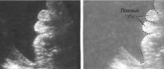
The taste buds have already fully formed, and the baby begins to recognize the food that the mother eats. The genitals are formed and the sex becomes distinguishable.
The rings of the umbilical cord or the baby's arms are sometimes perceived as a boy's penis, and therefore errors may occur in determining the sex of the baby, which are corrected during the next ultrasound.
The heart pumps about 28 liters/day of blood, its beat is about 150 beats/min. The gallbladder secretes bile, which then becomes the baby's first stool. The kidneys actively secrete urine.
It is excreted directly into the amniotic fluid. The respiratory organs are improved and the lungs are intensively trained. The baby actively makes breathing movements, due to which lung tissue is formed.
By this time, the gap is formed and open.
There is an active process of development of the child’s musculoskeletal system. The legs of the fetus become longer than the arms. His skin is still thin and translucent, but its appendages are actively developing (his hair is becoming thicker).
At the 15th week of pregnancy, an important stage begins - the formation of the baby’s brain structures. In addition, his heart, lungs, and excretory system begin to work more actively. If the baby’s position is favorable, an ultrasound can reveal its gender, but sometimes the data requires subsequent confirmation
The state of the structures that support the vital functions of the fetus
During pregnancy at 15 weeks, an ultrasound diagnostic doctor evaluates the functioning of the uterus-fetus system. He carefully studies the structures and functional state:
The uterus is easily palpated through the peritoneum at the level from the navel to the pubis. At this stage, the doctor can assess the attachment of the placenta, its condition, the thickness of the myometrium (about 2.5 cm normally), its structure (normally homogeneous).
Preparatory activities for the procedure
Ultrasound at 15 weeks does not require special preparatory measures. For this procedure, it is important to be in a good mood and rested. Since the reaction to a stress factor can cause tension in the myometrial muscles, which will be perceived as a threat of pregnancy failure.
Some ultrasound scanners require that the bladder be full for clear images. You should check this point with your doctor in advance, and if necessary, drink up to 0.5 liters of clean, still water. A few days before the examination, it is better to exclude foods that cause gas from your diet.
Also, don’t get carried away with fatty, fried and smoked foods.
Some parents do not want to find out the sex of the baby before birth. It is worth warning the ultrasound diagnostic doctor about this, because at 15 weeks, a good specialist is able to recognize the sex of the fetus. Most ultrasound machines allow you to take a photograph of your baby in the womb. If you want to see the baby on the monitor or take a picture, you need to notify the doctor before the examination begins.
a disposable sheet with you and cover the couch. And disposable dry wipes to wipe off excess special gel.
Most often, an ultrasound of the 15th week of pregnancy is performed transabdominally - this method does not cause inconvenience to the expectant mother. Sometimes the doctor requires filling the bladder before the study - you need to find out about this in advance and drink 0.5 liters of clean water first
Ultrasound at 15 weeks
This is a painless procedure and is comfortable for the patient. During the process, you just need to lie comfortably and not worry. The procedure is performed transabdomnally. The doctor lubricates the skin at the site of contact with the sensor with hypoallergenic acoustic gel.
Then, using a transducer (sensor), it performs diagnostics. Ultrasound is considered one of the safest methods of examining a mother and her baby at any stage of pregnancy.
Even at the very early stage of pregnancy and at 15 weeks, when the baby’s brain structures are intensively formed.
Structures assessed by ultrasound at 15 weeks
The results of the diagnostic procedure at 15 weeks give the doctor the opportunity to assess the situation and begin treatment in time if necessary. Ultrasound allows the obstetrician to:
- get acquainted with the state of affairs in real time;
- diagnose pathological processes at the very beginning of their inception;
- recommend adequate and timely treatment to the expectant mother.
During the diagnostic process, the doctor assesses the condition of the fetus, its main structures and their functionality. Compares data with existing standards.
Even if an ultrasound was already performed before the 15th week, by this time the baby has changed so much that the mother will be interested in seeing the changes, and the doctor will be able to obtain informative data about the baby’s main body systems.
The period of 15-16 weeks is a favorable period for the use of ultrasound diagnostics:
- Firstly, when pathology is detected at this stage, the process is easier to correct than at a later stage.
- Secondly, if a severe or incompatible pathology is identified and a decision is made to terminate the pregnancy, this period allows minimizing the loss of the woman’s psychological state to the body.
- Thirdly, the baby’s organs and systems are working actively and it is possible to assess their functional state, which was previously impossible.
- Fourthly, it is possible to find out the sex of the child during an ultrasound diagnostic procedure at 15-17 weeks.
Source: https://zdorovo.live/beremennost/mozhno-li-opredelit-na-15-nedele-beremennosti-pol-rebenka.html
Methods of conducting the examination
During this period of intrauterine development, research can be carried out in different ways. The most commonly used method is transabdominal examination. To do this, the doctor examines the expectant mother's belly using a special sensor.
For better refraction and reflection of ultrasonic waves, a special diagnostic gel is used. It is applied to the skin of the pregnant woman's abdomen. The hypoallergenic chemical composition of the gel is not capable of causing any allergic reactions in women.
If the obstetrician-gynecologist recommends a transvaginal examination, then in this case the doctor inserts an ultrasound probe into the vagina. This ultrasound method is usually chosen in the early weeks of pregnancy.
The examination is carried out in a regular ultrasound room. Before the procedure, the pregnant woman is placed on a special couch. In some cases, the ultrasound technician may ask the expectant mother to turn on her left side. In this case, the uterus can be better visualized.
If an expectant mother goes to a regular clinic for examination, she should take paper napkins and a towel with her. Using a handkerchief you can remove any remaining gel from the skin. A towel is placed on the couch before the procedure. If the study is carried out in a private clinic, then in this case all consumables are provided and included in the cost of the procedure.
The duration of the study may vary. This is largely determined by the experience and qualifications of the specialist who conducts the examination. On average, the procedure lasts 30-40 minutes. If additional Doppler sonography is required, the duration of the study in this case can be about an hour.
What structures can be identified?
At the 15th week of pregnancy, the fetus is already clearly visualized in the uterus. Modern devices allow you to get a fairly clear image. It is displayed on a special screen. With the help of such a monitor, the expectant mother can examine her unborn baby together with the doctor.
If during the examination the doctor reveals any serious pathologies, he can immediately tell the pregnant woman about it. Also, these violations are entered into a special medical report form, which is given to the patient. It is permissible to take photographs during such a study. They are also handed over and attached to the medical card.
Modern 3D and 4D techniques make it possible to obtain three-dimensional images in which even the smallest structures of the baby’s body are visualized. The expectant mother can see how her baby moves or bends his fingers. Also, during the examination, spontaneous movements of the fetal chest are often visible.
At this stage of intrauterine development, a doctor can determine some heart defects in the fetus. Normally, a baby's heart consists of 4 chambers. Between them there are special holes that are closed with valves. The presence of abnormalities in the valvular apparatus of the heart leads to the formation of heart defects.
Week 15 is the time for mandatory assessment of the pregnant woman’s cervix. Gynecologists identify several types of pathologies that can develop in an expectant mother if there are defects in this reproductive organ. One of these dangerous pathological conditions is the development of isthmic-cervical insufficiency.
Also, during the study, an examination of the ovaries is required. This diagnosis is especially important for women who, before pregnancy, were diagnosed with various cysts or neoplasms in these reproductive organs. The ovaries play a very important role during pregnancy. They provide optimal hormonal levels, which are necessary for the proper development of the fetus.
How to determine the gender of a child according to folk signs
Whether traditional methods are trustworthy or not is up to you to decide. But although some of them seem like an absurd fiction, most of the signs have a completely logical scientific explanation.
Here are the most popular folk signs by which an expectant mother can determine the gender of her baby.
You may be having a girl if:
- you want something sweet;
- first trimester with severe toxicosis;
- the stomach is covered with pigment spots;
- the right breast is smaller than the left;
- you have gained noticeable weight;
- you have pimples;
- you cannot live without fruits and juices;
- your belly is round and raised;
- your mood is too changeable;
- the color of the urine has become dull;
- you often feel cold, chills;
- Do you prefer to eat bread crumbs?
- your heart rate is more than 130 beats per minute;
- It is more convenient for you to sleep on your right side.
You may be having a boy if:
- the stomach has a pointed shape;
- in the first weeks you did not feel sick;
- you want meat and cottage cheese;
- your feet are cold;
- you like sour and salty foods;
- hair on legs grows quickly;
- good mood, not changeable;
- clumsiness and frequent headaches appeared;
- legs swell;
- urine turned bright yellow;
- the skin of the hands and face has become drier;
- it is more convenient for you to sleep on your left side;
- you eat bread crusts.
You can also determine gender by remembering the process of conception. If the father was more active in the process of intimacy, then a daughter will be born, if the mother is a son.
Norms of indicators
Week 15 is very close to the second screening. This determines the fact that not many studied parameters are used to assess this period of intrauterine development of the fetus.
The size of the fetus at this stage of pregnancy is usually 10 cm. The weight of the baby is 60-70 grams. It is immediately worth noting that in case of multiple pregnancy, these parameters may be different. Quite often at this stage of pregnancy, future twins or triplets weigh less than 50 grams.
To visualize what a baby looks like at this stage, you just need to remember an orange. The weight of the fruit at this stage, as a rule, corresponds to the size of this fruit.
At the 15th week of intrauterine development, the baby already resembles a real person. His fingers, hairs and even nails are visible.
To determine uteroplacental blood flow, blood flow in the blood vessels of the placenta is determined. An experienced specialist conducting the study can identify pathological narrowing or even torsion of such vessels feeding the fetus. If such a situation does arise during pregnancy, then this requires an urgent consultation with a gynecologist.
The inner layer of the uterus is also examined. It is important that there are no myomatous nodules or any neoplasms on the endometrium. The thickness of the muscular layer of the uterus at this stage is usually 2-3 cm.
During the study, the position of the placenta is also determined. It is usually located either on the anterior or posterior wall of the uterus. There should be no excess growth of the placenta. If it grows to the lower corner of the uterus at this stage, then careful monitoring of its growth is required in the future.
The results obtained must be interpreted by an obstetrician-gynecologist. If the presence of certain pathologies is suspected, hCG is also determined. This biochemical marker allows you to identify various disorders in a variety of periods of pregnancy. The conclusion of one ultrasound examination cannot in any case become a diagnosis.
Changes in a woman’s body at 16 weeks of pregnancy
Now a woman who is 16 weeks pregnant does not feel any particularly strong changes in her physiological state. Unless he may be worried about the arrow on the scales, which is steadily moving in the direction of increase. Forget about a slim figure here. Until the baby is born, the weight will tirelessly grow, as will the tummy. However, control is also necessary in this matter: excessive obesity negatively affects a woman’s body. Excessive appetite and obesity usually cause severe edema, shortness of breath, and surges in blood pressure (BP).
At this stage, the expectant mother can already clearly see her rounded tummy
, and this sight clearly pleases him.
Now the volume of the uterus has already increased significantly. The volume of amniotic fluid is approximately 250 ml.
Another unpleasant thing: now the intestines are still not working well, so constipation is a fairly common occurrence.
Breasts continue to grow
– She is preparing to feed the baby.
However, there are some unpleasant moments here. Since blood flow increases significantly in the body, a venous pattern and the so-called “Montgomery tubercles” appear on the chest. stretch marks
may appear on the skin - not a very pleasant aesthetic sight.
You should worry about getting rid of stretch marks on your chest and stomach now. There are a lot of cosmetics that will prevent these unsightly scars from appearing on the skin. You can start using creams and ointments against stretch marks now. As they say, it is easier to prevent than to treat.
Is it harmful?
The second trimester is a time when conducting many studies is not at all dangerous. However, expectant mothers should remember the possible risk of adverse consequences if ultrasound is overused. Frequent use of this method without special medical indications can lead to various neurological disorders in the unborn baby.
In the normal course of pregnancy, there are no strict indications for an ultrasound examination at this stage. In this case, it would be reasonable to wait for the second screening. It is usually performed from 16 to 20 weeks of pregnancy. In this case, it is much easier to identify any pathologies.
To see what changes occur at 15 weeks of pregnancy, watch the following video.
Here is our photo from the ultrasound)
Oh, well, you are already so big)))
This is at 16 weeks)
Apparently there is a big difference between 15 and 16)
We have a boy, but I couldn’t catch him in the photo so that I could take a photo right between his legs. In the end, she gave me a photo, but it’s not clear to me, she showed me two little things there, like testicles))) my son didn’t want to show himself to me, although he immediately appeared to the uzistka, but then he began to toss and turn and turn away and I never managed to see with my own eyes pisyun))) in the end, an incomprehensible photo in which she said that there were balls