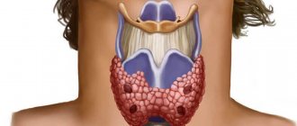
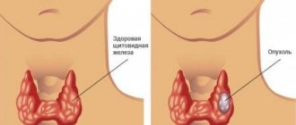
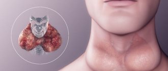
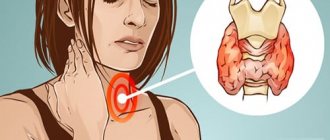
A structural feature of the thyroid gland is the presence of follicles that are filled with colloid. It contains precursors of thyroid hormones - thyroxine, triiodothyronine. Under unfavorable conditions (external or internal), the formation of colloid and hormones increases to provide the body with energy. If the speed of this process exceeds the outflow capabilities, the follicles stretch, forming nodes and cysts. A thyroid cyst is a cavity containing fluid inside.
Provoking factors : iodine deficiency; environmental pollution; sudden climate change; stress; inflammation, often of autoimmune origin; disruption of other endocrine organs; injuries; congenital developmental disorders, genetic predisposition; radiation; poisoning by toxic compounds; smoking; viral infections; chronic, long-term diseases of the ENT organs, especially tonsillitis.
Symptoms of the presence of formations are determined by size. Small formations are painless, are not accompanied by discomfort, and do not disrupt the synthesis of hormones. An endocrinologist often discovers them during an examination or ultrasound in connection with another pathology, during a routine medical examination.
The reason for a visit to the doctor is a visual deformation of the neck when the cyst grows and is easy to notice. This situation occurs when the diameter exceeds 3 cm. Patients during this period also note : unpleasant sensations (“lump”) when swallowing; cough, sore throat, hoarse voice; painful sensations in the cervical region; difficulty breathing. With large volumes, the formation can compress the carotid artery, which is manifested by dizziness, and fainting is possible.
Types of thyroid cysts
- Follicular. They are dilated follicles lined with a flat or cubic inner layer (epithelium). They can transform into malignant neoplasms.
- Colloidal. Colloid cysts arise inside nodular formations and are a stage in the progression of nodular goiter. It may not appear clinically for years, sometimes it suddenly begins to grow or decrease in size.
- Small, multiple. Even ultrasound cannot always detect them, especially with a size of up to 1.5 mm. Such cysts are dilated follicles containing an excess amount of colloid. They may disappear when the provoking factor is eliminated.
- Malignant. Only 5-6% of cysts degenerate into cancer. The vast majority of them are larger than 4 cm and cause compression of surrounding organs.
Colloid cyst of the thyroid gland
The most common complication with the development of a cyst of any location is the tendency of such formations to suppurate. The patient develops neck pain, fever, headache, nausea, body aches, and the lymph nodes become enlarged and inflamed. Large formations compress blood vessels and nerves, the esophagus and larynx. The most dangerous consequence is cancer.
Diagnosis of the patient: external examination, palpation, ultrasound, puncture biopsy, blood test for thyroxine and triiodothyronine. If cancer is suspected, CT and MRI are prescribed. To analyze compression of adjacent tissues, pneumography with X-ray control, angiography with the introduction of a contrast agent into the vessels, or examination of the esophagus with a barium suspension.
Treatment of the formation: if the size of the cyst is up to 1 cm, then no special therapy is performed. When the size of the puncture cyst is restored within a week during observation, surgery is recommended. Small cysts are treated with levothyroxine (Euthyrox) and iodine. In case of suppuration, antibiotics are prescribed after culture and determination of a suitable drug.
If the cystic formation is present in only one lobe, then the affected part is removed - hemithyroidectomy. The remaining share completely takes over the functions of the affected one. If the cysts are in two lobes, then almost all of the gland is removed .
Read more in our article about thyroid cysts, its manifestations and therapy.
Causes of thyroid cysts
A structural feature of this organ is the presence of follicles that are filled with colloid. It contains precursors of thyroid hormones - thyroxine, triiodothyronine. Under unfavorable conditions (external or internal), the formation of colloid and hormones increases to provide the body with energy.
If the speed of this process exceeds the outflow capabilities, the follicles stretch, forming nodes and cysts. A thyroid cyst is a cavity containing fluid inside.
The provoking factor for its formation may be:
- iodine deficiency;
- environmental pollution;
- a sharp change in climatic living conditions;
- psycho-emotional stress;
- inflammation, often of autoimmune origin;
- disruption of the functioning of other endocrine organs (pituitary gland, hypothalamus, adrenal glands);
- traumatic injuries;
- congenital developmental disorders, genetic predisposition;
- radiation;
- poisoning by toxic compounds;
- smoking;
- viral infections;
- chronic, long-term diseases of the ENT organs, especially tonsillitis.
We recommend reading the article about the symptoms and treatment of hypoparathyroidism. From it you will learn about the causes of hypoparathyroidism, classification, forms, symptoms in children and adults, as well as methods for diagnosing and treating this pathology. Find out more about the treatment of hyperparathyroidism here.
Cysts
Among all thyroid neoplasms, this pathology occupies a small segment, ranging from 3 to 5%.
The macroscopic structural element of glandula thyreoidea is a pseudolobule, consisting of follicles (also called acini or vesicles) surrounded by a capillary network. The inner surface of each follicle is lined with thyrocytes, and its cavity is filled with colloid in which protohormones of the thyroid gland are deposited.
The pathogenesis of the cyst briefly occurs in three stages:
- Impaired outflow of liquid contents of the follicular cavity, which can develop for various reasons.
- Colloid accumulation.
- Overstretching of the follicle walls and further growth of its size.
As a rule, the cyst does not affect the preservation of the functional abilities of the thyroid gland. The symptom complex is formed by other diseases of this organ, developing parallel to its growth, or triggering its development. As for the course of the pathological process, it is often benign, very rarely malignant, and then the cyst reaches an extremely large size.
As for the clinical manifestations of cysts, they can occur in completely different scenarios: in some cases, their sizes remain stable for many years, sometimes these neoplasms demonstrate very rapid growth and, conversely, cases of spontaneous disappearance of such pathological formations are observed.
Symptoms of the presence of formations
The clinical manifestations of the cyst are determined by its size. Small formations are painless and not accompanied by discomfort. They, as a rule, do not disrupt hormone synthesis. An endocrinologist often discovers them during an examination or ultrasound in connection with another pathology, during a routine medical examination. Such structures have a predominantly benign course.
The reason for a visit to the doctor is a visual deformation of the neck when the cyst grows and is easy to notice. This situation occurs when the diameter exceeds 3 cm. Patients during this period also note:
- unpleasant sensations (“lump”) when swallowing;
- cough, sore throat, hoarse voice;
- painful sensations in the cervical region;
- difficulty breathing.
With large volumes, the formation can compress the carotid artery, which is manifested by dizziness, and fainting is possible.
Nodes
Interesting! Representatives of the weaker sex suffer more from them (from 1:4 to 1:8 compared to men).
These pathological neoplasms are classified according to three main parameters:
- Quantity
(there are both solitary (single) and multiple). - Features of the course
(can be malignant and benign). - Ability to produce hormones
(there are autonomous toxic ones (actively releasing biologically active substances) and calm non-toxic ones).
The incidence of pathology increases with age.
Types of thyroid cysts
Depending on the structure of the cyst and its prevalence, several clinical variants of the disease have been identified. To accurately determine the type of formation, instrumental diagnostics is required.
Follicular
Such cysts are dilated follicles lined with a flat or cubic inner layer (epithelium). In most cases, they are found against the background of a diffuse goiter (enlarged tissue) of the gland. They are formed due to stagnation of colloidal substance. If the course is unfavorable, pathologies can transform into malignant neoplasms.
Colloidal
Its development is preceded by nodular goiter. Colloid cysts occur inside nodular formations and are a stage in the progression of the pathology. It may not appear clinically for years, sometimes it suddenly begins to grow or decrease in size.
Small, multiple
Even ultrasound cannot always detect them, especially with a size of up to 1.5 mm. When examining the tissue (histology), it is discovered that such cysts are dilated follicles containing an excess amount of colloid. They may disappear when the provoking factor is eliminated. They belong to the most favorable type of cystic changes in the thyroid gland.
Ultrasound of the thyroid gland (multiple thyroid cysts)
Malignant
Only 5-6% of cysts degenerate into cancer. The vast majority of them are larger than 4 cm and cause compression of surrounding organs. A biopsy is performed to distinguish benign from malignant lesions. More often, the diagnosis is confirmed during surgery with urgent histological analysis of a tissue sample.
Both shares
The course of the disease is determined by the type of cystic change in the thyroid gland. For small cavities, clinical signs are mild. If the size exceeds 3-4 cm, then such cysts lead to dangerous compression of the neck organs, which manifests itself in disruption of the passage of air and food, and a decrease in blood flow to the brain. Therefore, if a growing cyst is detected in both lobes, surgical treatment as early as possible is recommended.
Features of formations in the pelvic area
Based on the ultrasound, it can be assumed that the patient has a solid ovarian tumor. Their features are listed below:
- With incomplete torsion, the appendage itself appears as a solid neoplasm, which is caused by tissue edema.
- looks like a solid tumor with reduced sound conductivity due to the volume of connective tissue.
- Cystadenofibromas have a specific structure, which is due to the presence of areas with calcification in them.
- Other foreign inclusions of the ovaries are metastases from oncological structures of the gastrointestinal tract, lymphomas.
Differential diagnosis of formations is carried out after micro- and macroscopic excision of the tumor. Based on their appearance, they are divided into mucinous and cystic. Dermoids stand apart.
Most often, a cystic-solid formation of the ovary is a Brenner tumor. Sometimes it has a heterogeneous structure. On a section, such a tumor is represented by numerous chambers, inside of which there is liquid or mucous exudate. The inner lining is smooth or strewn with papillary growths, loose.
Patient diagnosis
With an external examination and palpation, the doctor can only detect an enlargement of the thyroid gland and the presence of a compaction in its structure. To clarify its origin, an examination is necessary. The recommended minimum includes:
- Ultrasound – cavity in the tissue, dimensions;
- puncture biopsy - contents mixed with blood, red or brown in color, containing red blood cells and destroyed follicles. With a congenital cyst, the fluid is clear and has a yellow tint. A bacterial infection produces pus. After puncture, some cysts may subside;
- blood test for thyroxine and triiodothyronine - to determine the functional activity of the thyroid gland.
Thyroid-stimulating hormones affect basic metabolic processes.
If there is a suspicion of malignant degeneration or for large formations, tomography is indicated - CT, MRI. If there are signs of compression, it is necessary to examine the vocal cords and respiratory tract (laryngoscopy and brochoscopy). To clarify the spread to adjacent tissues (typical of cancer), pneumography with x-ray control, angiography with the introduction of a contrast agent into the vessels, or examination of the esophagus with a barium suspension is used.
What is more important than nodes?
Very often, all the attention of patients is focused only on the identified nodes. As a rule, for them nothing is more significant in relation to the thyroid gland itself than nodes. Not infrequently, the entire consultation conversation begins and comes down to knots at the initiative of the patient.
Please tell us about what worries you, I usually suggest to the patient during a consultation.
“I have a nodule in my thyroid gland,” she replies.
How exactly does this node manifest itself? — I clarify, trying to find out the peculiarities of my well-being.
No way. I did an ultrasound. And they found a knot there,” I hear in response.
Sooo? — I try to use my intonation to encourage a further story about myself.
So, we found a node... So tell me, should it be removed? Is there any way to do this without surgery?
As a result, it is possible to find out that the patient, for example, is worried about weakness, hair loss, dry skin, chilliness and discomfort in the neck. After clarifying the patient’s well-being, I conduct an examination and find out the nature of the node based on ultrasound, scanning, thermography of the gland and the results of a cytological examination of the contents of this node. I am also determining the functional state of the thyroid gland. If I discover that the nodule is benign, colloidal, then I explain how it formed and what awaits it in the future without surgical removal.
I’m talking about whether you can expect a reverse transformation of the node, or whether its state will change in accordance with the stages already familiar to you. At the same time, I always pay attention to a more important circumstance - the reason and reason for the formation of knots! There are no causeless changes in the gland. And it is very important not only to deal with the consequence - the knot, but also to restore the normal functioning of the organ. But, unfortunately, these words are not perceived by the patient’s consciousness, which is absolutely focused on the node.
Often we have to consider cases of new nodes appearing. For example, there was one, and after 2-3 years three more were discovered. There are also frequent cases when, after removing one node, after some time nodes reappear in the place of the gland where they were not previously present. Such cases should make you think!
If the nodule is benign and its appearance is caused by a dysfunction of the gland, then first of all you should think about restoring normal functioning of the thyroid gland. And if such a node is capable of producing hormones, leave it under observation. It is not risky and is better than taking hormonal medications daily.
Let me remind you that the appearance of nodes is caused by functional overload of the gland. Removing nodules does not eliminate the causes of their formation. Without restoring the optimal activity of the thyroid gland, without replenishing its compensatory and adaptive capabilities, we can expect the appearance of new nodes.
The presence of nodes in the gland should be assessed as an adaptive restructuring of the gland tissue in response to a lack of hormone supply to the body. Therefore, restoring the functional ability of the thyroid gland by compensating for conditions in the body allows not only to improve the condition of existing nodes and prevent the appearance of new ones, but also to provide the help the body needs.
A solid ovarian mass is a benign or malignant tumor. To identify pathology, ultrasound of the pelvic organs and histological examination are performed.
Solid foreign inclusions of the ovaries are less common than fibroids of the reproductive organ. Often they are thecomas and fibromas of the appendages. According to ultrasound results, rapidly growing epithelial tumors (cystadenofibromas) are similar to solid formations. When fibroids appear, the volume of fluid in the peritoneal area increases, i.e., benign ascites occurs.
Ultrasound image. Cystic-solid formation of the ovary. Click to enlarge
Treatment of education
If the size of the cyst is up to 1 cm, then no special therapy is performed. Such a formation is monitored, and as its volume increases, it is punctured. If it is known for sure that the cyst does not have signs of malignant degeneration or suppuration, then the puncture is repeated.
To improve the adhesion of the walls after emptying, ethyl alcohol is injected. When the size of the cyst is restored within a week after this manipulation, surgery is recommended.
Small cysts that do not compress neighboring structures are treated with levothyroxine (Euthyrox) and iodine . Patients should have their hormone levels checked with a blood test at least once a month and undergo an ultrasound scan once a quarter.
After 30-40 days, the presence of antibodies to thyroid cells is examined to exclude autoimmune thyroiditis, which can progress under the influence of iodine. In case of suppuration, antibiotics are prescribed after culture and determination of a suitable drug.
Diagnosis of cystic solid formation
There are several methods for recognizing a mixed node type. Its diagnosis is based on several studies.
Only a specialist can decide which one to resort to.
- Ultrasound. Ultrasound, first of all, helps to identify the structure of the formed cavity and the nature of its contents. This is the most proven and accurate method for diagnosing pathologies associated with nodular thyroid defects. Using ultrasound, a specialist will be able to see the presence of tissue material and a liquid component and, accordingly, draw a conclusion about the presence of a mixed node. But this study is not enough to make a diagnosis, much less for adequate treatment, since it is necessary to find out what type of pathology, malignant or benign, is.
- Fine needle biopsy. With the help of an aspiration biopsy, a specialist can understand what type of tumor he is dealing with and prescribe appropriate treatment. The procedure itself, despite the seriousness of its name, is not difficult or painful for the patient. To take the material, a needle is used so thin that the patient does not even need local anesthesia.
- When diagnosing a mixed nodule, it is impossible to do without a blood test aimed at identifying dysfunctions of the thyroid gland. The endocrinologist examines the level of hormones T3, T4, TSH.
- CT scan. It is carried out only as a result of detection of a malignant tumor and if the cystic solid tumor is large. This study is necessary in order to obtain more accurate and valuable information about the nature of the pathology before surgery.
Operation to remove
Surgical intervention is considered imminent if:
- rapid growth or initially large volume;
- compression of the larynx, esophagus, blood vessels or nerve bundles;
- rapid accumulation of fluid after puncture.
If the cystic formation is present in only one lobe, then the affected part is removed - hemithyroidectomy.
The remaining share completely takes over the functions of the affected one. If the cysts are located in two lobes, then almost all of the gland must be removed. Sections of tissue are left near the recurrent nerve and parathyroid glands - subtotal thyroidectomy. If there is a risk of malignant degeneration, the entire gland is removed, as well as nearby fatty tissue and lymph nodes.
Tumor in the thyroid gland
If the cyst is presented in the form of a colloidal scar, this indicates its structural structure. At first it doesn't show up at all. When it reaches a sizeable size of 10 mm, the cystic formation of the thyroid gland becomes noticeable not only visually, but also noticeable by characteristic signs. The person has difficulty swallowing.
Other systems also suffer, since abnormalities in the functioning of the thyroid gland are manifested by a feeling of constriction in other internal organs. The presence of pathology is characterized by influxes of heat. A woman may often get angry, irritated for any reason and complain about a bad mood. The level of hormones in the blood increases, which indicates the development of thyrotoxicosis.
Tumor on the right side
A cyst of the right lobe of the thyroid gland is a fairly common occurrence among women. The right part is larger in size than the left, perhaps this plays an important role in the formation of a tumor. The structure of the organ is laid down at the physiological level. If the neoplasm is fixed in the right lobe, in most cases it is benign. Pathological changes in size in a larger direction are extremely rare.
If the tumor was not detected and there is no treatment, its size can reach from 5 to 7 mm. In this case, the patient feels the following symptoms:
- feeling of squeezing in the neck;
- the presence of a lump in the throat;
- difficulty swallowing;
- labored breathing.
Prevention of development
To prevent the formation of cysts you must:
- ensure the supply of a strictly defined physiological norm of iodine and vitamins;
- prevent sunbathing;
- Physiotherapy on the neck and massage are not recommended.
Such precautions are especially important if you have a hereditary predisposition or live in an area where goiter is often found (endemic). When the gland is removed or the cyst is cured in another way, patients are observed for a long time by an endocrinologist.
We recommend reading the article about nodular goiter of the thyroid gland. From it you will learn about the reasons for the formation of nodular goiter of the thyroid gland, why it is dangerous, classification, types and degrees, as well as methods for diagnosing and treating nodular goiter of the thyroid gland. Find out more about thyroid cancer here.
A thyroid cyst is formed by exposure to unfavorable factors on hormone-producing cells. Small cavities do not manifest themselves clinically, and as their sizes increase, signs of neck compression appear. Complications include suppuration and malignant degeneration. To make a diagnosis, ultrasound and fine-needle biopsy are performed.
Treatment is conservative only for small sizes. For radical disposal, surgery is prescribed to remove a lobe, almost the entire gland, or a complete thyroidectomy.
Diagnostic methods and the role of surgery
The formation of follicles, in contrast to cystic manifestations of a different nature, is characterized by a wave-like character. At various points in time, the cyst can change its size and shape, up to its complete disappearance, and after that it can grow again with renewed vigor. Due to the absence of symptoms, determining the presence of the disease is quite difficult.
In general, the following methods can be used to clarify the diagnosis:
clarification of complaints and identification of symptoms;- visual inspection and palpation of tissues;
- Ultrasound diagnostics;
- taking histological samples of cyst particles and studying them;
- scintigraphic study using a radiotracer.
Histological tests show the greatest accuracy and reliability of results among similar methods. All other diagnostic methods provide only an approximate result, since they are carried out exclusively superficially.
The only effective treatment for this pathology is surgery, called thyroidectomy. This procedure involves the complete removal of the thyroid gland, and not just the removal of a tumor or affected area of tissue. Surgical intervention is more than justified and advisable. In most cases, the presence of a follicular cyst is determined only at an advanced stage, when the tumor area reaches enormous sizes and affects one of the lobes of the thyroid gland almost completely.
Partial resection of the gland does not prevent the subsequent development of pathology, due to the high activity of the malignant process in the cells. As a result, the remaining part of the organ is also exposed to their influence, and the tumor develops again, this time in the remaining tissue areas. In turn, complete removal of the thyroid gland makes it possible to avoid re-development of the tumor and, accordingly, a new surgical intervention.
If the cystic formation did not have time to reach large sizes and was diagnosed in time, it is possible to avoid complete removal of the organ. However, such a development of events is possible only in the initial stages of its formation. In this case, only the tumor itself and part of the neighboring tissues are eliminated, and the remaining areas remain untouched.
The formation of a neoplasm can also be caused by cystically dilated follicles, which increase in size and appear as small nodules. Their formation occurs under the influence of some pathological process that affects the tissue.
Follicles are the functional and structural units of the thyroid gland, forming its structure.
Moreover, their expansion can lead to the development of a follicular cyst, which arises at the site of the affected structural unit.
The reasons for the expansion of follicles are generally similar to the factors that serve as a catalyst for the development of cystic formations, and consist in a lack of iodine. As a result of a deficiency of the main nutrient, the thyroid tissues cannot function fully, which leads to a weakening of their cellular structure.
Yakutina Svetlana
Expert of the ProSosudi.ru project
about the author
Did the article help you?
Let us know about it - rate it
( 2 ratings, average: 5.00 out of 5)
Treatment of thyroid cysts with folk remedies: effectiveness and feasibility of use
Thyroid cyst: symptoms and treatment
Cyst of the left lobe of the thyroid gland: etiology, clinical picture, diagnosis, therapy and prevention
Why can a cyst appear on the thyroid gland in a child and how to treat it?

