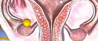
Every month, cyclical processes occur in a woman’s body aimed at developing, maturing and preparing the egg for release from the ovary, where it should be fertilized and give rise to a new life. Thanks to these processes, a woman is able to become a mother. If pregnancy does not occur, the egg dies, but if fertilization occurs, then a hormonal change begins in the woman’s body, aimed at preserving the developing fetus.
Unfortunately, not every doctor considers it necessary to explain ultrasound results to patients, without thinking that many entries in the examination sheet simply frighten women and make them nervous. The corpus luteum during pregnancy is often perceived by women as some kind of deviation, especially in cases where the entry is accompanied by the word “cyst”.
What is it - the corpus luteum of the ovary? And how is it formed?
Ovulation in a woman of childbearing age occurs every month, on average every 21–35 days, when a mature egg leaves the follicle. If fertilization does not occur, then a new egg matures next month and the cycle repeats. But ovulation is not the only preparatory stage for the possible development of new life in a woman’s body.
Once the mature follicle ruptures and the egg begins to move from the ovary to the uterus, the follicle cells, which have a granular texture, begin to form a body called yellow due to the color of the substance found inside it.
During pregnancy, the corpus luteum is formed in the body of every woman, and is a temporary gland related to the endocrine system, the main function of which is the production of the necessary hormones - progesterone and estrogen.
Main characteristics
The size of the corpus luteum in the ovary during pregnancy can vary depending on the phase of the cycle and the period of the “interesting position”. After ovulation, the gland measures no more than 2 cm. If conception does not occur, it begins to fade and disappears. The size of the corpus luteum will change during pregnancy, but it should not exceed 3 cm. With the beginning of the second trimester, the placenta takes over the role in the production of necessary hormones and nutrition of the fetus; the corpus luteum begins to dissolve and then disappears, having fulfilled its function.
The importance of the corpus luteum during pregnancy
Progesterone is often called the pregnancy hormone because it is necessary for the proper formation of the fertilized egg, its attachment to the uterine wall and the further development of the child. It is the normal level of progesterone that creates favorable conditions for the onset of pregnancy and its development.
In addition, progesterone prepares the endometrium in the uterus for further growth, and also suppresses the contractile functions of the uterine muscles and the formation of new eggs.
It is progesterone that further stimulates the preparatory processes and the production of milk by the mammary glands and prepares the woman for the process of bearing and giving birth to a baby, exerting the necessary influence on her nervous system.
Lack of progesterone in the body usually leads to some form of female infertility, since even a successfully fertilized egg cannot attach to the wall of the uterus. A decrease in hormone levels after pregnancy can lead to early miscarriage.
The production of progesterone is carried out precisely by the corpus luteum and for this purpose it is formed every month in the woman’s body after the release of the egg from the ovary. The adrenal glands are also capable of synthesizing a small amount of the hormone, but it is not enough for the onset and development of pregnancy.
After formation, the corpus luteum develops quickly, but its fate depends entirely on whether the egg is fertilized or not:
- If fertilization does not occur, then the corpus luteum continues to function for about two weeks, after which it dies and it is at this time that the woman begins her next menstruation.
- When pregnancy occurs, the active development of the corpus luteum and hormone production continues until 12–15 weeks, after which all functions of this gland are transferred to the already formed placenta. Gradually, the activity of the corpus luteum stops and after the end of its existence, a small scar with a whitish color forms in its place in the ovary.
During an ultrasound, some women, having heard about the presence of a corpus luteum in the right or left ovary, perceive this as a kind of sign of pregnancy, believing that its formation is possible only in this case.
But the fact that there is a corpus luteum in the ovary only means that the egg has matured and the body is ready to conceive and bear a child. If there is no corpus luteum in the ovary, this is an indication that there was no maturation of the eggs in this cycle, and, therefore, pregnancy is impossible.
The presence of a corpus luteum can become a sign of pregnancy only if, during an ultrasound scan before the start of the next menstruation, approximately 1–2 days before, this gland is clearly visible in a certain area, for example, in the right ovary, and has a pronounced size that does not decrease.
The size of the corpus luteum during pregnancy varies; in the early stages its diameter does not exceed 15–20 mm, but gradually it increases to 27–28 mm, maintaining this figure during the normal course of pregnancy up to 15 weeks. After this, a gradual cessation of the main function and a decrease in size begins.
Diagnostics
View gallery
Diagnosis is made during an ultrasound examination of the ovaries, which can be performed using two methods.
- Transabdominal. In this case, ultrasound is performed through the abdomen. The bladder should be full.
- Transvaginally. A vaginal sensor is used. In this case, the bladder should be emptied.
The gland looks like a heterogeneous round formation located next to the ovary. There are cases in which the corpus luteum is not detected during an ultrasound, which can be a serious pathology that requires treatment. If an ultrasound examination does not reveal the corpus luteum in the early stages of pregnancy, it is too early to judge the pathology. It happens that the culprit is a low-quality ultrasound machine or an unqualified ultrasound doctor. In this case, you must undergo tests prescribed by your doctor.
- If the test shows the presence of pregnancy and a fertilized egg, but the corpus luteum cannot be seen, this may indicate a high risk of miscarriage. In this case, you should immediately begin therapy with drugs containing progesterone.
- If the menstrual cycle is delayed and the fertilized egg is not visible, the presence of a progressing gland indicates successful conception.
Many people wonder if a corpus luteum was diagnosed in the ovary, whether there is a pregnancy. Sometimes its presence in the body only indicates past ovulation, after which it will disappear if pregnancy does not occur.
It is impossible to draw conclusions about the condition of the woman and fetus only from the results of ultrasound diagnostics. A number of other tests need to be carried out to help clarify the causes of the concerns.
The size of the corpus luteum during pregnancy on ultrasound should be within the stated norms, otherwise this may indicate pathologies in the development of the gland.
Pathologies in the development of the corpus luteum
There is also the concept of a corpus luteum cyst, when the size of the gland during pregnancy exceeds 30 mm, which is a pathology. But do not worry if, during an ultrasound, the doctor reports that there is a corpus luteum cyst in the ovarian cavity. Its presence will not in any way affect the course of pregnancy and the woman’s health, since even with an increased size of the iron it produces progesterone.
The appearance of a cyst can be caused by various reasons, which most often include:
- constant stress;
- presence of bad habits or harmful working conditions of the expectant mother;
- extreme diets;
- infectious diseases of the genital organs;
- taking hormonal medications before pregnancy;
- the presence of functional disorders of the circulatory or lymphatic system of the ovaries.
As a rule, a corpus luteum cyst does not require special treatment, but a pregnant woman who has one will need additional monitoring of the condition of the gland and strict limitation of physical activity.
There is no need to fear that the cyst may degenerate into a malignant formation, since such cases have never been encountered in medical practice. The cyst usually does not cause discomfort or pain.
However, if the doctor's requirements regarding limiting physical activity and sexual intercourse are not followed, the corpus luteum cyst may rupture, which will require urgent surgical intervention to remove it. The rupture of a cyst is always accompanied by severe pain and severe bleeding, and if it is not removed, this can lead to infection of the internal organs. Read more about corpus luteum cyst →
Surgical intervention will also require torsion of the cyst stem, since this causes severe compression of the tissue, which can lead to necrosis.
In some cases, when performing an ultrasound on a woman with an established pregnancy, doctors do not detect the presence of a corpus luteum. This happens when outdated equipment is used to conduct ultrasound in the early stages, or when the study is carried out by a doctor who does not have the necessary qualifications.
In this case, the woman needs to undergo an additional ultrasound in another clinic and, if the diagnosis is confirmed, the pregnant woman will need urgent medical attention in the form of hormonal correction.
Another pathology is insufficiency of the corpus luteum, which often leads to spontaneous termination of pregnancy (miscarriage). With insufficient iron development, it cannot produce progesterone in the amount necessary for the normal course of pregnancy, and in this case, the uterus does not allow the fertilized egg to implant and rejects it.
When pregnancy has already occurred, insufficient progesterone levels greatly increases the risk of rejection of the developing fetus, as well as the subsequent occurrence of placental insufficiency.
Today, doctors successfully solve the problem of insufficient development and functioning of the corpus luteum, replenishing the missing amount of the hormone with special drugs, which makes it possible to save many pregnancies. It is important to remember that seeing a doctor in the earliest stages of pregnancy is the key to bearing a healthy baby.
Author: Irina Vaganova, doctor, especially for Mama66.ru
Deviation from the norm in the functioning of the gland
View gallery
Specialists very carefully examine the corpus luteum during pregnancy, especially in the early stages, when the life and health of the fetus depends on its work. Deviations noticed in time will help avoid miscarriage and frozen pregnancy.
There are only two pathologies that are associated with the work of the corpus luteum - its insufficiency and a cyst.
If, during ultrasound examination, the size of the corpus luteum during pregnancy is less than 10 mm, then this indicates hypofunction of the gland. This is a very serious disorder that can lead to termination of pregnancy, because if the corpus luteum is insufficient during this period, the required amount of progesterone necessary for the normal development and maintenance of pregnancy is not produced. Diagnosis is carried out not only using ultrasound, but also through hormone tests. If the diagnosis is confirmed, hormonal drug therapy is prescribed.
Insufficiency of the corpus luteum may also indicate an ectopic pregnancy. In this case, progesterone will be produced in very small quantities. If such a pathology is likely, a dynamic hCG analysis is prescribed.
During a frozen pregnancy, progesterone ceases to be produced completely. Additional symptoms will be:
- Absence of toxicosis if previously present.
- A condition in which the chest no longer hurts.
- Pain in the lower abdomen.
- Lack of fetal growth and heartbeat according to ultrasound results.
- Spotting.
How is it formed?
The formation of a temporary gland occurs in accordance with certain stages. The entire lifespan of this temporary formation is called the corpus luteum phase. Here are the main stages of the life of the gland in non-pregnant women:
- Proliferation - follicular membranes, the integrity of which was disrupted at the time of ovulation, begin to “group” into a characteristic fold, and a gland is formed.
- Vascularization - gland cells divide, it begins to be actively supplied with blood due to the germination of the blood network.
- Heyday is the period of maximum productivity of the temporary gland, when the production of hormones occurs at an active pace in the highest concentrations.
- Regression - dystrophic changes begin inside the gland. It gradually decreases, reduces and gradually stops the production of hormones altogether, becoming whitish scar tissue, which eventually resolves on its own. The gland disappears, only to return in the next menstrual cycle.
If a woman has conceived a baby in the current cycle, then after the flowering stage there is no regression, and the temporary formation itself begins to be called the gravidate (corpus luteum graviditatis), or corpus luteum of pregnancy. It becomes such immediately after the embryo attaches to the endometrial layer of the uterus.
Immediately after implantation, the chorionic villi begin to produce a special active substance - human chorionic gonadotropin (hCG). The task of this hormone is to maintain the functionality of the corpus luteum, since the need for progesterone during pregnancy is high.
HCG keeps the temporary gland in working order until a full-fledged placenta is formed, which is capable of taking over the function of producing progesterone and a number of other active substances necessary to continue pregnancy.
Thus, outside of pregnancy, iron in the fair sex lives 10-13 days after the release of the egg. During pregnancy, the lifespan of the gravid corpus luteum increases; it is active until 11-14 weeks of pregnancy. At the end of the first trimester, the young placenta begins to work and already at the beginning of the second trimester the stage of regression for the gravid corpus luteum begins. It proceeds in the same way, the gland begins to decrease, the secretion of progesterone by it is reduced, a whitish body is formed and gradually no trace remains of it.
The calculator will help you estimate the size of the corpus luteum (CL) during ultrasound by week of pregnancy.
VT – corpus luteum CVT – corpus luteum cyst
CL – corpus luteum CLC – corpus luteum cyst
The corpus luteum forms after ovulation at the site of the ruptured follicle from which the egg was released. When pregnancy occurs, the corpus luteum continues to function and support the pregnancy.
A corpus luteum cyst is often detected on ultrasound from early pregnancy (as an anechoic, homogeneous, round-shaped formation with clear, even contours). Its dimensions can reach 40-50 mm, and sometimes 60-90 mm.
What it is?
The corpus luteum is a temporary formation similar to an endocrine gland that forms in the right or left ovary after a woman ovulates. During the first half of the female cycle, a vesicle - a follicle - matures on the surface of the reproductive gland, inside which the female reproductive cell develops. By the middle of the cycle, under the influence of certain hormones, the follicle membranes become thinner and rupture.
The egg, ready for fertilization, is released and captured by the villi of the fallopian tube. Fertilization occurs in the tube, after which the egg, which has become a zygote, begins its journey to the uterus. If within 24-36 hours the oocyte is not fertilized, then it dies.
- Menstruation
- Ovulation
- High probability of conception
Ovulation occurs 14 days before the start of the menstrual cycle (with a 28-day cycle - on the 14th day). Deviation from the average value occurs frequently, so the calculation is approximate.
Also, together with the calendar method, you can measure basal temperature, examine cervical mucus, use special tests or mini-microscopes, take tests for FSH, LH, estrogens and progesterone.
You can definitely determine the day of ovulation using folliculometry (ultrasound).
- Losos, Jonathan B.; Raven, Peter H.; Johnson, George B.; Singer, Susan R. Biology. New York: McGraw-Hill. pp. 1207-1209.
- Campbell NA, Reece JB, Urry LA ea Biology. 9th ed. - Benjamin Cummings, 2011. - p. 1263
- Tkachenko B. I., Brin V. B., Zakharov Yu. M., Nedospasov V. O., Pyatin V. F. Human physiology. Compendium / Ed. B. I. Tkachenko. - M.: GEOTAR-Media, 2009. - 496 p.
- https://ru.wikipedia.org/wiki/Ovulation
Regardless of whether the fateful meeting of the sperm and the egg occurred or not, on the surface of the gonad after the release of the oocyte, a corpus luteum invariably forms on the site of the former follicle and remains there as long as it is needed. The main function of the temporary gland is to produce the sex steroid - the hormone progesterone, which is necessary for the normal functioning of the body of the fair sex in the second half of the menstrual cycle.
If a woman does not become pregnant in the current cycle, then the temporary gland disappears and menstrual flow begins at the appointed time. But everything changes during pregnancy . The gland itself was invented by nature in order to help the embryo survive at first, while there is no placenta.
Progesterone produced by the gland is very important for prolonging pregnancy - it carries out preparatory changes in the endometrium for the upcoming implantation of the embryo, “builds up” it, ensures the absence of menstruation, under the influence of this hormone the cervical mucus thickens in the cervical canal, forming a mucus plug that reliably protects the cavity uterus from outside penetration. Progesterone during pregnancy relaxes the uterine muscles, preventing its contraction and possible miscarriage.


