What kind of disease is this
Antiphospholipid syndrome, or APS, is a pathology that is characterized by repeated thrombosis of the venous, arterial, microvasculature, the pathology of pregnancy with fetal loss and the synthesis of antiphospholipid antibodies (aPL): cardiolipin antibodies (aCL) and/or lupus anticoagulant (LA), and/or antibodies to beta2-glycoprotein Ⅰ. APS is a variant of often acquired thrombophilia.
The ICD 10 revision code is D68.8.
The basis of the pathogenesis of antiphospholipid syndrome is the attack of cell membranes by antibodies. Most often, antiphospholipid syndrome develops in women - 5 times more often than in men.
The syndrome manifests itself through the occurrence of thrombosis and miscarriage. Often, before the development of gestation, women were not aware of the presence of this pathology and the presence of antibodies in the blood.
What it is
Antiphospholipid syndrome, or Hughes syndrome, is an autoimmune disease. That is, it occurs due to malfunctions of the entire immune system or its parts. With Hughes syndrome, the body produces antibodies to phospholipids (the substance that makes up cell structures) and the proteins that bind them. Antibodies interact with phospholipids and damage cell membranes. Problems arise in the blood coagulation system. This, in turn, can have such unpleasant consequences as thrombosis (blockage) of veins and arteries, miscarriage and the appearance of other obstetric pathologies, and a decrease in the level of platelets in the blood (thrombocytopenia). According to statistics, about 5% of the world's inhabitants suffer from this disease. There are more women than men among the sick people.
It is difficult to say exactly why such disruptions in the immune system occur that trigger antiphospholipid syndrome. Medical science names possible provoking factors. These include genetic predisposition, previous bacterial or viral diseases, as well as cancer, long-term use of potent medications (psychotropic, hormonal). Hughes syndrome is often a precursor to systemic lupus erythematosus (a severe autoimmune disease) or may develop simultaneously with it.
Antiphospholipid syndrome (APS) can be asymptomatic or present with characteristic symptoms. The most common symptom of APS is venous thrombosis. The deep veins in the legs are often affected; this condition may be accompanied by swelling of the extremities and fever. Sometimes non-healing ulcers appear on the legs.
Often, with APS, the superficial veins and vessels of the liver and other organs are affected. In this case, a serious complication may develop - pulmonary embolism. Its signs are shortness of breath, severe coughing, coughing up blood, and severe chest pain. As the syndrome develops, the heart may suffer. It is rare, but it happens that APS is manifested by deterioration of vision (due to damage to the retinal vessels) and the development of renal failure.
With Hughes syndrome, spider veins can often be seen on the skin of different parts of the body, most often on the lower legs, feet, and thighs.
Classification
There are several variants of antiphospholipid syndrome. Their main classification is as follows:
- Primary – associated with hereditary hemostasis defects.
- Secondary APS arose against the background of autoimmune diseases (rheumatoid arthritis, systemic lupus erythematosus), vasculitis, organ-specific pathologies (diabetes mellitus, Crohn's disease), oncological processes, drug exposure, infections (HIV, syphilis, malaria), and end-stage renal failure.
- Other AFS options:
- seronegative
- catastrophic
- other microangiopathic syndromes (DIC syndrome, HELLP).
Three types of antiphospholipid syndrome
Primary antiphospholipid syndrome
Primary APS is an independent disease, in 70% of cases caused by malfunctions in the genetic makeup of the carrier. It is not a consequence of other autoimmune disorders and coexists with them in the body. The risk factor is the genetic marker HLA-DR7.
At the same time, relief of symptoms and complications of the syndrome as such in primary APS is easier than in secondary APS, since it is not complicated by the course of another disease.
Secondary antiphospholipid syndrome
Secondary APS is a companion and complication of another disease. As a rule, it is also autoimmune and most often - systemic lupus erythematosus. But it is also possible to make a diagnosis for infections, diabetes, and malignant tumors. Previously, it was very dangerous, but now it stops by 90% and often goes into remission.
In combination with lupus, for which a complete cure has also not been found, the priority becomes to relieve the carrier of its symptoms. Treatments for autoimmune diseases are similar and often overlap, allowing for simultaneous therapy.
In addition to systemic lupus erythematosus and other diseases, typical for secondary APS, there are also genetic reasons for its development - markers HLA-B8, HLA-DR2 and DR3-HLA.
Catastrophic antiphospholipid syndrome
Catastrophic APS is a rapidly developing form of antiphospholipid antibody syndrome that is life-threatening to the carrier. Arising suddenly, antiphospholipid antibodies cause the blood to clot in several organs at once and this causes a corresponding deficiency in body functions. Failure to promptly treat catastrophic APS results in death in 50% of cases.
Causes of miscarriage
Pathogenesis of the development of obstetric pathology in APS.
The influence of APS in the development of such pregnancy complications has been proven:
- infertility of unknown origin;
- early preembryonic losses;
- failed IVF;
- miscarriages at different stages;
- intrauterine fetal death;
- postpartum fetal death;
- fetal growth retardation syndrome;
- preeclampsia and eclampsia;
- thrombosis during pregnancy and after childbirth;
- fetal malformations.
In the postpartum period, the child also experiences consequences of antiphospholipid syndrome: thrombosis, neurocirculatory disorders with the development of autism in the future. 20% of children born to mothers with APS have antiphospholipid antibodies in their blood without symptoms, which indicates intrauterine transmission of aPL.
The pathogenetic basis for the development of all manifestations of APS during pregnancy is placental decidual vasculopathy, which is caused by a lack of prostaglandin production, placental thrombosis and a violation of the implantation mechanism. All these mechanisms prevent pregnancy.
Diagnostics
APS can be confirmed by detecting the following elements in the blood:
- lupus anticoagulant;
- anticardiolipin antibodies;
- antibodies to phospholipids.
Antiphospholipid syndrome is said to occur if these substances were detected in a woman’s blood two or more times in a row. Studies are carried out at intervals of 6-8 weeks. A single detection of antibodies is not indicative. Such substances can occur transiently, that is, for a short period of time. The transient presence of antibodies does not lead to infertility or the development of pregnancy complications.
Indications for testing:
- examination for infertility;
- preparation for pregnancy after miscarriage or regression;
- suspicion of APS during pregnancy;
- cases of thrombosis in the past (heart attacks, strokes, cerebrovascular accidents);
- complicated heredity (thrombosis in close relatives under the age of 45 years).
Blood for antibody determination is taken from a vein in the morning on an empty stomach. On the eve of the study, it is recommended to refrain from eating for 8-12 hours. You can drink clean water before donating blood.
Risk factors
One of the factors in the occurrence of APS is a genetic predisposition to this pathology. Thus, in patients with APS, antigens of the HLA system are found more often than in the population. Familial cases of APS are also known, accounting for up to 2% of cases. Another important factor is the presence of a bacterial and/or viral infection, which does not exclude the possibility of developing thrombotic complications as part of APS.
For the pathological process to occur, it is necessary to have in the body not only antibodies to phospholipids, but also so-called cofactors, upon binding with which true antigen-antibody complexes are formed. As a result of the action of various external and internal environmental factors (viral infection, malignant neoplasms, the effect of drugs), APA interacts with cofactors, which leads to serious disturbances in the blood coagulation system. In this case, first of all, microcirculation processes are disrupted and changes in the vascular wall occur.
Due to the fact that antiphospholipid syndrome is one of the most common types of pathology of the blood coagulation system, its recognition should be included in the diagnostic process in all cases of early and, especially, recurrent venous and arterial thrombosis, thromboembolism, dynamic cerebrovascular accidents and ischemic strokes , including those occurring with migraine syndromes, memory impairment, paresis, visual impairment and other manifestations, as well as persistent miscarriage (intrauterine fetal death, miscarriages).
Types of antiphospholipid syndrome
There are primary and secondary APS. The presence of secondary APS is caused by autoimmune diseases (with systemic lupus erythematosus, periarteritis nodosa, etc.), cancer, infectious diseases, as well as exposure to a number of drugs and toxic substances. Accordingly, in primary APS, the listed diseases and conditions are absent.
In some cases, the so-called catastrophic APS is isolated, which is characterized by sudden and rapidly developing multiple organ failure, most often in response to factors such as infectious diseases or surgical interventions. Catastrophic APS is manifested by acute respiratory distress syndrome, impaired cerebral and coronary circulation, stupor, disorientation, and the possible development of acute renal and adrenal failure, thrombosis of large vessels.
Symptoms and complications of the disease
One of the main and most dangerous clinical manifestations of APS is recurrent thrombosis. Most often, venous thrombosis occurs, localized in the deep veins of the legs, which is associated with the risk of developing thromboembolism of the branches of the pulmonary artery. However, cases of thrombosis of the renal and hepatic veins are not uncommon. Thrombotic lesions of the portal, subclavian, inferior vena cava, cerebral vessels, arteries and veins of the retina, large vessels of the lower extremities, and various parts of the aorta may occur. Clinical manifestations of arterial thrombosis are peripheral gangrene, aortic arch syndrome, blindness, cerebrovascular accidents, etc. The danger of thrombotic complications increases with the course of pregnancy and the postpartum period.
It is known that APS leads to non-developmental pregnancy
, intrauterine growth retardation of the fetus, up to fetal death in the 2nd and 3rd trimesters. In the first trimester of pregnancy
APA can have a direct damaging effect on the fertilized egg, followed by spontaneous termination of pregnancy.
From the early stages of pregnancy, there is an increase in the functional activity of platelets, and the protein-synthesizing and hormonal functions of the placenta decrease. In the absence of appropriate treatment, an increase in the activity of the blood coagulation system occurs. In this case, thrombosis occurs in the vessels of the placenta, placental insufficiency, chronic hypoxia and often fetal death due to lack of oxygen develop.
Diagnosis criteria
There are criteria by which the diagnosis of “Antiphospholipid syndrome” is established. Among the clinical criteria, the following are highlighted:
- Vascular thrombosis of any location: both venous and arterial, confirmed by visual examination methods. When using histological examination, biopsy specimens should show no signs of inflammation of the vascular wall.
- Complications of pregnancy:
- one or more episodes of death of a normally developing fetus after 10 weeks of gestation, or
- one or more episodes of preterm birth before 34 weeks due to significant preeclampsia, eclampsia, placental insufficiency, or
- three or more consecutive cases of spontaneous abortions in less than 10 weeks, in the absence of pathologies of uterine anatomy, genetic mutations, or sexually transmitted infections.
Laboratory criteria are:
- Antibodies to cardiolipin, immunoglobulins of classes G and M, were detected in the blood in medium and high titers, at least 2 times in 12 months.
- Antibodies to b2-glycoprotein I classes G and/or M in medium or high titers, at least 2 times a year.
- Plasma lupus anticoagulant VA was determined in 2 more laboratory studies over an interval of at least 12 months. The presence of VA in the blood can be suspected if the aPTT in the coagulogram increases by 2 or more times.
An antibody test is considered highly positive - 60 IU/ml, an average positive response - 20-60 IU/ml, low positive - less than 20 IU/ml.
Read also: Can endometritis lead to infertility?
To make a diagnosis of Antiphospholipid Syndrome, one clinical and one laboratory criterion must be present.
How does antiphospholipid syndrome work?
Due to the high mortality rate due to APS, scientists have spent a lot of effort and money studying it. Now we cannot say with certainty why the body of patients with antiphospholipid syndrome behaves this way, but research continues.
Causes of antiphospholipid syndrome
How antiphospholipid syndrome occurs is currently unknown. The body in this state produces antibodies to its own cells or tissues, which has disastrous consequences. The natural work of the immune system turns into a formidable weapon against itself.
A person may never know for the rest of his life that he had antiphospholipid antibody syndrome if nothing makes it work. Research shows that the trigger for a bright manifestation can be any intervention in the body: infection, surgery, hormonal contraceptives, oncology. Or the course of another autoimmune disease. Based on these characteristics, APS is divided into primary and secondary.
Moreover, all types of APS have symptoms in addition to thrombotic ones: a lack of platelets in the blood and anemia.
The mechanism of action of antiphospholipid antibody syndrome
In antiphospholipid syndrome, proteins are found that affect the cells lining blood vessels and the outer lining of blood cells. At the same time, the walls of the veins and arteries thicken, and the clots, blocking the blood flow, stop the supply of necessary nutrients to the organs and increase the pressure; lead to ischemia, stroke, impaired thinking and memory, miscarriages and premature birth.
Any body system can be affected, but the most common consequences are deep vein thrombosis of the lower extremities, neurological disorders and obstetric pathologies. As a rule, testing for the presence of antibodies is recommended after several repeated early miscarriages or recurrent thrombosis.
Prevalence of antiphospholipid antibody syndrome
Even women who are completely healthy according to clinical and laboratory indicators can have this disease without any manifestations. They have the opportunity to learn about the disease only after making an extremely unpleasant diagnosis of “recurrent miscarriage” or detection of blood clots. Moreover, the disease is selective: as a rule, within one organism, either veins or arteries are affected, but not all vessels at the same time.
A low level of antibodies is detected quite often compared to a high level, which entails a diagnosis: out of 20-50 fixations of antiphospholipid antibodies, only 5 require close attention from doctors. At the same time, men encounter this problem much less often - only 1 case out of 6. And 85% of the total incidence occurs in women aged 15-50 years.
Symptoms
The main symptom of antiphospholipid syndrome is thrombosis. In women, this pathology manifests itself as miscarriage. In addition to these obvious signs, women may exhibit additional clinical criteria:
- livedo reticularis;
- a history of migraines, chorea;
- trophic ulcerative defects of the lower extremities;
- endocarditis and so on.
The catastrophic form of antiphospholipid syndrome is very difficult. It is accompanied by a clinical picture of acute renal failure, respiratory distress syndrome, liver failure, impaired cerebral blood flow, and thrombosis of large vessels, including the pulmonary artery. It is impossible to live with this form for a long time without urgent help.
Causes
Antiphospholipid syndrome is detected in 2-4% of all pregnant women. The exact causes of this pathology are still not known. Specific antiphospholipid antibodies are found in a wide variety of conditions, including some infectious diseases. Why in some women this phenomenon leads to the development of pregnancy complications, while in others it goes unnoticed, it is not possible to find out.
APS is considered a hereditary disease. It is known that in women suffering from this pathology, some specific genes of the HLA system are detected much more often. It is these genes that cause the immune system to malfunction. As a result, the body begins to produce aggressive antibodies that destroy its own cells.
Specific antibodies act directly on phospholipids - components of cell membranes. The endothelium (inner lining) of blood vessels suffers the most. Developing endothelial dysfunction leads to disruption of various processes in the hemostasis system. Blood clotting increases and the risk of thrombosis increases. Thrombosis in the blood vessels of the placenta can lead to miscarriage, placental abruption and other serious complications of pregnancy.
Risk factors for developing APS:
- autoimmune diseases (systemic lupus erythematosus, scleroderma, Sjogren's syndrome and others);
- infectious diseases (viral hepatitis, HIV, Epstein-Barr virus);
- oncological processes (ovarian tumors, blood cancer);
- taking certain medications (hormonal drugs and others).
Treatment
Many specialists treat APS: rheumatologists, hematologists, obstetricians and gynecologists, cardiologists, cardiac surgeons and others.
First group of patients
Patients who do not have laboratory-defined signs or clinical symptoms do not require constant laboratory monitoring and continuous anticoagulant therapy. In this group of patients, standard prophylaxis of venous thrombosis is carried out.
Second group
In patients with a high titer of lupus anticoagulant and/or antiphospholipid antibodies more than 10 IU/ml without thrombosis, specific prophylaxis is required - Aspirin at a dosage of 75-100 mg once a day.
Third group
These people test negative for antibodies, but there are confirmed cases of thrombosis and a high risk of their formation. These patients are treated with anticoagulants of low molecular weight heparin in therapeutic doses. Immediately after diagnosis, use:
- Dalteparin 100 IU/kg 2 times a day;
- Nadroparin 86 IU/kg or 0.1 ml per 10 kg 2 times a day subcutaneously;
- Enoxaparin 1 mg/kg 2 times a day subcutaneously;
- From the second day, Warfarin is prescribed at 5 mg per day.
Patients in this group undergo heparin therapy for at least 3 months. At the beginning of therapy, the INR is monitored every 4-5 days to maintain the target value of 2.0-3.0.
Fourth group
This group includes people in whom thrombosis occurs against the background of elevated titers of lupus anticoagulant and antiphospholipid antibody. In this category of patients, Warfarin and low doses (75-100 mg) of Acetylsalicylic acid are prescribed. High-risk patients should receive lifelong anticoagulation therapy.
Complications of pregnancy
Antiphospholipid syndrome can cause the following complications during pregnancy:
Spontaneous miscarriage
Termination of pregnancy with APS occurs either in the earliest stages or after 10 weeks. In the first case, the implantation of the embryo is disrupted, which leads to its rejection and death. Miscarriage occurs in the first 2-3 weeks of pregnancy, often before a missed period. The woman may not even know that she was pregnant. If you have been trying for a long time and unsuccessfully to conceive a child, you must undergo testing for APS.
Miscarriage after 10 weeks is associated with impaired blood flow in the developing placenta. The formation of blood clots in the mother-placenta-fetus system leads to chorion detachment, bleeding and miscarriage. Termination of pregnancy in the second trimester may also be associated with antiphospholipid syndrome.
Premature birth
Termination of pregnancy at 22-36 weeks is called premature birth. Antiphospholipid syndrome is one of the common causes of this pathology. The start of labor ahead of schedule is indicated by the appearance of the following symptoms:
- lower abdominal pain;
- pain in the lower back;
- opening and shortening of the cervix;
- discharge of the mucus plug;
- outpouring of water
Premature birth results in the birth of a premature newborn. The shorter the pregnancy, the more difficult it will be for the baby to adapt to existence outside the mother’s womb. Nursing of premature babies takes place in a specialized department. For some time, the newborn is in an incubator - a special device that supports the child’s life. Discharge home is possible only after the baby has fully adapted to the new living conditions.
Placental insufficiency
An increase in blood clotting inevitably leads to the formation of numerous blood clots in the placenta. As a result, blood flow in the mother-placenta-fetus system is disrupted. Placental insufficiency develops - a condition in which the baby suffers quite a lot. The blood of the fetus does not receive enough nutrients, which leads to a delay in its development. Significant developmental delays in the baby can cause serious health problems after birth.
Placental insufficiency inevitably leads to another complication of pregnancy - chronic fetal hypoxia. With this pathology, the baby does not receive a sufficient amount of oxygen necessary for its full development. Hypoxia primarily affects the fetal nervous system. Prolonged hypoxia can cause cerebral palsy and other diseases of the nervous system.
Preeclampsia
Preeclampsia is a specific pathology that occurs only during pregnancy. It is assumed that the main reason for the development of gestosis in APS is endothelial dysfunction and a natural disruption of the woman’s body’s adaptation to the onset of pregnancy. Increased thrombus formation leads to a sharp rise in blood pressure up to the development of eclampsia. Severe gestosis is one of the causes of premature birth and antenatal fetal death.
Premature abruption of the normally located placenta (PONRP)
PONRP is an extremely serious complication of pregnancy. The formation of blood clots and impaired blood flow in the placenta after 20 weeks can lead to its detachment from the uterine wall and massive bleeding. This condition is dangerous for the life of a woman and her baby. In case of severe blood loss, an emergency caesarean section is performed regardless of the stage of pregnancy.
HELLP syndrome
A rare and extremely dangerous pathology in obstetrics, in which the probability of death of the woman and fetus is very high. HELLP syndrome occurs in the third trimester, most often after 34 weeks. With this pathology, blood thickening occurs, blood clots form, followed by bleeding. HELLP syndrome is considered an extreme degree of multiple organ failure, which occurs when the body’s adaptation to pregnancy is disrupted.
Signs of HELLP syndrome:
- nausea and vomiting;
- pain in the epigastric region;
- pain in the right quadrant;
- swelling;
- headache;
- jaundice;
- vomiting blood;
- hemorrhages at injection sites.
Symptoms are quite nonspecific and can occur in a wide variety of diseases. As the pathology progresses, severe liver failure, seizures and coma develop. HELLP syndrome is a direct indication for emergency caesarean section and intensive care.
Preconception preparation
Preparation for pregnancy with APS is carried out in 2 successive stages. At the first stage, the coagulogram is assessed, the antigenic components of the blood are determined, and infectious foci are removed and sanitized.
The second stage is the immediate preparation for pregnancy and its management. This requires anticoagulant therapy. It is carried out individually during 1-2 menstrual cycles. To do this, you need to place the woman in one of the following groups:
- A seronegative variant of APS with a history of obstetric manifestations of the syndrome. Only antibodies to beta2-glycoprotein I can be detected in the serum. In this group, preparation is carried out using the following drugs:
- one of the low molecular weight heparin preparations 1 time/day subcutaneously (dalteparin (Fragmin) 120 antiXa IU/kg or enoxaparin (Clexan) 100 antiXa IU/kg;
- fish oil 1-2 capsules 3 times a day;
- folic acid 4 mg/day;
- If lupus anticoagulant is absent, but APLA is present without thrombosis and obstetric clinical manifestations:
- with a moderate titer of APLA, Aspirin 75-100 mg/day is prescribed, and if pregnancy develops, it is discontinued and replaced with dipyridomole 50-75 mg/day;
- with a high and moderate titer of antiphospholipid antigen, combine Acetylsalicylic acid 75 mg/day and low molecular weight heparin once a day subcutaneously;
- fish oil 1-2 capsules 3 times a day;
- folic acid 4 mg/day.
- If there is no lupus anticoagulant in the blood, but there is a high or moderate amount of antiphospholipid antigen and there is a clinical picture of thrombosis and obstetric complications:
- one of the LMWHs (Clexane, Fragmin, Fraxiparine) 1 time per day subcutaneously;
- Aspirin 75 mg/day with its discontinuation during pregnancy and the prescription of Dipyridamole 50-75 mg/day;
- fish oil 1-2 capsules 3 times a day;
- folic acid 4 mg/day.
- APLA was detected in the woman's plasma and the lupus anticoagulant VA was determined from 1.5 to 2 conventional units. Until VA normalizes, you should abstain from pregnancy. To normalize VA to less than 1.2 conventional units, the following is used:
- Clexane 100 antiXa IU/kg or Fragmin 120 antiXa IU/kg once a day subcutaneously;
- recommended intravenous human immunoglobulin 25 ml every other day, 3 doses, repeat administration of the drug at 7-12 weeks of pregnancy, at 24 weeks and the last administration before childbirth;
- after VA is established within normal limits, Acetylsalicylic acid 75 mg/day is prescribed until pregnancy;
- Clexane or Fragmin once a day subcutaneously in the same dosages;
- fish oil 1-2 drops. 3 times a day;
- folic acid 4 mg/kg.
- If VA in the blood is more than 2 conventional units, then conception is delayed for at least 6-12 months. The risk of developing thrombosis in such women is very high. The target value of VA is 1.2 conventional units. Therapy is carried out for at least 6 months.
Read also: Is infertility caused by masturbation?
Laboratory diagnostics and examination when planning pregnancy necessarily includes the following blood clotting indicators:
- platelets – 150-400*109/l;
- fibrinogen – 2-4 g/l;
- INR – 0.7-1.1;
- degradation products of fibrinogen and fibrin – less than 5 μg/ml;
- d-dimers – less than 0.5 μg/ml;
- soluble fibrin monomer complexes should be absent;
- protein C – 69.1-134.1%;
- antithrombin Ⅲ – 80-120%;
- platelet aggregation activity with adenosine diphosphate salt – 50-80%, with adrenaline hydrochloride – 50-80%;
- anticardiolipin antibodies – all classes of immunoglobulins less than 10 IU/ml;
- VA – negative or less than 0.8-1.2 conventional units;
- hyperhomocysteinemia – negative;
- mutation FV (Leiden) of the gene responsible for the synthesis of factor V, or mutation G20210A of the gene responsible for the synthesis of factor II – absent;
- general urine test to determine hematuria;
- control over the development of infectious diseases: lymphocytes, ESR.
Antiphospholipid syndrome: features of the course in pregnant women and treatment options
Pregnancy significantly affects the mother's immune system: depression of the cellular immune system, increased secretion of immunoglobulins, decreased lymphocyte function due to the expression of special proteins PSP (pregnancy-specific proteins). All these transformations are aimed at the survival of the fetus. Processes of changes in the cytokine profile of type 2 T helper cells are dominant in maintaining “immunotolerance” during pregnancy and can affect various autoimmune diseases. There are a number of phenomena that can be used to see the effect of rheumatic pathology on pregnancy, and vice versa. These processes are multidirectional: on the one hand, there may be both the onset of a systemic autoimmune disease (SAD) and an exacerbation of an existing pathology (for example, an outbreak of lupus nephritis), on the other hand, numerous cases of pregnancy-induced remission have been described in patients with rheumatoid arthritis. In addition, the autoimmune dysfunction characteristic of SAZ and the presence of antiphospholipid antibodies (APAs) may lead to an increased risk of miscarriage, fetal death, and preeclampsia. Transplacental transport of pathological macromolecules, especially anti-Ro/La or SS-A, SS-B antibodies, directly affects the fetus and increases the likelihood of developing neonatal lupus. Finally, high immunoinflammatory activity and damage to internal organs within the SAZ can have a significant impact on maternal and fetal mortality rates. Pregnancy causes many physiological changes in the mother's body in addition to immune system dysfunction. Thus, there is a significant increase in the volume of circulating blood (up to 40–45%), which can aggravate the course of kidney or cardiovascular diseases. Glomerular filtration rate (GFR) increases by approximately 50% during normal pregnancy, so a patient with preexisting proteinuria will almost certainly have some increase in the amount of protein in the urine. As a result of changes in the coagulation component of hemostasis, platelet activity, fibrinolysis, venous stasis, vascular compression by the pregnant uterus, and forced bed rest, the likelihood of thrombotic complications increases. Swelling and bleeding of the gums, gastroesophageal reflux, significant bone loss due to pregnancy, lactation, and the possible use of glucocorticosteroids (GCS) are noted. Thus, even normal pregnancy can aggravate the course of SAZ. Physiological or pathological changes, including pregnancy-induced hypertension, can also mimic the activity of SAZ, which poses certain difficulties in making a differential diagnosis. For example, redness or hyperpigmentation of the face can easily be confused with a centrifugal malar butterfly rash. Palmar erythema in pregnant women may look like cutaneous vasculitis. Physiologic leukocytosis, anemia, and low platelet counts due to hemodilution, common in pregnancy, may mimic the hematologic manifestations of SAZ. As a result of increased fibrinogen levels and anemia, an acceleration of the erythrocyte sedimentation rate may be observed, and this parameter cannot be an objective marker of disease activity. Many women complain of diffuse arthralgia, muscle and bone pain, especially during their first pregnancy. Hypertension, proteinuria, renal failure and edema associated with gestosis can mimic various diseases or their exacerbation, including lupus nephritis, acute scleroderma nephropathy, relapse of vasculitis, necrotizing glomerulonephritis. HELLP syndrome is a variant of preeclampsia characterized by low platelet counts, elevated liver enzymes, hemolysis, abdominal pain, and can mimic systemic lupus erythematosus (SLE) or an exacerbation of systemic vasculitis. Finally, eclampsia, which involves seizures or cerebrovascular accidents, may be confused with central nervous system involvement in SLE or neurovasculitis. Antiphospholipid syndrome
In the early 1950s. Antiphospholipid syndrome (APS) has been described as a variant of SLE or lupus-like syndrome. However, it was very soon established that the connection between overproduction of APA and thrombotic disorders occurs in the absence of reliable clinical and serological signs of SLE or any other leading disease. To define this new nosological form, the term “primary antiphospholipid syndrome” was proposed. The development of radioimmunoassay (1983) and enzyme-linked immunosorbent assay methods for the determination of antibodies to cardiolipin contributed to the expansion of research concerning the role of APA in human diseases. It turned out that these antibodies are a serological marker of a unique symptom complex, including venous and/or arterial thrombosis, various forms of obstetric pathology (primarily recurrent miscarriage), thrombocytopenia, as well as other various neurological, skin, cardiovascular, and hematological disorders. In 1986, G. Hughes et al. proposed to designate this symptom complex as APS. In 1994, at the VI international symposium dedicated to the study of APS, it was proposed to call APS Hughes syndrome - after the English rheumatologist who first described it and made the greatest contribution to the development of this problem. The most recent revision of the criteria for this disease took place in Sydney in 2006. The interpretation of clinical manifestations was slightly changed; antibodies to beta-2 glycoprotein I (AB2GP) were added to the laboratory criteria (Table 1). Practical diagnosis of APS is currently being developed based on the Australian criteria.
The clinical spectrum of manifestations that are associated with APS is quite wide: migraine, arthritis/arthralgia, pulmonary hypertension, livedo reticularis, leg ulcers, etc. Although most of them were not included in the final diagnostic criteria for APS from 2006, the place of these phenomena is actively discussed in the literature. Pregnancy loss is a common complication of APS in obstetric practice; in addition, attention is drawn to the fact that preeclampsia and eclampsia often occur when APS and SLE are combined. HELLP syndrome in combination with ACPA circulation is more severe and often occurs in the second rather than third trimester. The risk of developing liver infarction in patients with HELLP syndrome associated with ACPA increases 30 times compared with that in the seronegative version of HELLP syndrome [2]. In addition, APS often develops other multiple thrombotic complications that require more aggressive treatment than in patients with the traditional course of HELLP syndrome. Pregnancy itself is a risk factor for the development of hypercoagulation, and with the appearance of APS, the likelihood of thrombosis in the mother increases significantly. In rare cases, catastrophic APS can develop during pregnancy: in the analyzed studies, 15 cases were identified, a characteristic feature of which was the fact that almost half of the patients previously had a history of latent APS [3]. Patients may have other hematological complications of APS, such as severe thrombocytopenia in the second and third trimesters of pregnancy. The most common adverse events associated with APS in pregnant women are preterm birth and intrauterine growth retardation. Preterm birth is most common in patients who have a combination of APS and SLE, and the incidence ranges from 10 to 40%. In one study, the authors attempted to determine the causes of adverse neonatal outcomes (preterm birth, intrauterine growth restriction, low Apgar scores). Such factors were the presence of VAC, ACLA, AB2GP antibodies, and a history of vascular thrombosis before pregnancy. In the absence of these factors (even in the presence of a previous burdened obstetric history), a more favorable neonatal outcome was noted [4]. In rare cases, thrombosis develops in the fetus or newborn due to transplacental transport of APA. In such cases, we may be talking about the presence of neonatal APS [5]. The risk of thrombosis in this cohort of patients decreases along with a decrease in the concentration of maternal APA, but later learning difficulties, a decrease in memory and other cognitive functions are noted. Currently, there are registries (European registry to babies born to mothers with antiphospholipid syndrome) to monitor the long-term neuropsychological consequences of neonatal APS [6]. VAC appears to be the most important risk factor for adverse pregnancy outcomes, including pregnancy loss. Currently, methods for determining the concentration of BAC are not standardized, in contrast to tests associated with the determination of anticardiolipins and beta-2 glycoprotein I. The PROMISSE multicenter prospective observational study is currently being completed, the main goal of which is to monitor APS-positive and SLE-associated pregnant women . A recent analysis of interim data found that BAC level is the only laboratory marker of APS associated with adverse pregnancy outcomes such as intrapartum fetal death, intrauterine growth restriction, and preterm birth [7]. On the other hand, there is evidence that only the combination of VAC, ACPA and AB2GP in a particular patient is a predictor of intrapartum fetal death [8]. This information is somewhat reassuring for those patients who have a low or moderate titer of ACLA, AB2GP. To timely assess the risk of thrombosis in patients with APS, it is necessary to conduct thorough screening for the presence of markers of hereditary thrombophilia (fibrinolysis genes: PAI-I, PLAT; platelet receptor genes: ITGA2, ITGB3, Gplba; blood coagulation system genes: Fl, F2, F5, F7 ), the presence of hyperhomocysteinemia [8–10]. Recently, evidence has emerged regarding the relationship between changes in serum complement levels during pregnancy and adverse outcomes. The interpretation of this phenomenon seems very difficult: with eclampsia and primary APS, an increase in titer is possible, and with SLE and secondary APS, hypocomplementemia is possible. Complement is an important predictor of adverse pregnancy outcome and mortality in a mouse model of APS [11], and it seems likely that activation of the complement system plays a similar negative role in the human population.
Recommendations for the management of pregnant women with APS
The timely development of treatment for obstetric APS dates back to 1980–1985, when patients with ACPA, VAC and obstetric failures began to receive GCS (prednisolone) and acetylsalicylic acid (ASA) in small doses. Moreover, the dose of corticosteroids was gradually increased until BAC or other APS markers reached acceptable levels. In the 1990s. studies have shown that low-dose heparin in combination with low-dose ASA was as effective as corticosteroids, but with much fewer side effects. Currently, the combination of antiplatelet drugs and direct anticoagulants is standard: the dosage of low molecular weight heparin (LMWH) is usually 40 mg enoxaparin daily, some experts use 30 mg twice a day, the dose for unfractionated heparin (UFH) is usually 5000 units twice a day. /day [12]. Studies show that the combination of low-dose heparin and low-dose ASA is more effective than ASA monotherapy [13], with a success rate of about 75% versus 40%. No fundamental differences were found between low and high doses of anticoagulants, as well as UFH and LMWH heparin [14, 15]. This type of therapy is first-line and is the most effective in preventing early pregnancy loss [16]. Second-line therapy includes the use of intravenous human immunoglobulin (IVIG). Some experts at this stage recommend increasing the dose of heparin to general therapeutic doses. The only controlled trial using IVIG did not show significant improvements in pregnancy outcomes. However, there is a significant number of uncontrolled studies, published clinical cases with an excellent effect with the combined use of IVIG, low doses of ASA and LMWH. With a further increase in the level of ACLA, VAC, plasmapheresis can be successfully used. The most promising methods for treating APS in the future are complement inhibition [11], as well as the use of genetically engineered biological therapy [17]. Treatment with medium/high doses of corticosteroids is currently practically not used due to the lack of evidence of their effectiveness and negative effects on the body of both the mother and the fetus. The use of GCS is justified only if APS develops against the background of some disease (SLE, Sjogren's disease, etc.). The use of GCS in these cases is aimed at treating not APS, but the underlying disease. In the postpartum period, anticoagulant therapy should be continued for a period of 6 to 8 weeks. even in patients without a history of thrombosis. The tactics for managing pregnant women with APS and the dosage regimen largely depend on the previous medical history (presence/absence of non-placental thrombosis, number of spontaneous abortions, previous therapy). In this regard, the following subgroups can be distinguished:
1. Patients with only serological markers of APS (no previous pregnancy, one episode of unexplained spontaneous abortion before 10 weeks of gestation), no history of thrombosis.
The tactics for managing this category of women is to use small doses of ASA, which is prescribed for the entire period of pregnancy and for 6 months.
after delivery. If pregnant women have highly positive ACPA (more than 65 GPL units), it is advisable to prescribe LMWH. The risk of developing thrombotic complications is high not only during pregnancy, but also in the postpartum period (within 6 months after birth). In case of vaginal delivery, it is advisable to resume LMWH treatment in the postpartum period. In the case of a cesarean section, the administration of LMWH is canceled 2-3 days in advance and resumed in the postpartum period, followed by a transition to taking indirect anticoagulants. 2. Patients with APS without a history of non-placental thrombosis and women with serological markers of APS and a history of two or more unexplained spontaneous abortions (before 10 weeks of gestation).
The management tactics for this category of pregnant women consist of the combined use of small doses of ASA (50–150 mg/day) from the moment of conception until delivery and UFH (enoxaparin, etc.) or UFH (5000–10,000 units every 12 hours) from the moment of documented pregnancy and before giving birth.
12 hours after birth, treatment with LMWH, UFH (or warfarin) should be resumed. Long-term heparin therapy in pregnant women can lead to the development of osteoporosis. As a result, all pregnant women receiving heparin therapy must take calcium supplements (1500 mg/day) and vitamin D3 (at least 1000 IU/day). 3. Patients with APS and a history of non-placental thrombosis (who received warfarin before pregnancy).
Required up to 6 weeks.
pregnancy, stop warfarin. Subsequently, the pregnant woman takes ASA in low doses in combination with UFH. 4. If standard therapy is ineffective during the next pregnancy,
immunoglobulin is used intravenously at 0.4 g/kg for 5 days every month of pregnancy.
Management of pregnancy with APS
To prevent thrombosis and fetal loss during pregnancy, prevention is necessary - non-drug and medicinal.
Non-drug:
- physical activity stimulates its own tissue plasminogen;
- elastic medical knitwear 1-2 compression class;
- a diet with plenty of vegetable oils, beets, prunes, figs, bananas, since these products have a laxative effect - it is important not to create increased pressure on the walls of the veins during bowel movements.
Drug prevention of thrombosis during pregnancy
There are several prevention options depending on the course of antiphospholipid syndrome.
- There are no serological markers of VA and anticardiolipin antigen, thrombotic complications, antibodies to beta2-glycoprotein I can be detected.
- In the first trimester, Clexane or Fragmin is prescribed in a dosage for optimal maintenance of d-dimers and folic acid 4 mg/kg.
- Second and third trimesters - Frigmin or Clexane until normal levels of d-dimers, fish oil, Aspirin 75-100 mg/kg with increased platelet aggregation, FFP 10 ml/kg or antithrombin concentrate with a decrease in antithrombin 3 less than 80%.
- Before birth, Aspirin is discontinued 3-5 days in advance, the evening dose of LMWH is changed to FFP 10 mg/kg with heparin 1-2 U for each ml of FFP.
- During delivery - normal level of d-dimers FFP 10 mg/kg, with a high level before surgery - FFP 5 ml/kg plus heparin 1 U per 1 ml FFP or antithrombin concentrate 3, during surgery FFP 5 ml/kg.
- If APLA is present in the blood with or without thrombosis, there is no lupus anticoagulant.
- 1st trimester – Klesan or Fragmin to maintain normal levels of d-dimers + folic acid 4 mg/day.
- 2nd and 3rd trimesters – Clexane or Fragmin in individual dosages + Aspirin 75 mg/day + fish oil 1-2 drops 3 times a day, with a decrease in antithrombin 3 less than 80% of activity – FFP 10 ml/kg or antithrombin concentrate Ⅲ – 10- 50 IU/kg, with an increase in d-dimers more than 0.5 μg/ml - increase the dosage of LMWH.
- Before childbirth, Aspirin is discontinued 3-5 days in advance, LMWH is replaced with FFP 10 ml/kg + UFH 1-2 units for each ml of FFP, if antiphospholipid antibodies increase, Prednisolone (Methylpred) 1-1.5 mg/kg intravenously is prescribed.
- During delivery, if D-dimers are normal - FFP 10 ml/kg; if d-dimers are elevated, then before surgery FFP 5 ml/kg + UFH 1 unit for each ml of CPG or antithrombin concentrate 3, during surgery - FFP 5 ml/kg, with a significant increase in antibodies - Prednisolone 1.5-2 ml/ kg intravenously.
- When VA increases from 1.5 to 2 conventional units.
- 1st trimester - basic intake of Fragmin or Clexane in the same dose as in the previous version + folic acid + human immunoglobulin 25 ml every other day, 3 doses every 7-12 weeks. If there is an increase in VA by more than 1.5 conventional units in the first trimester, then the pregnancy should be terminated.
- 2nd and 3rd trimester – Fragmin and Clexane in dosage for normal maintenance of d-dimers + Aspirin 75 mg + fish oil 1-2 drops 3 times a day, with reduced antithrombin – FFP 10 ml/kg or antithrombin concentrate Ⅲ 10-50 IU/ kg IV, if D-dimers increase, increase the dosage of LMWH, immunoglobulin 25 ml every 1 day 3 times every 24 weeks, if VA is increased from 1.2 to 2 conventional units - Prednisolone 30-60 mg/day IV , from 13 to 34 weeks it is possible to switch to Warfarin under INR control.
- Before birth, if there was Warfarin, then it is canceled 2-3 weeks before, transferred to LMWH, Aspirin is canceled 3-5 days before birth, FFP 10 ml/kg + UFH 2 units for each ml of plasma, Prednisolone - 1.5-2 ml/kg IV, with reduced antithrombin Ⅲ - antithrombin concentrate Ⅲ 10-30 IU/kg.
- During childbirth - before surgery FFP 500 ml + UFH 1000 units, during surgery - FFP 10 ml/kg, Prednisolone 1.5-2 mg/kg IV.
- If VA increases by more than 2 conventional units, pregnancy should be terminated.
Read also Psychological infertility
If a woman has developed catastrophic antiphospholipid or HELLP syndrome, plasmapheresis or plasma filtration may be prescribed.
Drugs for the treatment of APS during pregnancy
Antiphospholipid syndrome during pregnancy must be treated in order to carry and give birth to a healthy baby. There are a number of medications that the doctor prescribes:
• Glucocorticoids; • Aspirin in small doses; • Unfractionated heparin; • Low-dose aspirin + unfractionated heparin (effective); • Low molecular weight heparin (effective); • Low molecular weight heparin + aspirin in small doses (effective); • Warfarin; • Hydroxychloroquine; • Plasmapheresis (not recommended during pregnancy).
Author: E.M. Antokhina
Postpartum period
After delivery, thromboembolism prevention should be resumed after 8-12 hours with Fraxiparin (Nadroparin) - 0.1 ml/10 kg, Clexane (Enoxaparin) 100 IU/kg, Fragmin (Dalteparin) 120 IU/kg, if there is no bleeding.
If a woman has a history of thrombosis, then therapeutic doses of these drugs are prescribed: Fraxiparine - 0.1 ml/10 kg 2 times a day, Clexane - 100 IU/kg 2 times a day, Fragmin - 120 IU/kg 2 times a day .
The use of LMWH must be continued for at least 10 days. And if there was an episode of proven thromboembolism, then anticoagulants are used for at least 3-6 months.
An increase in the concentration of antigens in the blood requires consultation with a hematologist or rheumatologist to decide on hormonal therapy.
Symptoms
The two main symptoms characteristic of antiphospholipid syndrome are:
- arterial and venous thrombosis;
- thrombocytopenia.
Venous thrombosis most often occurs in the lower extremities, but can also occur in other parts of the venous system. Arterial thrombosis occurs primarily in the vessels of the brain, but can also appear in the arteries of other organs.
Depending on the location of the thrombosis, phospholipid syndrome leads to various complications: pulmonary embolism, heart attacks, renal infarctions and strokes. The exact mechanisms of blood clot formation are not fully understood.
Another common symptom, especially in the case of primary antiphospholipid syndrome, is thrombocytopenia - a decrease in the number of platelets, which is characterized by an increased tendency to bleed. Patients may experience paradoxical bleeding in the skin. Women with phospholipid syndrome have an increased risk of early miscarriage.
Visual signs of aPL include bluish discoloration of the extremities and skin ulcers that can occur in various parts of the body.
Antiphospholipid syndrome is a common cause of stroke in young patients. If a patient under the age of 45 years has a stroke in the absence of risk factors (arterial hypertension, lipid metabolism disorders), antiphospholipid syndrome should be excluded.
It is important to understand that not all patients with antiphospholipid antibodies suffer from thrombotic complications. In a large study in which 360 patients with phospholipid antibodies were followed over a 4-year period, only 9% had venous thrombosis. Other studies have reported higher rates of venous and arterial thrombosis.
Price for tests
To identify APS, you can undergo diagnostics for a fee. Many private laboratories offer a panel for the determination of antiphospholipid antibodies. In the Invitro laboratory in Moscow, prices at the end of 2020 are as follows:
- detection of immunoglobulins G and M to cardiolipin costs 1990 rubles;
- diagnostics of secondary APS – price 3170 rubles;
- detailed serological test for APS – 4200 rubles;
- laboratory criteria for APS – 3950 rub.
In the Sinevo laboratory in Moscow, prices for analyzes of this panel vary slightly:
- immunoglobulins G and M to cardiolipin – 960 rubles;
- antibodies to beta2-glycoprotein I – 720 rubles;
- class G antibodies to phospholipids – 720 rubles;
- class M antibodies to phospholipids – 720 rub.
Other private laboratories in Russian cities can offer approximately the same prices.
Causes
The main reason for the manifestation of such an autoimmune disease as primary antiphospholipid syndrome is considered to be a genetic predisposition, in other words, the presence of certain genes that are responsible for failures in the functionality of the immune system and the production of protective proteins against the membrane of their own cells. APS occurs in 5 percent of pregnant women. Moreover, in medicine there are facts of the family disease APS.
The development of the secondary form of APS is caused by a number of autoimmune pathologies, oncology, infections, and the influence of toxins.
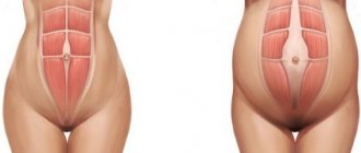
Clinical manifestations of antiphospholipid syndrome during pregnancy
It is possible to understand that a woman has APS by primary infertility of an unknown nature, which is manifested by a violation of implantation, both natural and extracorporeal. But it is not always possible to make the correct diagnosis immediately.
If a woman does become pregnant, as her gestational age increases, she experiences certain signs:
- Progressive gestosis, which in most cases progresses to the last stage - preeclampsia. The risk of this pathology in women with APS is more than 20%.
- Sudden premature separation of the placenta, which was fully functioning up to this point. The frequency of pathology is 10%.
- Severe thrombocytopenia (occurs in 20% of pregnant women with APS).
- Damage to veins by blood clots. This is the most common complication. The legs are predominantly affected.
Attention! Catastrophic APS occurs in 1% of women. It cannot be corrected, so it can be fatal.
As for the fetus, the negative impact of APS also extends to it. Stillbirth, embryonic death, developmental delay, and fetal thrombosis may occur.
Complications
Antiphospholipid syndrome is one of the relatively common autoimmune diseases. Complications of aPL mainly develop during pregnancy due to the development of placental vascular thrombosis. Such complications include:
- miscarriages and premature births;
- fetal death and intrauterine death;
- premature placental abruption;
- fetal development abnormalities;
- female infertility;
- eclampsia;
- gestosis.
Without treatment, pregnancy complications due to aPL occur in 80% of cases.
Clinical guidelines
Smoking is contraindicated for people with antiphospholipid syndrome
Regardless of the form of antiphospholipid syndrome, all patients with this diagnosis should lead a lifestyle that reduces the risk of thromboembolic complications: it is recommended to stop smoking and using other psychotropic drugs.
It is necessary to move more in the fresh air, take enough fluids and not abuse alcohol. Clinical recommendations largely depend on the patient's condition.
Patients with phospholipid syndrome should avoid using estrogen-containing contraceptives as they may contribute to the development of thrombosis.
Pregnancy should be carefully planned due to the increased risk of miscarriage. Treatment of the syndrome must be adjusted during pregnancy to prevent spontaneous abortions and not endanger the fetus. Women who want to become pregnant should be aware of the possible risks and treatment options during pregnancy.