In preparation for motherhood, a woman's body undergoes physiological changes. In the normal state, the woman’s pelvic bones are firmly fixed, but with the onset of pregnancy, a special hormone, relaxin, is produced, which relaxes the ligaments and cartilage in this area and makes the pubic, iliac and ischial bones more mobile to ensure the safe passage of the fetus through the birth canal. Excessive bone separation is accompanied by pain, which seriously affects the physical activity of a pregnant woman and worsens her quality of life.
Currently, work is underway on the website to change the price list; for current information, please call: 640-55-25 or leave a request, and an operator will contact you.
Prices for servicesServices
- Ultrasound examination (ultrasound) of the symphysis pubis repeatedly 1360a
- Ultrasound examination (ultrasound) of the symphysis pubis primary 1560a
The information and prices presented on the website are for reference only and do not constitute a public offer.
Our clinics in St. Petersburg
Medical center on Pionerskaya Primorsky district Polikarpov Alley 6k2
Medical center South-West Kirovsky district Marshal Zhukov Ave. 28k2
Medical center in Devyatkino Vsevolozhsk district Okhtinskaya alley 18
For detailed information and to make an appointment, you can call +7
Make an appointment
50% discount on all types of ultrasound in all Medicenter branches
50% until 04/30/2020 There are contraindications. Specialist consultation is required.
Gynecologists recommend ultrasound of the symphysis pubis during pregnancy as the most informative and safe non-invasive examination at any stage.
During an ultrasound of the symphysis pubis during pregnancy, the doctor receives reliable information about the condition of the pelvic bone tissue, soft tissues and ligaments of the area being examined. Based on these data, the doctor makes a decision about the origin of the nagging pain in the pubic region, determines the likelihood of rupture of the pubic symphysis, and also recommends an appropriate method of delivery.
Indications and contraindications
Symphysitis is an inflammation of the symphysis pubis. It occurs in the second half of pregnancy and is so serious that at the slightest suspicion it requires an ultrasound of the symphysis pubis. In the early stages, the disease is not observed due to the absence of load on the pelvis.
Indications for examination:
- symphysitis during a previous pregnancy;
- child's weight over 4 kg;
- pelvic pathologies of a congenital or acquired nature (consequences of injury);
- pain in the pubic area, which can radiate to the perineum, legs, and intensifies when walking up the stairs;
- inability to fully straighten your legs when lying down;
- swelling in the pubic area, determined by palpation and visually;
- change in gait to that of a duck (the woman waddles from side to side);
- clicking in the pubic area when walking, palpation;
- rapid weight gain;
- breech presentation of the fetus.
There are no contraindications to the examination. A woman’s reluctance to undergo an examination for personal reasons is the only reason why an ultrasound of the symphysis pubis is not performed. In this case, an official refusal from research is issued.
Watch a video on the topic of the disease:
How to do an ultrasound of the symphysis pubis
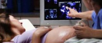
An ultrasound of the symphysis pubis can be done in any ultrasound diagnostic room. The duration of the study does not exceed 10 minutes. During the diagnosis, the condition of the tissues and bones of the symphysis pubis and adjacent areas is studied.
To carry out the diagnosis, a woman needs to take a comfortable position in a horizontal position and free the skin in the pubic area and pelvis. After this, the ultrasound doctor applies a special gel, which facilitates the movement of the sensor across the skin and improves the passage of ultrasound waves deep into the tissue.
Decoding the results
After performing an ultrasound of the symphysis pubis, the doctor compares the data obtained with established standards and draws a conclusion about the condition of the pubic symphysis. There are general norms; there are no gradations by week.
The most important thing is the condition of the symphysis pubis before childbirth, the distance between the bones, and the presence of an inflammatory process that determine the woman’s management tactics.
Signs of normality and pathology on ultrasound of the pubic symphysis:
- During pregnancy, bone divergence by a distance of up to 5 mm is considered normal, this is physiological;
- an increase in the gap from 6 to 9 mm indicates the 1st degree of symphysitis;
- 2nd degree is characterized by a discrepancy of 10-19 mm;
- if the bones of the symphysis have diverged by 20 mm or more, this is the 3rd degree of symphysitis;
- the presence of inflammation, tissue swelling, softening or separation of bones is a sign of the disease.
The pain syndrome does not always correspond to the degree of the disease; much depends on the pain threshold of a particular woman. When the bones diverge by 6–7 mm, some cannot walk, while others with a gap width of 12 mm give birth.
Basic recommendations for detecting signs of symphysitis during ultrasound of the symphysis pubis:
- wearing a bandage or bandaging the pelvis;
- decreased physical activity - lie more, walk less;
- when standing or sitting, distribute weight on both legs;
- do not sit cross-legged;
- do not stay in one position for more than an hour;
- correction of body weight;
- Use the elevator to get to the floor;
- a course of vitamins B, C, PP, etc.;
- a course of minerals containing calcium, phosphorus, etc.;
- performing a set of exercises prescribed by a doctor, the purpose of which is to reduce the load on the pubic symphysis, which reduces pain.
With the 1st degree of symphysitis, natural delivery is possible if the baby is not large, the pelvis is not narrow, and the birth canal is well passable. After the baby is born, the woman continues to take medications and wear a bandage.
A woman with stage 2 disease often has a natural birth. If there are complications, for example, an inflammatory process or a large child, a caesarean section is used.
During the pushing period and the birth process itself, the child’s condition is assessed using CTG. Based on the number of heartbeats, the doctor makes a conclusion about the well-being of the baby in the womb. This is important because there is a possibility of hypoxia.
3rd degree poses a danger to the health and life of a woman. Rupture of the symphysis pubis during childbirth is a severe traumatic situation that always requires treatment and a long period of rehabilitation. In rare cases, when the bones diverge by 30–50 mm, surgical treatment may be used to connect the pelvic bones. Symphysitis of the 3rd degree is an indication for a cesarean section.
After delivery, ultrasound of the symphysis pubis is performed several times.
This is necessary to assess the dynamics of reduction of the resulting gap. The pain syndrome may persist for several more weeks.
Conducting an ultrasound during pregnancy and obtaining results
In the ultrasound room, a pregnant woman lies on her back. The specialist takes a standard transvaginal probe and places it over the upper edge of the pubic symphysis. This is where scanning begins during pregnancy. Then the specialist gradually lowers the sensor down and takes appropriate measurements at certain points.
Reduced echogenicity of the joint indicates the presence of a sprain. That is why specialists during an ultrasound examination do not have difficulty determining the width of the pubic symphysis. The only disadvantage of ultrasound is that it is very difficult to assess changes in the pubic bones during scanning.
Based on the diagnostic results, the degree of divergence of the pubic symphysis is determined (the width of the symphysis pubis is indicated in parentheses):
- 1st degree, i.e. mild (from 5.1 to 8 mm);
- 2nd degree, i.e. moderate severity (from 8.1 to 11.0 mm);
- 3 degree, i.e. severe severity (more than 11.0 mm).
As a rule, mild severity is detected in women in the first trimester. As the gestational age increases, the disease progresses.
If symphysis pubis dysfunction is suspected, doctors prescribe more than just an ultrasound. MRI and X-ray pelvimetry are additional studies. They allow you to assess the size of the pelvis, determine the structure of the pubic symphysis and the structure of the bones that form it.
How to avoid the occurrence of symphysiopathy?
The occurrence of this disease is not fully understood. In order not to think about how the ultrasound procedure of the symphysis pubis is performed, it is worth familiarizing yourself with a number of general recommendations. These include the following:
- A pregnant woman needs to eat more foods that contain calcium;
- It is best to take vitamin complexes that will enrich the body with essential nutrients;
- you need to take medications with bifidobacteria and lactobacilli;
- moderate physical activity is indicated;
- A pregnant woman should take up swimming, which has a beneficial effect on the general condition of the body;
- It is useful to regularly walk in the fresh air;
- You cannot spend a lot of time in the same position, and you also need to monitor your posture.
If symphysitis has already manifested itself, you will need to follow a certain regime, including bedtime.
It might be useful to read:
- How to win the love of a Turk? ;
- Ruslan Narushevich lectures;
- Ruslan Narushevich Learning to love each other;
- information and analytical portal of independent parents! ;
- How to write the first message to a foreigner;
- Is it possible for a Muslim to marry a Christian?
- National characteristics;
- Sex in Germany: Seven Things Germans Do Differently;
How to avoid the occurrence of symphysiopathy?
Damage to the symphysis pubis will in no way affect the health of the newborn, but will cause a lot of problems for the mother.
Healthy eating to prevent symphysitis
For prevention, it is necessary to monitor your health even before conception. It has not yet been fully clarified how to avoid the occurrence of this disease, but general medical recommendations are as follows:
- eating foods rich in calcium;
- taking lacto- and bifidobacteria;
- taking vitamin complexes;
- playing sports (especially swimming, special gymnastics);
- regular walks in the fresh air;
- moderate physical activity;
- You should watch your posture and avoid sitting in one position for a long time (for example, at the computer).
Preventive measures are especially important for a woman who encountered an insidious pathology during a previous pregnancy.
During pregnancy, a lot of changes occur in a woman’s body, changes aimed at bearing a child, childbirth and feeding. The pelvis of a pregnant woman is a ring that includes the sacrum, coccyx, as well as the ischium, pelvic and pubic bones, which are located symmetrically. The pubic bones are connected to each other by the pubic symphysis, which is otherwise called the “articulation of the pubis.”
How is ultrasound diagnostics performed?
Carrying out an ultrasound of the symphysis pubis does not require any preliminary special preparation from the pregnant woman. As already noted, it is absolutely safe and will not cause any harm to either the mother or her unborn baby. The procedure is painless and does not cause any inconvenience during it.
Using an ultrasound, the doctor will be able to assess the condition of the symphysis pubis
The examination is carried out on a special couch in a lying position. The doctor applies a water-based gel to the area being examined so that there is no air layer between the body and the sensor. The ultrasound wave contacts the area of the body being examined and is reflected from the tissues without causing any harm to them. An image of the area being diagnosed appears on the device screen. The study takes no more than 15 minutes, while direct contact with the sensor lasts only 5-7 minutes.
The harmlessness of ultrasonic waves is confirmed by the fact that diagnostic doctors conduct consultations in the usual form, without any additional protection. But in just one shift they examine more than a dozen people. If necessary, ultrasound of the symphysis pubis is performed several times during pregnancy. You can also safely perform other diagnostic measures on the same day. No recommendations are required to be followed either before or after the examination. Immediately after the procedure, you can go about your normal activities.
What is revealed
Ultrasound examination makes it possible to determine the degree of stretching of the symphysis:
- first – 7-9 mm;
- the second – up to 20 mm;
- the third – more than 20 mm.
If dilation exceeds 10 mm, depending on the woman’s condition and the size of the fetus, the obstetrician-gynecologist may decide to deliver by cesarean section. Natural childbirth with severe deformation of the symphysis pubis is fraught with rupture of the pubis, requiring a long period of rehabilitation during the most critical period for a young mother.
If symphysitis was diagnosed on ultrasound
If during ultrasound diagnostics a significant discrepancy of the pubic bones, sprained ligaments or inflammation of the symphysis pubis is revealed, a certain regime must be followed in the future.
In the future, until the birth, it is necessary to limit physical activity, wear a special pelvic bandage, and perform gymnastics, which will be taught by exercise therapy instructors for pregnant women.
Therapeutic exercises during pregnancy will help correct problems
In more complex situations, periodic treatment in a hospital, in the pathology department for pregnant women, is indicated. In between courses of treatment, it is necessary to follow all the recommendations of the treating doctor, and also limit walking, especially on stairs, monitor weight gain, eat foods rich in calcium, try to keep your legs symmetrical and sit for no more than an hour.
Detection of symphysis pubis injuries
Dysfunction of the pubic symphysis is not the only pathology. During an ultrasound examination, a rupture of the connection may be detected. It occurs when injured due to compression of the pelvis in the anteroposterior direction or a strong blow. This often happens during road traffic accidents, falls from a height, or the body being crushed by something heavy (for example, a collapsed wall or a slab that fell on a construction site).
Symptoms of symphysis ruptures are vivid. The pelvis becomes expanded. In men it takes on a feminine form. The external genitalia also swell and swell. When palpating the pubis, a gap is felt. Women sometimes report dysfunction of urination, which is manifested by urinary incontinence.
If ultrasound and x-ray diagnostics confirm a rupture, the doctor prescribes appropriate treatment. If the discrepancy is less than 2 cm, the patient wears a special pelvic bandage. For ruptures of the pubic symphysis greater than 2 cm, surgical intervention is indicated.
Ultrasound diagnostics is a safe way to detect pubic symphysis dysfunction during pregnancy or injury. However, not all pathologies and injuries can be detected by ultrasound. That is why doctors, in addition to this study, prescribe X-ray diagnostics and MRI.
During pregnancy, a woman's body adapts to bearing and giving birth to a child. The female pelvis is a ring consisting of the sacrum, coccyx, as well as symmetrically placed ischial, pelvic and pubic bones. The latter are located in the center of the lower abdomen and are connected to each other along the midline by the symphysis pubis (the pubic symphysis).
Nature arranges it in such a way that during pregnancy the symphysis softens and the ligaments relax a little to facilitate the passage of the child during childbirth.
The softening is promoted by the hormone relaxin, which is produced by the ovaries of a pregnant woman and the placenta (baby place).