Classification
According to the time and mechanism of occurrence, two types of PN are distinguished
:
- Primary
(up to 16 weeks), associated with disruption of the processes of placentation and implantation; - Secondary
(after 16 weeks), develops under the influence of external factors affecting the fetus and/or mother in an already formed placenta.
According to the clinical course, PN is divided into
:
- Acute
– most often occurs against the background of abruption of a low or normally located placenta. Acute PN occurs mainly during childbirth, but its occurrence at any stage of gestation cannot be ruled out; - Chronic
– occurs at various stages of pregnancy. In turn, it is divided into:
A compensated form of PN – in which metabolic processes in the placenta are disrupted, but blood circulation in the “mother-placenta-fetus” system is not impaired. Due to the compensatory capabilities of the mother’s body, the fetus adapts to these changes and does not experience discomfort;
Subcompensated form of PN - if the causes of PN have not been eliminated, the body’s compensatory mechanisms are under stress and gradually begin to deplete, reaching the next stage;
Decompensated form of PN - progression of the pathological process leads to the occurrence of hemodynamic disturbances in the “mother-placenta-fetus” system at the level of fetal-placental and/or utero-placental circulation.
When using the Doppler method, 3 degrees of decompensated form of FPN
:
- I degree FPN I degree – hemodynamic disturbance occurs only in the uteroplacental blood flow;
- FPN degree Ib - the disorder occurs only in the fetal-placental blood flow;
- II degree – there is a disturbance of blood flow at two levels, but they do not reach critical values;
- III degree – the level of disturbances in the fetal-placental blood flow is critical, the life of the fetus is at risk.
In 60% of cases, FPN leads to intrauterine growth retardation (IUGR), so it is also divided into:
- Placental insufficiency with FGR;
- Placental insufficiency without FGR.
How does the placenta work?
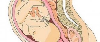
It is no coincidence that the placenta is called the “organ of pregnancy” - after all, it cannot be classified as either a woman’s organs or a baby’s organs. The placenta develops from the chorion, the villous membrane of the fetus. The formation of the placenta is completely completed by the 16-18th week. Externally, the placenta resembles a cake about 3 cm thick, 18 cm in diameter and weighing from 500 to 800 g. A mature placenta consists of 15-20 lobules, separated by connective tissue partitions. One side of the placenta is attached to the inner wall of the uterus; this surface of the placenta is called maternal. The other surface of the placenta - the fruit surface - faces the uterine cavity, towards the baby. The fetal surface of the placenta is covered with amnion - the fetal membrane (wall of the amniotic sac).
During pregnancy, an additional, third circle of blood circulation is formed in the body of the expectant mother - between the mother and the fetus. The most important component of this circulatory circle is the placenta, because it is in it that the blood vessels of the mother and baby meet. The new circulatory system is called the mother-placenta-fetus system.
The placenta consists of 80% blood vessels. These are mainly small vessels. The vascular network of the placenta consists of two systems: uteroplacental and fetal. The uteroplacental vessels bring blood from the vessels of the uterus, that is, from the maternal body, into the intervillous space, from where the blood flows back to the uterus through the veins. At the same time, the blood flow through the uteroplacental network is quite slow: the vessels are small, and the intervillous space of the placenta is extensive. The fetal vessels consist of branches of two umbilical arteries. One large branch of the artery approaches each lobule of the placenta. In the lobule, the vessel branches so that there is a vessel for each placental villi. These vessels of the villi break down into very small ones - capillaries. The ends of the arterial capillaries pass into the venous capillaries, and they merge into increasingly larger vessels, resulting in the umbilical vein.
Thus, each placental lobule consists of a rich vascular network. Moreover, the circulatory systems of the mother and fetus remain completely separate. But, despite the fact that the blood of the mother and fetus does not mix anywhere, a fairly intense exchange of substances occurs between the mother’s body and the baby - through the thinnest membrane of the capillary walls.
During the period of intrauterine development of the fetus, the placenta performs an extremely important mission - the functional life support of the baby. In fact, the placenta is a kind of external power supply that communicates between the maternal body and the fetus. The significance of this connection is enormous: before its birth, the baby is not capable of independent existence. Both oxygen and nutrients, so necessary for the normal growth and development of the child’s body, reach the fetus from the mother’s body through the placental bloodstream. It goes without saying that waste substances and metabolic products that are no longer needed by the fetus enter through the placenta back into the mother’s body for excretion through the kidneys of the expectant mother.
Here are the main functions of the placenta:
- gas exchange: oxygen necessary for fetal respiration is supplied from the maternal vessels to the fruit vessels, and carbon dioxide is transferred back;
- metabolism: proteins, fats, carbohydrates, vitamins, and beneficial microelements are supplied from mother to baby. Products of nutrient processing are removed back;
- protection: there is such a thing as the placental barrier. Such a barrier, a protective obstacle, is the contacting walls of the maternal and fetal capillaries. The barrier has selective permeability, allowing the passage of substances necessary for the baby (oxygen, proteins, fats, sugars, vitamins) and preventing the penetration of many harmful substances and pathogens. The placental barrier prevents the mixing of the blood of mother and baby, which may differ by group and Rh affiliation;
- hormone production: the placenta secretes chorionic gonadotropin, folliculin and progesterone, which play a huge role during pregnancy and childbirth, and the development of the baby.
Causes
The causes of fetoplacental insufficiency are multiple.
Primary PN occurs as a result
:
- Genetic disorders;
- After bacterial and viral infections suffered in the early stages of pregnancy;
- Endocrine pathologies (ovarian hormonal insufficiency, etc.).
Secondary FPN develops as a result
:
- Primary PN;
- Obstetric and gynecological diseases (menstrual irregularities, infertility, gestosis, miscarriage, uterine fibroids, multiple births, etc.);
- Extragenital diseases (diabetes mellitus, hypertension, blood, lung, kidney diseases);
- Congenital and hereditary pathology in the fetus or mother;
- Unfavorable social and everyday factors (mother's age is more than 30 or less than 18, psycho-emotional overload, heavy physical labor, harmful working conditions, smoking, drinking alcohol and drugs);
- Exposure to external environmental factors (ionizing radiation, use of medications).
The most significant risk factors for the development of FPN are the presence of extragenital pathology and gestosis, accompanied by vascular damage.
How to diagnose a problem with ultrasound of the uterus and fetus
Diagnostics is an integral part of the treatment of pathology. With the correct and complete diagnostic series of procedures, the risk of miscarriage or lack of oxygen in the fetus is significantly reduced. Doctors strongly recommend carrying out diagnostic procedures, even with significant deviations in the due date. The necessary manipulations are Doppler measurements and cardiotocography (CTG).
Dopplerography
This type of diagnosis is intended to assess blood circulation in the vessels of the umbilical cord and brain. The study clarifies the situation and assesses the degree of nutrient deficiency for the fetus in the 2nd trimester. If no obvious deviations are found, further forecasts are positive.
Fetal CTG
Cardiotocography helps to determine whether there are disruptions in the functionality of the heart of the intrauterine fetus. The procedure is carried out quickly, the results are visible on the monitor screen. If the result is positive, the patient is sent home, with the obligatory condition of visiting diagnostics every few days.
Symptoms
Information The compensated form of chronic FPN most often does not manifest itself at all. The woman’s condition is satisfactory; disturbances in the mother-placenta-fetus system can only be determined by additional studies (ultrasound, Doppler).
Symptoms of fetoplacental insufficiency begin to appear only in the decompensated form of PN. However, at first, a pregnant woman may only present complaints characteristic of extragenital and obstetric diseases, against the background of which FPN develops, for example, headache with hypertension or swelling with gestosis. But in parallel with this, she may note that the number of fetal movements has decreased. Remember that regular, but not too frequent movements of the fetus are an important sign of its condition!
If intrauterine growth retardation occurs during PN, then there is some discrepancy between the height of the uterine fundus and the gestational age (an indirect sign), which can be detected at the next visit to the antenatal clinic.
The appearance of bloody vaginal discharge at different stages of pregnancy is a dangerous symptom. It indicates that premature placental abruption has occurred, which can lead to acute FPN and fetal hypoxia. In this case, it is better to seek medical help immediately!
Clinical picture
Clinical manifestations of placental insufficiency depend on its form. In the case of the development of chronic compensated placental insufficiency, there are no characteristic symptoms of the disease, and the diagnosis is established only by ultrasound and Doppler ultrasound.
If there is acute or chronic decompensated placental insufficiency, then obvious clinical signs appear, primarily those that indicate the development of intrauterine fetal hypoxia.
- At first, the pregnant woman feels frequent and erratic movements of the fetus, and the doctor notes an increase in its heart rate (tachycardia).
- In the future, in the absence of treatment, movements become less frequent (normally, after 28 weeks of gestation, the expectant mother should feel at least 10 movements of the unborn baby per day), and bradycardia (decreased heartbeat) occurs.
As a rule, placental insufficiency is accompanied by gestosis and the threat of miscarriage, which is not only the cause of its occurrence, but also a consequence (the production of hormones by the placenta is disrupted).
- In the first trimester, the threat of miscarriage can result in a miscarriage or a frozen pregnancy.
- At a later date, due to the permanent threat of miscarriage, pregnancy often ends in premature birth,
- In the third trimester, due to impaired hormone-producing function of the placenta, pregnancy may be carried to term, which aggravates fetal hypoxia.
In addition, a disorder of the endocrine function of the placenta leads to the development of insufficiency of the vaginal epithelium, which creates favorable conditions for the activation of opportunistic vaginal microflora and the development of colpitis. Inflammatory processes in the vagina contribute to infection of the membranes, which can lead to chorioamnionitis and intrauterine infection of the baby.
In addition to the failure of the hormonal function of the placenta, fetoplacental insufficiency causes pathology and excretory function, as a result of which oligohydramnios develops, and in some cases (fetal hemolytic disease or maternal diabetes mellitus) polyhydramnios.
But the most characteristic manifestation of decompensated placental insufficiency is delayed fetal development, which is facilitated by progressive hypoxia. Clinically, developmental delay of the unborn child is established according to external obstetric examination (measuring the size of the abdomen).
Indicators such as the height of the uterine fundus and abdominal circumference lag behind the current stage of pregnancy. The form of intrauterine developmental delay of the unborn child is determined by ultrasound.
- The symmetrical form is characterized by a proportional lag in the weight and length of the fetus, that is, all indicators are reduced to one degree or another.
- Evidence of an asymmetrical form of developmental delay is a disproportionate lag in development of the fetus, that is, the baby’s body length is within normal limits, but its weight is reduced due to a decrease in the circumference of the chest and abdomen (due to a decrease in subcutaneous fatty tissue and a lag in the growth of parenchymal organs: lungs, liver and others).
Even more interesting:
Unidox solutab treatment
Eggworm photo of a child
Diagnostics
Particular attention should be paid to women at risk for developing FPN. Such pregnant women require regular clinical monitoring. At each obstetric examination, the doctor should pay attention to weight gain, measure the height of the uterine fundus (UFH) and abdominal circumference. The discrepancy between the difference in the numerical value of the gestational age and the gestational age, a decrease in the number of movements, a change in the frequency and dullness of the fetal heartbeat sounds is a signal for additional research.
FPN can be determined using 3 main methods
:
- Ultrasonography
. This method allows you to assess the condition of the placenta (thickness, location, degree of maturity, etc.) and the fetus (size, presence of malformations, compliance of growth rates with gestational age), as well as measure the amount of amniotic fluid; - Doppler
. Using this diagnostic method, the blood flow in various vessels of the mother and fetus is examined, which makes it possible to more accurately determine the degree and level of disturbances in the mother-placenta-fetus system, and also allows you to select rational therapy; - Cardiotocography
. A study that allows you to evaluate the cardiac activity of the fetus and indirectly judge its condition.
Timely diagnosis of FPN helps to recognize and respond with adequate treatment to abnormalities in the development of the fetus and the course of pregnancy.
Feto-placental insufficiency (FPI)
Feto-placental insufficiency (FPI)
is a symptom complex in which various disorders occur on both the placenta and the fetus due to various diseases and obstetric complications.
The variety of variants of manifestation of feto-placental insufficiency, the frequency and severity of complications for the pregnant woman and the fetus, the predominant violation of one or another function of the placenta depend on the duration of pregnancy, the strength, duration and nature of the influence of damaging factors, as well as on the stage of development of the fetus and placenta, the severity of compensatory — adaptive capabilities of the mother-placenta-fetus system.
Causes of FPN
Feto-placental insufficiency can develop under the influence of various reasons. Disturbances in the formation and function of the placenta can be caused by diseases of the heart and vascular system of the pregnant woman (heart defects, circulatory failure, arterial hypertension and hypotension), pathology of the kidneys, liver, lungs, blood, chronic infection, diseases of the neuroendocrine system (diabetes mellitus, hypo- and hyperfunction thyroid gland, pathology of the hypothalamus and adrenal glands) and a number of other pathological conditions. Feto-placental insufficiency in anemia is caused by a decrease in iron levels, both in the maternal blood and in the placenta itself, which leads to inhibition of the activity of respiratory enzymes and iron transport to the fetus. In diabetes mellitus, metabolism is disrupted, hormonal disorders and changes in immune status are detected. Sclerotic vascular damage leads to a decrease in the supply of arterial blood to the placenta. An important role in the development of feto-placental insufficiency is played by various infectious diseases, especially those occurring in an acute form or worsening during pregnancy. The placenta can be affected by bacteria, viruses, protozoa and other infectious agents.
Of no small importance in the formation of feto-placental insufficiency is the pathology of the uterus: endometriosis, myometrial hypoplasia, malformations of the uterus (saddle-shaped, bicornuate). Uterine fibroids should be considered a risk factor for fetoplacental insufficiency. However, the risk of fetoplacental insufficiency in pregnant women with uterine fibroids is different. The high-risk group includes primiparas aged 35 years and older with a predominantly intermuscular location of large myomatous nodes, especially when the placenta is localized in the area where the tumor is located. The group with a low risk of feto-placental insufficiency consists of young women under 30 years of age without severe extragenital diseases, with small myomatous nodes of a predominantly subperitoneal location in the fundus and body of the uterus.
Preeclampsia
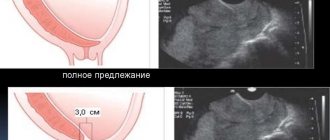
Among the pregnancy complications that most often accompany feto-placental insufficiency, the leading place is occupied by gestosis. The threat of miscarriage should be simultaneously considered both as a cause and as a consequence of feto-placental insufficiency. Due to the different etiology of feto-placental insufficiency with the threat of miscarriage, the pathogenesis of this complication has different options, and the prognosis for the fetus depends on the degree of development of protective and adaptive reactions. With a low location or placenta previa, vascularization of the subplacental zone is reduced. The thinner wall of the lower segment of the uterus does not provide the necessary conditions for sufficient vascularization of the placental bed and its normal functioning.
Placental abruption
Relatively often, with this pathology, placental abruption occurs, accompanied by blood loss. Multiple pregnancy represents a natural model of feto-placental insufficiency as a result of inadequate provision of the needs of two or more fetuses. The basis of feto-placental insufficiency with isoserological incompatibility of the blood of the mother and fetus most often lies in the processes of impaired maturation of the placenta. The fetus develops anemia and hypoxia, developmental delay occurs due to disturbances in protein synthesis processes and decreased enzyme activity. The functional state of the placenta is largely determined by the degree of its development in accordance with the gestational age and the preservation of protective and adaptive mechanisms. Correspondence of the maturity of the placenta to the gestational age is one of the most important conditions for ensuring adequate development of the fetus and its protection. There is no doubt that the late age of a pregnant woman (over 35 years old) or vice versa, young age (under 17 years old), a burdened medical history (abortions, inflammatory diseases), bad habits, exposure to adverse environmental factors, poor nutrition, social vulnerability and domestic instability also contribute to complicated formation of the placenta and disruption of its function.
Clinical practice and scientific research results indicate the multifactorial nature of feto-placental insufficiency. In this regard, it is almost impossible to identify any single factor in the development of this complication. The listed pathological conditions do not equally influence the development of feto-placental insufficiency. Most often, several etiological factors are involved in the development of this pathology, one of which may be leading. Pathological changes that occur with fetoplacental insufficiency lead to a decrease in uteroplacental and fetoplacental blood flow; decreased arterial blood supply to the placenta and fetus; limitation of gas exchange and metabolism in the feto-placental complex; disruption of placental maturation processes; decreased synthesis and imbalance of placental hormones. All these changes suppress the compensatory and adaptive capabilities of the mother-placenta-fetus system, slow down the growth and development of the fetus, and cause a complicated course of pregnancy and childbirth (threat of premature termination of pregnancy, gestosis, premature and late birth, labor anomalies, premature placental abruption, etc. ).
Fetal hypoxia
As a result of exposure to damaging factors and the implementation of pathogenetic mechanisms leading to feto-placental insufficiency, fetal hypoxia naturally develops. At its initial stages, vasopressor factors are activated in the fetus, the tone of peripheral vessels increases, tachycardia is noted, the frequency of respiratory movements increases, motor activity increases, and cardiac output increases.
Further progression of hypoxia leads to a change from tachycardia to bradycardia, arrhythmia appears, and cardiac output decreases. The adaptive response to hypoxia is the redistribution of blood in favor of the brain, heart and adrenal glands with a simultaneous decrease in blood supply to other organs. At the same time, the motor and respiratory activity of the fetus is inhibited.
Classification of FPN
It is advisable to classify feto-placental insufficiency taking into account the state of protective and adaptive reactions. A compensated form of feto-placental insufficiency, in which there are initial manifestations of the pathological process in the feto-placental complex. Protective and adaptive mechanisms are activated and experience a certain tension, which creates conditions for the further development of the fetus and the progression of pregnancy. With adequate therapy and management of labor, the birth of a healthy child is possible.
The subcompensated form of feto-placental insufficiency is characterized by an increase in the severity of the complication. Protective-adaptive mechanisms are under extreme stress (the capabilities of the feto-placental complex are practically exhausted), which does not allow them to be implemented sufficiently for an adequate course of pregnancy and fetal development. The risk of complications for the fetus and newborn increases.
Decompensated form of feto-placental insufficiency - there is overstrain and disruption of compensatory and adaptive mechanisms, which no longer provide the necessary conditions for further normal progression of pregnancy. Irreversible morphofunctional disorders occur in the feto-placental system. The risk of developing severe complications for the fetus and newborn (including their death) increases significantly. The clinical picture of feto-placental insufficiency is manifested in violations of the basic functions of the placenta.
Intrauterine growth restriction
Changes in the respiratory function of the placenta are mainly indicated by symptoms of fetal hypoxia. In this case, the pregnant woman first pays attention to the increased (erratic) motor activity of the fetus, then to its decrease or complete absence. The most characteristic sign of chronic feto-placental insufficiency is intrauterine growth retardation. The clinical manifestation of intrauterine growth retardation is a decrease in the size of the pregnant woman’s abdomen (abdominal circumference, height of the uterine fundus) compared to the normative indicators characteristic of a given period of pregnancy.
With a symmetrical form of intrauterine growth retardation, which develops from the early stages of pregnancy, there is a proportional lag in the length and weight of the fetus. At the same time, quantitative indicators of fetometry have lower values compared to individual fluctuations characteristic of a given stage of pregnancy.
The asymmetric form of intrauterine growth retardation is characterized by disproportionate development of the fetus. This form most often occurs in the second or third trimester of pregnancy and is manifested by a lag in fetal body weight with its normal length. The size of the abdomen and chest of the fetus is predominantly reduced, which is associated with a lag in the development of parenchymal organs (primarily the liver) and subcutaneous fatty tissue. The dimensions of the fetal head and limbs correspond to the indicators characteristic of this stage of pregnancy.
Placental dysfunction
A reflection of violations of the protective function of the placenta when the placental barrier is weakened is intrauterine infection of the fetus under the influence of pathogenic microorganisms penetrating the placenta. It is also possible for various toxic substances to penetrate the placental barrier, which also have a damaging effect on the fetus.
A change in the synthetic function of the placenta is accompanied by an imbalance in the level of hormones it produces and a decrease in protein synthesis, which is manifested by intrauterine growth retardation, hypoxia, and pathology of uterine contractile activity during pregnancy and childbirth (long-term threat of premature termination of pregnancy, premature birth, labor anomalies).
Prolonged and frequent increase in myometrial tone leads to a decrease in arterial blood flow to the placenta and causes venous stagnation. Hemodynamic disorders reduce gas exchange between the body of the mother and the fetus, which makes it difficult for the fetus to receive oxygen and nutrients, remove metabolic products, and contributes to an increase in fetal hypoxia.
Disruption of the endocrine function of the placenta can also lead to post-term pregnancy. A decrease in the hormonal activity of the placenta causes dysfunction of the vaginal epithelium, creating favorable conditions for the development of infection, exacerbation or the occurrence of inflammatory diseases of the urogenital tract. Against the background of a disorder of the excretory function of the placenta and amniotic membranes, a pathological amount of amniotic fluid is observed - most often oligohydramnios, and in some pathological conditions (diabetes mellitus, edematous form of hemolytic disease of the fetus, intrauterine infection, etc.) - polyhydramnios.
Diagnosis of feto-placental insufficiency
At the initial stage of development of feto-placental insufficiency, the listed clinical signs may be weakly expressed or absent. In this regard, methods of laboratory and instrumental dynamic monitoring of the state of the feto-placental complex in a group at high risk for the development of feto-placental insufficiency acquire significant importance. The dominant position in the clinical picture may be occupied by signs of the underlying disease or complication in which feto-placental insufficiency has developed. The severity of fetoplacental insufficiency and violations of compensatory and adaptive mechanisms are directly dependent on the severity of the underlying disease and the duration of its course. The most severe course of fetoplacental insufficiency occurs when pathological signs appear during pregnancy up to 30 weeks or earlier. Thus, the most complete information about the form, nature, severity of feto-placental insufficiency and the severity of compensatory and adaptive reactions can be obtained from complex dynamic diagnostics.
Taking into account the multifactorial etiology and pathogenesis of feto-placental insufficiency, its diagnosis should be based on a comprehensive examination of the patient. To establish a diagnosis of fetoplacental insufficiency and identify the causes of this complication, significant attention should be paid to the correct collection of anamnesis
. During the interview, the patient’s age (late or young age of a primiparous woman), her health characteristics, previous extragenital, neuroendocrine and gynecological diseases, surgical interventions, the presence of bad habits are assessed, and her profession, conditions and lifestyle are clarified.
Information about the characteristics of menstrual function
, the period of its formation, the number and course of previous pregnancies. Menstrual dysfunction is a reflection of the pathology of the neuroendocrine regulation of reproductive function. It is important to assess the course of this pregnancy, the nature of obstetric complications and, most importantly, the presence of diseases against which this pregnancy develops (arterial hypertension or hypotension, pathology of the kidneys, liver, diabetes mellitus, anemia, etc.). You should pay attention to the complaints of a pregnant woman: an increase or suppression of fetal motor activity, pain in the lower abdomen, increased tone of the uterus, the appearance of atypical discharge from the genital tract, which may be accompanied by an unpleasant odor and itching.
During an objective examination, the state of uterine tone
.
The height of the uterine fundus and abdominal circumference are measured, comparing them with the body weight and height of the pregnant woman, as well as the specified gestational age. Such measurements are important and, at the same time, the simplest indicators for diagnosing intrauterine growth retardation, oligohydramnios and polyhydramnios. During an external examination of the genital organs and when examining with the help of mirrors, it is necessary to pay attention to the presence of signs of inflammation, the nature of discharge from the genital tract
, take material from the vaginal wall, from the cervical canal and from the urethra for microbiological and cytological examination.
During echographic examination
determine the size of the fetus (dimensions of the head, torso and limbs) and compare them with standard indicators characteristic of the expected gestational age. The basis of ultrasound diagnostics to clarify the correspondence of fetal size to the expected gestational age and to identify intrauterine growth retardation is the comparison of fetometric indicators with normative data. An indispensable condition is the assessment of the anatomical structures of the fetus to identify abnormalities of its development. An echographic examination also includes placentography. In this case, the localization of the placenta, the thickness of the placenta, the distance of the placenta from the internal os, the correspondence of the degree of maturity of the placenta to the gestational age, pathological inclusions in the structure of the placenta, the location of the placenta in relation to myomatous nodes or the scar on the uterus are determined. During the study, the volume of amniotic fluid, the structure of the umbilical cord and the location of the umbilical cord loops are assessed.
Dopplerography
is a highly informative, relatively simple and safe diagnostic method that can be used for comprehensive dynamic monitoring of the state of blood circulation in the mother-placenta-fetus system after 18-19 weeks of pregnancy, since by this time the second wave of cytotrophoblast invasion has completed. The nature of hemodynamics in the umbilical cord arteries allows us to judge the state of feto-placental blood flow and microcirculation in the fetal part of the placenta. To diagnose disturbances in uteroplacental blood flow, studies are performed in the uterine arteries on both sides.
Cardiotocography is an important part of a comprehensive assessment of the fetal condition.
(CTG), which is a method of functional assessment of the condition of the fetus based on recording the frequency of its heartbeats and their changes depending on contractions of the uterus, the action of external stimuli or the activity of the fetus itself. CTG significantly expands the possibilities of antenatal diagnosis, allowing one to resolve issues of rational pregnancy management tactics.
The final tactics for managing the patient should be developed not only on the basis of an assessment of individual indicators of the state of the feto-placental complex, but also taking into account the individual characteristics of a specific clinical observation (term and complications of pregnancy, concomitant somatic pathology, results of an additional comprehensive examination, condition and readiness of the body for childbirth etc.).
Treatment of pregnant women with FPN
If feto-placental insufficiency is detected, it is advisable to hospitalize the pregnant woman in a hospital for in-depth examination and treatment. An exception may be pregnant women with a compensated form of feto-placental insufficiency, provided that the treatment initiated has a positive effect and the necessary conditions are available for dynamic clinical and instrumental monitoring of the nature of the pregnancy and the effectiveness of the therapy. The leading place in the implementation of therapeutic measures is taken by the treatment of the underlying disease or complication in which feto-placental insufficiency occurs. At present, unfortunately, it is not possible to completely relieve a pregnant woman of feto-placental insufficiency using any therapeutic interventions. The means of therapy used can only help to stabilize the existing pathological process and maintain compensatory and adaptive mechanisms at a level that allows for the continuation of pregnancy until the possible optimal date of delivery.
Treatment of fetoplacental insufficiency should be aimed at improving uteroplacental and fetoplacental blood flow; intensification of gas exchange; correction of rheological and coagulation properties of blood; elimination of hypovolemia and hypoproteinemia; normalization of vascular tone and contractile activity of the uterus; strengthening antioxidant protection; optimization of metabolic and metabolic processes. A standard treatment regimen for fetoplacental insufficiency cannot exist due to the individual combination of etiological factors and pathogenetic mechanisms for the development of this complication. The selection of drugs should be carried out individually and differentiated in each specific observation, taking into account the severity and duration of the complication, etiological factors and pathogenetic mechanisms underlying this pathology. The dosage of drugs and the duration of their use require an individual approach. Attention should be paid to correcting the side effects of certain medications.
Treatment of feto-placental insufficiency begins and is carried out in the hospital for at least 4 weeks, followed by its continuation in the antenatal clinic. The total duration of treatment is at least 6-8 weeks. To assess the effectiveness of the therapy, dynamic monitoring is carried out using clinical, laboratory and instrumental research methods. An important condition for the successful treatment of feto-placental insufficiency is that the pregnant woman adheres to the appropriate regimen: proper rest for at least 10-12 hours a day, elimination of physical and emotional stress, and a rational, balanced diet.
One of the leading pathogenetic mechanisms for the development of fetoplacental insufficiency is disturbances of the uteroplacental blood flow (UPB) and fetoplacental blood flow (FPF), accompanied by an increase in blood viscosity, hyperaggregation of erythrocytes and platelets, a disorder of microcirculation and vascular tone, and arterial circulatory insufficiency. Therefore, an important place in the treatment of fetoplacental insufficiency is occupied by drugs with antiplatelet and anticoagulant action, as well as drugs that normalize vascular tone.
It should be taken into account that a periodic and prolonged increase in uterine tone contributes to impaired blood circulation in the intervillous space due to a decrease in venous outflow. In this regard, in the course of treatment of fetoplacental insufficiency in patients with symptoms of threatened miscarriage, the prescription of tocolytic drugs (β-mimetics) is justified. The progression of hypoxia occurs against the background of intensified lipid peroxidation, the formation and accumulation of peroxidation products that damage mitochondrial and cellular membranes. Activation of this process is due to weakening of antioxidant defense mechanisms. Of great importance in the treatment of fetoplacental insufficiency is the normalization of antioxidant protection, which has a positive effect on the transport function of the placenta. Considering the most important detoxification function of the liver, as well as its decisive role in the production of proteins and procoagulants, it is advisable to use hepatoprotectors in the complex therapy of fetoplacental insufficiency.
An integral part of the complex of therapeutic measures is the use of drugs aimed at improving metabolic and bioenergetic processes, which also helps to improve hemodynamics, gas exchange and other functions of the placenta. The presence of unfavorable and worsening signs of feto-placental insufficiency, which indicate decompensation of the feto-placental complex, accompanied by a pronounced combined reduction of utero-placental and feto-placental blood flow, a significant violation of CTG indicators and a low level of compensatory and adaptive capabilities of the fetus, serve as indications for emergency delivery . In this situation, caesarean section is most preferable. If there are no signs of decompensation of the feto-placental complex, the therapy is effective and the current clinical situation allows, then delivery is possible through the natural birth canal if the following conditions are met: cephalic presentation of the fetus; full proportionality of the fetal head and maternal pelvis; sufficient readiness of the body for childbirth.
Management of childbirth during FPN
For the successful start and further development of normal labor, one of the most important conditions is the presence of a “mature” cervix. Labor management must be carried out with adequate and gradual pain relief, especially if hypertensive syndrome occurs during childbirth. One effective method of pain relief is epidural anesthesia, which should be performed during the active phase of labor. During childbirth, patients with fetoplacental insufficiency are advised to undergo venipuncture for corrective infusion therapy, aimed at maintaining not only an adequate level of uteroplacental and fetoplacental blood flow, but also metabolic processes in the fetoplacental complex, as well as preventing abnormal contractile activity uterus.
During labor, labor weakness may develop, which will require the use of uterotonic drugs. In this regard, the most appropriate for labor stimulation in women with feto-placental insufficiency is the use of drugs based on prostaglandins E2 and F2α. If, with the introduction of uterotonic drugs, no dynamics of cervical dilatation is observed within 2 hours or the condition of the fetus worsens, then their further administration is not advisable. In this situation, the issue should be resolved in favor of operative delivery, the method of which depends on the obstetric situation. If labor is weak in the first stage of labor, combined with any other pathology (breech presentation of the fetus, complicated obstetric history, old age of the woman in labor, etc.), a caesarean section should be performed.
In the second stage of labor, exit forceps should be applied or vacuum extraction should be performed. You should not delay the second stage of labor, which should be completed in no more than 6-8 attempts. It is necessary to minimize the mechanical impact on the fetal head. It is better to conduct childbirth without “protecting” the perineum, to exclude forced traction on the fetal head. The indications for perineal dissection should be expanded. If the therapy is ineffective and signs of fetal hypoxia increase during labor, as well as if additional complications arise that aggravate the obstetric situation, the tactics of labor management should be reconsidered in favor of cesarean section.
Indications for reviewing labor management tactics may be: deterioration of the condition of the woman in labor or the fetus; the occurrence of labor abnormalities (weakness or incoordination of contractile activity of the uterus); disruption of the biomechanism of childbirth; identifying signs of disproportion in the size of the fetal head and maternal pelvis; signs of progressive feto-placental insufficiency (fetal hypoxia).
Preventive actions
For intrapartum assessment of the state of the feto-placental system, it is necessary to use complex diagnostics, the results of which are assessed in conjunction with the initial state (on the eve of birth) of the mother and fetus. One of the main preventive measures is the early identification and registration of pregnant women at risk for the development of feto-placental insufficiency. With the onset of pregnancy, the patient’s daily routine should be properly organized with proper rest, including sleep for at least 8-10 hours, including daytime sleep for 2 hours, as well as spending 3-4 hours in the fresh air.
A rational, balanced diet with an adequate content of proteins, fats, carbohydrates, vitamins and microelements is important for the health of the mother and the proper development of the fetus. Fluid intake (in the absence of edema) to prevent hypovolemia should be 1-1.5 liters. It is important to monitor weight gain during pregnancy. By the end of it, the increase in body weight should average 10.4 kg.
It is advisable to carry out preventive measures with the use of medications in pregnant women at risk at 14-16 weeks and 28-34 weeks. The duration of each of these courses should be 3-4 weeks. Drug prevention enhances the compensatory and adaptive reactions of the mother and fetus, prevents dyscirculatory disorders of the uteroplacental and fetoplacental blood flow and morphological disorders in the placenta.
An integral part of preventive measures is determining their effectiveness, assessing the development of pregnancy, placental formation, growth and development of the fetus based on the results of ultrasound and laboratory screening. A pregnant woman should be promptly hospitalized in an obstetric hospital to prepare the body for childbirth and decide on the timing and method of delivery.
Make an appointment with specialists by calling a single call center:
+7(495)636-29-46 (metro stations “Schukinskaya” and “Street 1905 Goda”). You can also make an appointment with a doctor on our website, we will call you back!
Treatment
In the compensated form of FPN, outpatient treatment is allowed subject to constant monitoring (the number of visits to the antenatal clinic increases). In other cases, hospitalization is necessary. Unfortunately, all existing treatment methods do not completely restore the normal functions and structure of the placenta. They only help stabilize the process and prevent its further progression.
Additionally , since the main reason for the development of this pathology is impaired blood circulation in the vessels, all actions should be aimed at eliminating them.
Therefore, FPN should be treated using the following drugs: Trental, Curantil, Pentoxifylline, Eufillin. Actovegin and Troxevasin are used to improve and activate metabolic processes in fetal tissues. FPN is often accompanied by increased uterine tone; to reduce it, medications such as Ginipral, No-shpa, and Magnesium Sulfate are used.
All treatment measures are carried out exclusively in a hospital, under the strict supervision of doctors and constant monitoring of Doppler measurements, ultrasound and CTG, for at least 2 weeks.
Diagnosis and treatment of fetoplacental insufficiency
Diagnostic tests for suspected FPN include ultrasound, Doppler (measurement of the level of blood flow in the vessels of the placenta, umbilical cord and uterine arteries using ultrasound), cardiotocography (hardware measurement of heart rate and fetal movements for 40-60 minutes), urine and blood tests to detect infection, hemostasiogram (blood test for clotting), etc.
If the development of FPN is suspected, the expectant mother is advised to be hospitalized in the department of pathology of pregnant women of the maternity hospital. The reason for hospitalization is, as a rule, the pathology that led to FPN, for example, an acute viral infection, gestosis, exacerbation of a chronic urogenital infection (cytomegalovirus, herpes, ureaplasmosis, etc.), DIC syndrome (increased blood viscosity), as well as the consequences FFU—intrauterine growth restriction. In the hospital, diagnostic studies are carried out to confirm the diagnosis and identify the degree of development of the pathology. Based on the research results obtained, the pregnant woman is selected for therapy aimed at eliminating the cause of FPN and preventing the development of complications.
If a viral infection is detected, antiviral drugs and agents that strengthen the immune system are prescribed. These drugs include Viferon, Interferon, Vilprofen, immunoglobulins. The drugs are prescribed in the form of vaginal suppositories (suppositories), tablets and intravenous injections at the discretion of the attending physician.
For FPN that has developed against the background of gestosis, the first stage of treatment will be the use of antihypertensive therapy, that is, drugs that lower blood pressure. The most applicable medications in this group are Atenolol and Lakren (tablet forms).
If the cause of the development of FPN is increased blood viscosity (DIC), treatment uses anticoagulants - drugs that reduce blood viscosity. During pregnancy, Fraxiparine is prescribed for this purpose. This medicine is given as injections (shots) under the skin.
To treat FPN itself, agents are used that improve the rheological properties of blood and placental blood flow - Actovegin, Curantil. The dosage and method of administration are selected by the doctor individually, taking into account the duration of pregnancy and the severity of the disturbance of placental blood flow. These drugs are included in any treatment regimen for FPN, regardless of the cause that caused it.
If treatment for FPN is ineffective and there is significant growth retardation in the fetus in the third trimester of pregnancy, delivery is performed by cesarean section after preliminary preparation.
Risks and consequences
Placental insufficiency during pregnancy often leads to the following complications:
- Termination of pregnancy;
- Fetal hypoxia;
- Delayed development and growth of the fetus;
- There is a risk of antenatal (intrauterine) fetal death.
important The presence of FPN during pregnancy does not mean that the child will be born with FGR or prematurely. With a small degree of hemodynamic disturbances in the mother-placenta-fetus system, early detection and adequate treatment, the prognosis for pregnancy and the fetus is favorable. Of course, with severe hemodynamic disturbances, the positive outcome of the disease is questioned.
Features of the pathology
The placenta is an organ that forms during pregnancy. Its main functions are the delivery of nutrients from mother to fetus, gas exchange and protection of the baby from various negative factors.
What is FPN during pregnancy? This is what medicine calls a set of changes in the placenta. In other words, this is a violation of blood circulation in the organ, as a result of which the baby may not receive the amount of oxygen or nutrients he needs.
If this pathology progresses, intrauterine growth retardation or even fetal death are possible.
There are no specific symptoms of placental insufficiency
The only thing you should pay attention to is a sharply increased or, conversely, decreased fetal activity
Know! Normally, it is believed that during the day you should feel its movements about 10 times. If the movements are practically inaudible or, on the contrary, they have become too active, then you need to consult with your doctor.
A doctor may suspect FPN during an examination of a pregnant woman. If the weight gain or increase in abdominal volume is too small and does not correspond to the specific stage of pregnancy, the woman will be advised to undergo an ultrasound, where it will be possible to determine the condition of the placenta and the degree of development of the child.
Prevention
Prevention of FPN during pregnancy should be aimed at eliminating unfavorable risk factors and include:
- Maintaining a healthy lifestyle: complete rejection of bad habits;
- Full sleep;
- Rational and proper nutrition;
- Taking vitamins;
- Elimination of heavy physical labor and stress;
- Treatment of chronic diseases and sexually transmitted infections at the stage of pregnancy planning.