An easy gait, beautiful posture, good health - all this can be ruined by a seemingly ordinary lump under the skin on the leg. Its appearance often indicates the beginning of the development of a disease in the body. Checking this tumor and eliminating its cause gives a chance to prevent the disease and maintain health.
Lump on feet
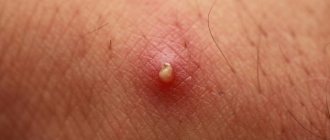
Lumps under the skin on the legs are a common occurrence. Their appearance initially does not cause concern to humans. Only a rapid increase in size of the lump, redness, severe pain, and an unaesthetic appearance of the legs make you come to see a doctor.
Such neoplasms can be different in size, origin, location on the legs, and appearance. They can be multiple and single, soft and hard, painful and painless, ulcerative and inflamed, malignant and benign.
Often, some of their types, when treatment is neglected, develop into serious complications: inflammation, suppuration, and the acquisition of a malignant nature.
Prevention measures
Preventing pathology is not difficult if you follow a few simple rules. It is recommended to carefully monitor your diet and diet, give up bad habits, and normalize your work and rest schedule.
Reducing the load on your hands will also have a beneficial effect on the condition of your joints. If the first signs of a disorder appear, it is recommended not to postpone a visit to a specialist, since this will be the only chance to cure the bumps forever. Otherwise, the pathology progresses.
In advanced stages, it is impossible to completely get rid of seals, but there are many ways to alleviate a person’s condition and prevent damage to other joints.
Common types of cones
There are many diseases that lead to the formation of a subcutaneous lump. Let's name the most common ones.
Gout
A disease that develops due to metabolic, metabolic and hormonal disorders. With it, uric acid salts begin to rapidly deposit in the joints. Pain and swelling appear, redness near the joint affected by the pathology, shine of the skin, temperature rises, and weakness is present. As gout progresses to the chronic stage, red bumps (tophi) form under the skin, which soften slightly during attacks. They can appear in any part of the body, including the hands.
Bursitis
This is the development of inflammation in the synovial joint sacs. There are acute and chronic forms. The first is the result of an injury in the area of the periarticular bursa, and may also be a consequence of previous influenza, furunculosis, or osteomyelitis. The knee, elbow joints, and less commonly the hip joints are affected. A soft elastic ball forms under the skin in the area of the affected joint. He constantly hurts and his temperature rises. If you do not consult a doctor in a timely manner, the disease will progress and become chronic.
It develops with an advanced acute form, regular exposure to the periarticular bursa. The pathology does not lead to impairment of motor function, but causes some limitations. Neglect of treatment leads to the fact that such a “ball” hurts, forms a long-term non-healing hole under the skin in the form of a fistula, and arthritis develops.
Varicose veins
Varicose veins are an increase in the volume of veins located close to the surface of the skin. The disease most often manifests itself on the legs, but it is possible that it may appear on the walls of the esophagus, rectum and bladder, vagina, and arms. Pathology provokes the development of inflammation in the veins. They gradually harden and form aneurysm-like local expansions - red nodes or bumps.
The main signs of varicose veins are:
- swelling of the ankles and legs;
- the appearance of a venous subcutaneous network;
- formation of ulcers, calluses;
- development of eczema, pigmentation on the lower leg and ankles;
- edema.
The causative factors of the disease are considered to be: age-related transformation of the walls of the veins, a sedentary lifestyle, prolonged sitting, pregnancy, and postural defects.
Valgus deformity
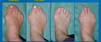
If a lump appears on the thumb with a curvature of this finger and the middle one, this is a hallux valgus deformity. It externally represents a rounded ball from the inside of the foot. The lump is hard, constantly hurts, there is redness and swelling. The root cause of the appearance is weak tendons, endocrine disorders, osteoporosis, arthrosis, flat feet, and uncomfortable shoes.
Subcutaneous cyst
This is a benign cavity tumor filled with pus or fluid. It can form not only on the legs, but also on the arms. The lump feels like a medium-density small ball. It is formed due to infection, closure of the sebaceous glands, or the entry of a foreign body. It has the following symptoms: it does not hurt, it increases slowly, and when pressed it moves slightly to the side.
Dermatofibroma
Harmless red, round growths that form subcutaneously on the legs and arms. The exact reasons for its appearance are unknown. Their main features are:
- purple, brown, or red growths;
- their diameter varies in the range of 0.3-0.6 cm;
- in rare cases they cause itching, burning and pain.
Lipomas
These neoplasms are red balls formed from soft subcutaneous tissue. To the touch, lipomas are elastic and soft lumps. They grow slowly and are not harmful to health. Both single and group cones appear. The size of most is within the 5 cm range; they do not cause discomfort or unpleasant sensations. Pain occurs only when lipomas press on nerve endings.
Enlarged lymph nodes
A small ball (up to 0.5 cm), located on the back of the foot or sole. When palpated, the lymph nodes are dense and hot. The formation of such a “bump” is combined with infectious symptoms: general weakness, fever.
Advice
If subcutaneous lumps appear on your leg, you should immediately consult a doctor. Timely diagnosis is the key to successful treatment and preventing the development of complications.
Reasons for appearance
The exact cause of ganglion cysts is unknown. They are believed to occur as a result of excessive physical exertion, pressure, or trauma. The mechanical impact causes the joint tissue to break down, forming small cysts, which then combine to form a larger mass.
The reasons for the appearance of hygroma on the hands are still not well understood.
“If the hygroma has a typical localization and is superficial, its diagnosis, as a rule, is not difficult.”
Doctor-surgeon Kletkin M.E.
- Most experts agree that there is a connection between constant high load on the joint, or repeated traumatization.
- Hygromas often appear as a result of chronic bursitis, inflammation of the mucous bursae of the joint or chronic tendovaginitis, inflammation of the tendon sheath due to thinning of the connective tissue and sweating of protein-rich fluid from small capillaries.
- In a third of all cases, hygroma on a finger joint develops as a result of a single injury, less often in the case of repeated injury to the joints, or untreated chronic damage.
Hygromas on a child’s arm rarely develop and appear due to a genetic predisposition.
How is the treatment carried out?
A therapist, rheumatologist, dermatologist, oncologist, and infectious disease specialist will help you cope with the pathology. After studying all the obtained tests, an accurate diagnosis is made and the causative factor is determined.
Each type of lump has its own treatment method.
- If the appearance of a lump is a consequence of the transition of gout to the chronic stage, then treatment consists of preventing attacks, relieving pain and swelling. Drugs that reduce uric acid levels, decongestants, painkillers, and anti-inflammatory drugs are used. Additionally, the patient is recommended to follow a special diet and a course of physical therapy.
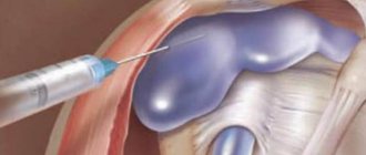
- When a lump forms as a result of bursitis progression, the synovial sac is washed and injected with antibacterial and anti-inflammatory agents. Physiotherapy is carried out, compresses and contrast lotions are prescribed. The patient is required to comply with hygienic requirements and limit physical activity. The advanced stage of bursitis is not amenable to drug treatment. The patient undergoes surgery.
- If a lump on the leg under the skin has formed due to the progression of varicose veins, treatment is carried out using non-surgical methods: sclerotherapy, laser, medication. Treatment of damaged veins by surgical excision is carried out in severe forms of the disease.
- If a lump appears due to hallux valgus, doctors recommend regular wearing of special shoes and insoles. Anti-inflammatory non-steroidal and corticosteroid (rare) drugs are prescribed. But to completely eliminate the “ball”, surgery will be required.
- If the subcutaneous “ball” is a cyst, then treatment is prescribed in extreme cases. It usually resolves on its own over time. If the cyst is inflamed and its growth progresses, a therapeutic course is carried out followed by surgical removal.
- The dermatofibroma lump does not require removal, but if the patient wishes, it can be removed surgically. To reduce its size and make it flat, cryotherapy is used - freezing with liquid nitrogen.
- A lipoma ball does not require surgical treatment, since adjacent tissues are not damaged. Its removal is carried out only at the request of the patient or in the case when it is a visible cosmetic defect.
- The formation of a lump due to inflammation of the lymph nodes is treated with anti-inflammatory drugs. To avoid further development of inflammation, heating and warming compresses should not be used!
Advice
Any of the bumps that appear on the leg cannot be ignored. Even if it does not bother you, you still need to see a doctor.
You should be careful about your health. The appearance of any type of compaction is a strong argument in favor of visiting a specialist.
Physiotherapy and pain relief
The review of folk remedies and solutions on this topic is replete with abundance: these include all kinds of compresses, and the application of cabbage leaves, and wrapping with burdock, and iodine nets. Of course, these procedures cannot correct a toe deformity. Roughly speaking, lotions have no effect on bone structures.
| Important! | |
| Surgery to restore the anatomical structure of the foot gives a more lasting effect, usually ridding the patient of bunions forever. However, if flat feet progress, the bunions may return. So after surgery, the described prevention methods are very important. | |
Physiotherapy is recommended in the early stages of Hallux Valgus. As a comprehensive treatment to relieve inflammation and pain, courses of magnets, lasers and other procedures are prescribed. At home, baths with sea salt are suitable for this purpose (1 tablespoon of sea salt per 1 liter of water), and it is important to observe the temperature regime - no more than 36–36.8 degrees. For the first 20 days, baths are done every day, and then 3 times a week. Patients with the first and second degree of hallux valgus are also recommended exercise therapy, special exercises for the foot (elementary walking on toes, on heels, on the outer and inner sides of the foot), and massage.
