What it is
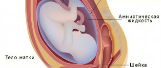
The placenta is an organ of embryonic development of mammalian organisms.
Man also belongs to this group of living organisms, and during pregnancy, this formation is formed in a woman’s body. In everyday life it is called a “children's place” and not by chance. It surrounds a certain area of space inside the mother’s body where the baby’s growth and development will occur.
Protects him and ensures a relationship with his mother.
The quality of the placental membrane determines how the fetus will grow and form. Therefore, it is very important to monitor her condition during pregnancy.
Methods for studying the condition of the placenta and their effectiveness
Examination of the placenta is an important part of the medical examination during pregnancy, which is performed in order to determine the condition of the child's place and, accordingly, the development of the baby.
The main method for examining the placenta is ultrasound diagnostics (ultrasound). This method makes it possible to study the correspondence of the thickness and maturity of the placenta to the gestational age, as well as the place of placenta attachment. In addition, ultrasound allows you to determine the number of amniotic sacs, the structure of the umbilical cord and possible pathological neoplasms in the child's place. During a normal pregnancy, the young mother will have to undergo one routine ultrasound in each trimester. If pathologies are present, their number can increase significantly.
The type of placenta previa during pregnancy can be determined by ultrasound
But unfortunately, this method is not ideal, since the results of its research do not always reliably reflect the state of fetal development. Therefore, to increase the efficiency of medical research, another method is used - Doppler. With its help, the doctor determines the ability of the placenta to supply the baby’s body with oxygen and nutrients, that is, the speed of blood flow in the vessels of the placenta, umbilical cord and fetus is carefully examined. This method allows you to carefully examine each blood vessel.
Doppler ultrasound helps to study the speed of blood flow in vessels
There are cases when ultrasound results show “premature aging” of the placenta, but Doppler ultrasound does not reveal any deviations from the norm and vice versa.
Dopplerography is similar in nature and principle to the ultrasound examination method. Doppler is prescribed only in the third trimester of pregnancy and is usually performed together with an ultrasound examination.
Tracking hormonal levels is also an integral part of research during pregnancy. To assess the level of hormones that are responsible for the normal course of pregnancy and prevent miscarriage and premature birth, laboratory methods are used. The main pregnancy hormones include: hCG (human chorionic gonadotropin), prolactin, lactogen, estriol, oxytocin, progesterone. To determine the level of these hormones, blood from a finger prick and urine are taken from a pregnant woman.
Another method that helps determine the condition of the baby in the womb, and therefore the development of the child’s place, is cardiotocography. Listening to the baby's heartbeat occurs through two sensors fixed on the surface of the mother's abdomen using belts. In this case, one sensor monitors the baby’s heart rate, the other monitors the frequency of uterine contractions, and the pregnant woman holds in her hand a kind of remote control with one button, which she presses every time the baby moves. This method is based on recording changes in heart rate, which depend on uterine contractions and the sex of the child.
During cardiotocography, special sensors are placed on the mother's abdomen.
During treatment in the pathology department, CTG (cardiotocography) had to be done almost every day. I would like to note that the procedure took at least half an hour, or even more. Moreover, it was not always possible to obtain results the first time. And despite the whole “bouquet” of my pathologies, the CTG results did not show any deviations from the norm.
All of the above methods for examining the placenta are not 100% effective if done separately. To obtain a complete and reliable picture of the condition of the child's place, it is necessary to conduct a full comprehensive examination using all methods that harmoniously complement each other.
Formation, structure and development
After fertilization of the egg by the sperm, the placenta immediately begins to form.
The place of its initial development is the mucous membrane of the uterus. As a rule, this occurs on the back wall of the muscular organ.
Structure
The amniotic membrane has a very complex structure. There are 8 layers in it:
- decidua - the first layer from the uterine wall, is a modified endometrium that contains a large number of cells with glycogen;
- Langhans layer - consists of a loose type of connective tissue, contains vessels of the circulatory and lymphatic systems;
- trophoblast - a layer that prevents the contractility of arterial vessels;
- lacunae - cavities that are filled with blood;
- multinuclear simplast - a layer of multinuclear muscle fibers;
- cytotrophoblast - a layer of special cells responsible for the secretion of active substances;
- stroma – a connective tissue layer with a large number of vessels;
- amnion is a layer that performs the function of synthesizing amniotic fluid.
For what period and when is it formed?
The completion of the formation of the “baby place” structures occurs at the end of the 4th month of pregnancy, at approximately 15-16 weeks.
A month later, active metabolic processes begin between the placenta and the embryo.
The baby begins to actively receive oxygen and nutrients, and fetal waste products are removed from the amniotic fluid.
From the 22nd week of pregnancy, she begins to actively gain weight. This is due to the child’s ever-increasing needs for nutrition and breathing.
Only by the 36th week of the perinatal period does it reach full maturity.
Development is accompanied by an increase in mass and thickness.
By the 40th week of pregnancy, the weight of the “baby place” is about 0.5 kg. Thickness up to 3 cm.
Placenta previa
Another serious complication of pregnancy associated with the location of the placenta in the lower parts of the uterus. In general, when such a placenta attachment site is fixed at the beginning of pregnancy, this does not mean anything: the size of the uterus changes greatly, its walls stretch, so that in the second half of pregnancy the placenta, together with the walls, rises up. However, sometimes it partially or completely covers the area of the internal pharynx and, accordingly, prevents the child from being born naturally.
The condition of the placenta is assessed using ultrasound examination; it provides a wealth of useful information about the size, thickness, internal structure and attachment site of the placenta. Based on the results of the ultrasound examination, the doctor makes certain decisions regarding the management of pregnancy.
Functions
During pregnancy and fetal development, the placenta plays a very important role and performs a large number of functions:
- ensures the supply of oxygen to the baby and the removal of carbon dioxide from the amniotic fluid;
- transports all the nutrients necessary for the child’s development;
- fetal waste products are removed through the vessels;
- protects the developing baby from attacks by the mother's immune system;
- participates in the synthesis of active biological substances necessary for the proper development of the fetus.
Thus, the placenta provides the functions of gas exchange, excretion, protection, nutrition, as well as the synthesis of substances. Without it, proper development of the fetus in the womb is impossible.
How the placenta works
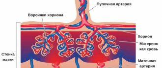
Between the fetal and maternal parts of the placenta (thus the decidua layer) there are “cups” filled with maternal blood. They are formed by the umbilical blood vessels of the fetus reaching towards them - crushing and branching, they form a plexus of the finest villi, which form the wall of the “calyx”.
Maternal and fetal blood flow is not directly communicated!
Nutrients come under the influence of osmotic pressure; they seem to “leak” through the walls of the blood vessels. This is why a “placental barrier” arises - something “passes” from mother to child, but some substances remain only in the mother’s blood. So, what is sent to the child?
First of all, gas exchange occurs through the placenta: oxygen dissolved in the blood passes from the maternal blood to the fetus, and carbon dioxide returns back from the child to the mother.
Secondly, through the placenta the baby receives the nutrients necessary for growth.
Unfortunately, the placental barrier is easily overcome by substances harmful to the development of the child: nicotine, ethyl alcohol (alcohol), many medications (therefore it is important to use only those medications that are recommended by the attending physician who knows about the patient’s pregnancy), as well as viruses (the child will get sick with your mother).
Fortunately, the child also receives maternal antibodies that protect the fetus from infections. At the same time, the placenta delays cells of the mother's immune system that could recognize the fetus as a “foreign object” and trigger a rejection reaction.
Finally, the placenta synthesizes a number of hormones necessary to maintain pregnancy - human chorionic gonadotropin (hCG, the level of which in the blood determines the fact of pregnancy), placental lactogen, prolactin and many others.
The placenta (afterbirth) is born within an hour after birth. Her condition is important to the doctor, so she is carefully examined: it can be used to judge the course of the pregnancy, determine whether there were detachments or infectious processes.
One of the complications of childbirth is incomplete birth of the placenta, when it (in whole or in part) grows tightly into the wall of the uterus. This can cause life-threatening bleeding for the mother in labor, and a small piece of placenta missed by the doctor and left in the uterus can cause an infectious disease.
Mother-placenta-fetus system
After the fusion of the germ cells of the mother and father, the “mother-placenta-fetus” system immediately begins to form. It includes:
- the body of a pregnant woman;
- developing fetus;
- children's place as a connecting link.
The connecting link can be conditionally divided into 2 parts:
- maternal;
- children's room
The maternal part contains a large number of lacunae with blood and vessels. It is a modified mucous membrane. The children's part is represented by the villous chorion.
Blood flow in the maternal part is slow. This makes it possible to thoroughly cleanse the blood that comes from the fetus. In the lacunae, the exchange of substances between the woman’s body and the fetus occurs.
What happens to the placenta after delivery?
There are a huge number of myths about what happens to the placenta after the baby is born. Some believe that the afterbirth is used in pharmacy and cosmetology, illegally obtaining the consent of a woman who, in a state of clouded reason, signs consent to its use. Others believe that the placenta must be eaten or buried. What actually happens to the baby's place after childbirth?
In fact, in 96% of cases, while still in the delivery room, the midwife places the placenta in a plastic bag, on which the gynecologist writes the first, last and patronymic name of the woman in labor, the weight, height and time of birth of the baby, as well as the characteristics of the course of pregnancy and complications. Then this bag is placed in a special refrigerator, where the afterbirths of other women in labor are also stored. During the first 24 hours, a car arrives and takes the material for histological examination to a pathologist, where after the examination it is disposed of by special services.
The placenta is not examined and is immediately disposed of if the pregnancy proceeded just perfectly, without any complications, including acute respiratory viral infections. But this happens very rarely.
Back in 2000, the Kiev Institute of Cell Therapy offered a paid placenta storage service - cryopreservation.
Everyone knows that stem cells from umbilical cord blood have beneficial properties, which is why there are specialized cord blood storage banks. But the procedure for collecting and storing stem cells is very expensive and not all women can afford such a “pleasure”. Also, the placenta, being a biological tissue, contains a large number of stem cells that are used in medicine:
- in the treatment of cancer;
- when correcting immunity;
- in the treatment of infectious diseases;
- in the treatment of chronic fatigue.
Moreover, stem cells are widely used in cosmetology for the preparation of creams, lotions and the like.
I never thought about the “fate” of a child’s place after childbirth and the thought that it could be stored and somehow used did not even occur to me. Even during my first pregnancy, my husband and I thought about preserving stem cells from umbilical cord blood, but after seeing the exorbitant price of this procedure, we decided to abandon this idea until the next pregnancy and childbirth. But at the moment I really regret that I refused. Still, raising a child as a worthy person is much more difficult than carrying and giving birth to him, although pregnancy and childbirth were very difficult for me. Therefore, we are not planning a second baby yet.
Video: Olga Pryadukhina, obstetrician-gynecologist, about pathologies of the placenta
Norms for placental thickness at different stages of pregnancy
Thickness is an important criterion for a woman’s pregnancy. During routine ultrasound examinations, this indicator must be monitored.
Deviations in values, both upward and downward, signal possible pathologies and complications.
Thickness is taken into account to determine the degree of maturity of the “children’s seat”.
The first measurement of this value occurs during a routine ultrasound examination at 20 weeks. The thickness norm at this time falls within the range of 16.7 to 28.6 mm.
Until the 36th week of pregnancy, an increase in thickness occurs. This figure increases by about 1 mm per month.
At 25 weeks, normal is considered to be from 20.3 to 34.0 mm, at 30 weeks from 23.9 to 39.5 mm.
Starting from week 36, it gradually becomes thinner. At 40 weeks of pregnancy, normal thickness ranges from 26.7 to 45.0 mm.
Monitoring the development of the placenta
Changes occurring in the placenta are monitored using ultrasound. To do this, use the classification of organ maturity:
- Stage 0 – up to the 30th week;
- Stage 1 – from 30th to 34th;
- Stage 2 – from 34th to 37th;
- Stage 3 – from 37th to 39th;
- Stage 4 – before delivery.
By monitoring the rate of organ maturation, our doctors can promptly determine the pathology:
- too early development indicates disturbances in the placental blood supply (if the woman does not have a genetic predisposition to this phenomenon);
- delayed maturation does not affect the child’s condition and does not pose a threat to him.
Research is carried out not only using ultrasound, but also by determining the level of lactogen, a hormone that indicates normal maturation. Normally, it should be at least 4 mcg/ml.
Pregnancy management in Nizhny Novgorod also includes daily monitoring of estrogen concentrations. A low level may indicate renal failure of the expectant mother, severe liver pathologies, or be a consequence of taking antibiotics.
The most common pathologies
Modern medical science divides all possible pathologies into 3 groups:
- deviation from normal values of size and shape;
- modification and malfunction of the chorion;
- pathological changes in the maternal part.
Pathological changes in size and shape
Normally, the placenta is round in shape. However, it may change under the influence of certain factors.
Causes of deformation:
- the presence of postoperative scars on the uterine walls;
- frequent abortion;
- an increase in the internal uterine surface during pregnancy.
In women, the most common pathology is called placenta accreta.
In this case, the “baby spot” is attached to the uterine walls and is very difficult to remove after the birth of the child.
In addition to changing shape, the placenta can change significantly in size, going beyond the acceptable limits.
Reasons for increasing the “children’s space”:
- the mother has diabetes mellitus;
- tendency to develop edema of varying severity;
- individual characteristics of the expectant mother’s body;
- the presence of infectious diseases during the perinatal period;
- overweight woman.
Causes of thinning:
- pathologies of the cardiovascular system;
- maternal hypertension;
- pathologies of kidney function.
Insufficient thickness can cause serious complications in fetal development.
Modification and malfunction of the chorion
Possible pathologies of the children's part:
- inflammatory processes that occur in the chorion;
- development of cysts;
- remnant of the yolk sac from the embryo;
- ingrowth of embryonic hair into the chorion;
- oligohydramnios for a long period.
In obstetric practice, cases of chorionic hemorrhage are often encountered. They provoke complications such as:
- early birth;
- intrauterine developmental delay of the child;
- stopping the child's development;
- internal bleeding.
Pathological changes in the maternal part
The maternal part of the placenta can be subject to such pathologies as:
- hemorrhage;
- placental abruption;
- placental infarction;
- intervillous thrombus formation;
- development of chorioangioma neoplasm.
These pathological conditions can cause the following consequences:
- weakening of the protective function and intrauterine infection of the fetus;
- intrauterine growth retardation and development of the child;
- child hypoxia;
- change in the amount of amniotic fluid;
- increased uterine tone;
- hydatidiform mole.
“Children’s place” is the most important structure during the development of a child. The health of the unborn baby and the condition of the pregnant woman depend on the quality of her work.
To minimize the risk of complications, it is important to regularly monitor the condition of this organ.
The expectant mother should know what the weight of the placenta depends on, what factors provoke the development of pathologies and how to diagnose them in time. As a rule, a timely visit to a doctor makes it possible to freely correct the problem.
Stages of organ maturation
The degree of maturity of the placenta is 0, i.e. The initial stage of organ formation normally occurs within 30 weeks after conception. During this period, the placenta has a fairly homogeneous structure and develops from a fuzzy amorphous system to the first signs of maturation. The main functional development is observed starting from 11-12 weeks, when the growth of the placenta and thickening of the membrane are actually observed. One of the main indicators of the zero stage is surface smoothness.
Degree of placental maturity 1 begins with the manifestation of signs of maturation on the placenta, expressed in a violation of the smoothness of the surface - slight waviness, inclusions. Ultrasound reveals individual zones of echogenicity. The normal course of pregnancy involves the development of this stage in the period 27-34 weeks.
The degree of maturity of the placenta 2 is characterized by the appearance of a noticeable relief on the surface of the organ, obvious convolutions of the membrane are recorded, ultrasound shows the presence of numerous changes in echogenicity. The average duration of this period is 34-39 weeks.
The placenta of the 3rd degree of maturity is a finally matured organ that has performed all the necessary functions and is preparing for childbirth. The transition stage to this degree normally begins at 38 weeks of pregnancy.
The main external characteristic is the appearance of a pronounced lobular structure and significant tortuosity of the membrane. This period expresses the natural aging of the placenta. This degree of maturity indicates that the fetus is actually mature, and therefore childbirth after 37 weeks of pregnancy is considered completely acceptable. Premature onset of stage 3 placenta maturity is very dangerous and is fraught with premature birth and insufficient development of the child.
An important characteristic of the maturation of the placenta is its thickness. It is this parameter that is often used to monitor placental development. The table shows the norms for this parameter by week of pregnancy.
The danger of rapid maturation of the placenta
Accelerated development of the organ entails such threats as: fetal hypoxia, insufficient supply of microelements and vitamins to the child, blood flow of the uterus or fetus, etc.
Early maturation of the placenta is caused by a number of reasons:
- Poor nutrition;
- Tobacco products, alcoholic beverages and narcotic substances;
- Viral diseases;
- Diabetes and other hormonal diseases;
- Late toxicosis;
- Abortions performed before the current pregnancy;
- Low physical activity, lack of walks in the fresh air;
- Specificity of the woman's body.
Despite these data, if the expectant mother is diagnosed with stage 2 placenta maturity at less than 32 weeks, there is no need to worry too much. Additional examination for embryo bleeding will definitely be carried out.
If no violations of the fetal development processes are detected, then you can be calm. In the opposite situation, treatment is prescribed in a day hospital. It will be of a restorative nature to the functionality of the placenta for good maintenance of the child’s vital functions. Stage 2 of maturation in a period that is not close to normal does not always mean that the mother has abnormalities.
If no real threat is identified, then it is possible to prescribe a course of Curantil or similar medications at home. But a woman should always listen to the recommendations and advice of her doctor and regularly attend appointments.
Thus, there are 4 stages of placenta development, at each of which significant changes occur in the woman’s body. This is why constant monitoring by a doctor is necessary.
Structural violations
What is the placenta formed from? It includes:
- decidual tissues;
- embryoblast;
- trophoblast.
The main component of this organ is the villous tree. As mentioned earlier, the placenta is necessary for the life support of the child. It is very important that there is no mixing of the blood of mother and fetus, since there is a placental barrier. This protection is very important, as it prevents Rh conflict.
During a normal pregnancy, the weight and size of the placenta increases in proportion to the development of the fetus. But until about the fourth month, the placenta develops a little faster than the baby. If the child dies for any reason, the placenta stops functioning and also dies. In this case, an increase in dystrophic changes can be detected.
As mentioned earlier, the mature placenta has the shape of a disk, the thickness of which is up to three and a half centimeters, and the diameter is about twenty centimeters. The weight of the organ is about six hundred grams. Both sides of the placenta have some differences.
- The maternal side faces the uterus. It is rough and formed from the basal component of the decidural membrane
- The fruit surface faces the child. It is covered with the amniotic layer. Blood vessels can be clearly seen underneath.
Now let’s briefly examine the question of when the placenta forms during a twin pregnancy. It is important to note that the appearance of her (or them) will directly depend on the implantation of the eggs.
Dizygotic twins are implanted separately. Based on the fact that they are found in the uterine cavity almost simultaneously, they can be implanted both in opposite corners and nearby. If implantation occurred nearby, then the placenta may seem like a single whole, but in fact this is not the case, each of them has its own vascular network and membranes. When implanted at a considerable distance, two placentas can be easily detected using ultrasound.
The fetuses of dichorionic twins are separated by a septum. It is important to note that this shell has virtually no blood vessels. Consequently, they receive nutrition from amniotic fluid.
With monozygotic twins, there is one placenta, but the babies are separated from each other by a thin transparent film. In most cases, babies have vessels that connect blood circulation throughout the entire placenta, which is not very good. In this case, there is a danger of transfusion syndrome.
There are also monoamniotic monochorionic twins, when there is no septum between the fetuses at all.
The structure of the placenta may have the following abnormalities:
- the presence of only two shares;
- presence of an additional share;
- fenestrated placenta.
Such violations cannot harm the baby, but they slightly complicate the process of passing the placenta. The doctor should be warned about this pathology, as measures will be taken to force the passage of the placenta. This will help prevent bleeding or infection.
Functional Features
The baby's place appears and is attached to the back wall, closer to the bottom of the uterus. This arrangement is determined by better blood flow and, accordingly, better nutrition.
The placenta plays an important role in the gestation process:
- transporting oxygen from mother to baby;
- delivery of nutrients and vitamins;
- removal of decomposition products;
- organizing a protective barrier against the influence of infections;
- synthesis of hormones (estrogen, progesterone, hCG);
- formation of immunity.
In order to ensure that the functioning of the organ does not decrease, constant monitoring of development and subsequent activities is necessary. The life of the fetus depends on the performance of the membrane, so doctors regularly conduct ultrasound examinations to monitor not only the child, but also the child’s place.