What is hemangioma in children?
The term "hemangioma" comes from the Greek words haema (blood), angeio (vessel) and -oma (tumor).
Hemangiomas are the most common benign tumors of infancy and occur in approximately 5-10% of children. They have a characteristic course, marked by early proliferation and subsequent spontaneous involution.
Infantile hemangiomas are proliferative lesions that usually develop shortly after birth - the first 4-6 weeks of life. They are different from vascular malformations, which are usually present at birth and are less common.
Hemangioma occurs in all racial groups, but is more common in the white race. They also appear more often in girls, premature babies, and low birth weight babies.
The causes of infantile hemangiomas are not entirely clear. The tumor is formed by the proliferation of endothelial cells. Studies have shown the role of fetal hypoxia as a trigger signal that initiates proliferation and abnormal formation of blood vessels by activating certain genes.
More than 80% of hemangiomas in children appear in the head and neck area. Some lesions are small and barely noticeable, while others are large and easily noticeable. All hemangiomas exhibit a characteristic evolution with early rapid growth (proliferation) followed by a period of stabilization and slow spontaneous involution. They will grow to 80% of their maximum size in the first 3 months, and most stop growing after about 5-6 months. However, they can continue to grow for up to 18 months.
Histopathology of proliferative infantile hemantiomas
They then undergo regression or involution (reverse development). Engagement is slow and may take 3-10 years. Almost all flat hemangiomas eventually disappear without treatment. However, regression of bulky hemangiomas is usually incomplete, and they may leave atrophic scars, pigment changes, and residual telangiectasia in at least 50% of cases.
Diagnosis is based on physical examination, skin biopsy, laboratory testing, and imaging.
Most pediatric hemangiomas do not require medical or surgical intervention. Treatment options for clinically significant cases include drug therapy, laser surgery, and surgical excision.
Despite their benign and self-limiting nature, some hemangiomas can cause complications such as ulceration, bleeding, and permanent disfigurement. In addition, some may impair the function of a vital organ.
Symptoms of the disease
The tumor can be benign - hard on palpation and very rarely malignant (a soft structure is a sign of the possibility of further growth of the tumor). Characteristic manifestations of venous hemangiomas include:
With this pathology, the formation may hurt.
- tissue inflammation;
- bluish or red tint of the tumor;
- dryness and roughness of the surface;
- pallor on palpation;
- pain syndrome;
- outlined above the skin level.
Hemangioma on the lip is detected in the presence of such signs as:
- various infections in the oral cavity;
- bleeding in the tumor area;
- having difficulty eating or talking;
- damage to nearby soft tissues;
- sensation of foreign objects in the nasal cavity.
Return to contents
What is the cause of hemangioma in a child?
Neither the cause nor the origin of infantile hemangiomas has been fully elucidated.
There are several theories and evidence supporting conflicting hypotheses about the origin of cells, including placental tissue, endothelial progenitor cells, and mesenchymal stem cells.
A group of tissue-specific markers, especially glucose transporter 1 (GLUT-1), are uniquely coexpressed in hemangiomas and placental microvessels, indicating a connection between hemangiomas and placental microvessels. Placenta and hemangioma have a similar life cycle of increased vascular growth.
Mesenchymal stem cells may also play a role in the formation of infantile hemangiomas. These cells were identified in hemangioma tissue. Mesenchymal stem cells retain the ability to differentiate into several mesodermal cells, including adipocytes, indicating that these cells may be the source of the adipose tissue found in invasive hemangiomas.
Hypoxia (insufficient oxygen to the skin) is considered a likely cause of blood vessel proliferation. Endogenous progenitor cells circulate in the fetus and induce the formation of new blood vessels in response to hypoxia. Endogenous progenitor cells usually disappear after birth, but they may still exist in low birth weight or premature newborns. As endogenous progenitor cells disappear later in life, the hemangioma may regress.
The cause of hemangiomas is not yet clear, but it is known that they are not related to medications that may have been taken during pregnancy, nor are they related to environmental exposures that may have occurred during this time.
Evidence to support a hereditary/genetic component in the development of most childhood hemangiomas is minimal. Most cases are sporadic. Rarely, in some families, several family members have been affected for several generations and are therefore considered to be associated with a genetic abnormality. There have been cases of autosomal dominant transmission of infantile hemangiomas. These hemangiomas have been observed in association with an increase in the number of vascular malformations (mainly capillary malformations) in different family members.
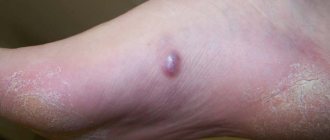
Photo of hemangioma in children
Most hemangiomas are round or oval in shape, but larger lesions may follow the shape of the affected body part.
The size of hemangiomas varies. Some of them are very small (1 mm), while others are very large (20 cm or more).
Each hemangioma differs in how quickly and for how long it grows before regression occurs.
What does a hemangioma look like in childhood?
Most hemangiomas are round or oval in shape, but larger lesions may follow the shape of the affected body part.
The size of hemangiomas varies. Some of them are very small (1 mm), while others are very large (20 cm or more).
Each hemangioma differs in how quickly and for how long it grows before regression occurs.
Dangerous dimensions
The first sign of hemangioma at an early stage is pain in the spine. As the tumor grows, the pain begins to intensify. Back pain appears at night, after excess physical activity. Reaching large sizes, the pain becomes unbearable. If the tumor becomes 1 cm or more in size, then the hemangioma becomes dangerously large.
If the hemangioma is located in the vertebrae in the chest area, then the symptoms appear:
- vertebral pain;
- feeling of numbness in the arms and legs;
- paresis and paralysis;
- the heart rhythm, digestive function, and pelvic organ function are disrupted.
Headaches, decreased mental performance, insomnia, dizziness, hearing and vision impairment are symptoms of cervical hemangioma.
With hemangioma of the lumbar region (in the body l1, l2, l3, l4), pain is felt in the groin and lower back, numbness of the limbs, paresis and paralysis of the legs. In its aggressive form in adults, infertility in women and impotence in men are possible.
Vertebral hemangioma grows gradually, but if the volume is critical, the person’s spine is destroyed, which is extremely dangerous, since the risk of fractures increases. If the formation has reached a volume of 15 mm or more, specific treatment is required. If you delay a visit to the doctor, the following signs may bother you:
- the limbs cease to respond normally to stimuli, as a result of which the legs become insensitive, and the risk of developing paralysis of the lower extremities is high;
- Normal urination is disrupted, urinary incontinence develops, and the organs of the genitourinary system become inflamed.
Diagnosis of hemangiomas in a child
In most cases, infantile hemangioma is usually diagnosed clinically based on history and physical examination. Most superficial lesions do not require any testing. Deep or segmental hemangiomas are usually examined using imaging techniques.
If there is any uncertainty regarding the differentiation of hemangioma from other lesions or vascular malformations, then ultrasound usually provides a definitive answer. In some cases, nuclear magnetic resonance imaging or computed tomography may also be needed to diagnose and determine the size of the lesion. Histological examination may be performed to confirm the diagnosis or if there is any doubt about the possibility of malignancy.
Children with complex lesions are best evaluated by a panel of experts, including a pediatrician, dermatologist, radiologist, ophthalmologist, vascular surgeon, and plastic surgeon.
Laboratory tests
There are no laboratory tests that are routinely done to diagnose hemangiomas in childhood.
However, vascular endothelial growth factor (VEGF) as well as beta-fibroblast growth factor and matrix metalloproteinase (MMP) can be studied as markers of hemangioma proliferation and differentiation.
Visualization
The imaging study to determine the location and distribution of cutaneous and extracutaneous hemangiomas is MRI with or without the use of gadolinium contrast agent. It also helps in differentiating hemangiomas from other vascular lesions.
MRI of infantile hemangioma
Ultrasound is useful in distinguishing hemangiomas from other deep dermal or subcutaneous structures such as cysts or lymph nodes.
Radiography has limited use but may be useful in evaluating hemangiomas that affect the airway.
Biopsy
If the diagnosis is not yet complete after a detailed history and physical examination, skin biopsy may be helpful in distinguishing atypical hemangiomas from other vascular lesions. Tissue samples can be analyzed by histological examination and specific staining techniques.
Classification of angiomas
First of all, angiomas can be divided into two large groups depending on the type of vessels involved in the pathological process:
Tumors can be:
The terminology “vascular cancer” is incorrect because angioma is a soft tissue tumor that can be classified as a sarcoma.
Hemangiomas can be capillary, arterial, cavernous and venous - each type is discussed in detail in the following table.
In appearance it is a lumpy area of the skin of a dark burgundy color.
Problems or complications
Most childhood hemangiomas will go through phases of growth and regression without causing any problems. However, about 25% of cases have complications. Your doctor will help determine whether your child's hemangioma is likely to cause complications based on size, location, and growth rate.
Possible complications of childhood hemangiomas include:
Organ dysfunction
As the hemangioma grows, it can impair organ function.
The most common case is when the hemangioma affects the eyelids or periorbital tissues. Hemangiomas can lead to amblyopia through three mechanisms: physical obstruction of the visual axis, astigmatism from direct pressure on the anterior segment of the eyelid lesion (more often than the upper eyelid), and unilateral myopia. Strabismus may be secondary to amblyopia or paralysis of the extraocular muscles infiltrated by the orbital hemangioma.
If a hemangioma around the eyes grows quickly, it can block a child's vision, which can lead to permanent vision loss.
If your child has a hemangioma in the periorbital area, monitor him closely and consult with your pediatrician. Other affected areas that may need urgent treatment are the genital area and around the mouth.
Airway obstruction is a rare complication of hemangioma. Injuries to the upper lip very rarely obstruct the two nasal passages.
Hemangiomas located on the lower back and spine may be associated with vertebral defects.
Hemangiomas affecting the genital area can cause abnormalities in the pelvis and urinary system.
Several cutaneous hemangiomas are often associated with hemangiomas of internal organs, including the liver, lungs, and intestines. Rarely, even the brain may be affected. Extensive hepatic hemangiomas may be associated with heart failure and hypothyroidism.
Ulcer formation
The skin over the lesion may be broken. Explication occurs in 10-15% of hemangiomas in children. The cause of ulceration is unclear, but may be the result of blood flow to the skin over the lesion or the action of certain cytokines.
Explication usually occurs with rapidly growing hemangiomas. Hemangiomas that are located around the mouth, nose, ears, or anogenital area have a higher risk of ulceration.
The ulceration(s) can be very painful for the child, which can lead to irritability, poor feeding and sleep problems. Ulcers may result in scarring after curing, which may take several months. Secondary infection is possible, but cellulitis, abscess, and bacteremia are rare.
Ulcers heal slowly, so your doctor may recommend treatment to speed up the process and prevent infection and scarring. Treatment for ulcers may include topical or oral antibiotics, dressings, laser surgery, becaplermin gel (recombinant platelet-derived human growth factor), and topical compression therapy (especially useful for lesions in the extremities).
Bleeding
The skin above the lesion protects the hemangioma from bleeding. If the hemangioma is damaged, it may bleed or develop a crust. The blood vessels that make up the hemangioma are abnormal.
When hemangiomas bleed, they usually bleed quickly, but only for a short time. Bleeding can be stopped by applying light, direct pressure to the wound for 15 minutes. If bleeding occurs again or does not stop with this method, contact your child's doctor.
Treatment of neoplasm
Active methods of therapy are used when there is an immediate threat to normal human life. Effects on the organs of smell, vision, the appearance of soft tissue ulcers, decreased functionality of organs, rapid transformation of the tumor are a number of indications of the negative impact of a nevus. Constant monitoring of the development of education is one of the foundations of therapy. In children under 7 years of age, under favorable circumstances, hemangioma disappears on its own. However, timely treatment prescribed by a doctor will contribute to the rapid elimination of venous hemangioma.
Medicines that help eliminate the pathology include cytotoxic drugs, corticosteroids, and beta-blockers. The growth of the formation stops with hormonal therapy, and beta blockers contribute to the death of the nevus. In case of injury and infections, antimicrobial therapy is carried out. Applying a pressure bandage to the tumor also helps to reduce it. In folk medicine, decoctions of walnuts, aloe, celandine, compresses from kombucha and copper sulfate solution are used.
You should not self-medicate, this can lead to negative consequences.
Psychosocial problems
The psychosocial problems associated with deformed facial hemangiomas can be significant.
In early childhood, parents often react with panic. Parental feelings of mistrust, panic or fear are often associated with the rapid growth of these lesions. Variability in the natural course and treatment time add to parental concerns. Parenting stress is increased by strangers who stare, intimidate, or raise questions about causes, such as injury, infection, or cancer. Psychosocial burden can be problematic for both parents and patients with deforming facial hemangiomas.