List of reasons
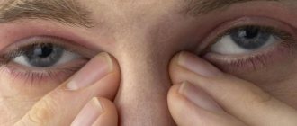
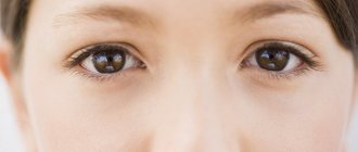
When a person experiences dual vision of objects, it means that one eye does not fixate on the object in sync with the other.
The causes of diplopia are:
- Weakness or paralysis of the muscles involved in eye movement.
- The optic nerve is affected by an aneurysm.
- Head injuries leading to items 1 and 2.
- Tumors and hematomas, due to which the eyeballs cannot move freely.
- Intoxication with drugs or alcohol.
- Mental neuroses.
- Consequences of surgical treatment of the brain or eyes.
The condition of double vision in one eye can be permanent or temporary. In the second case, the symptoms disappear on their own. This usually occurs after abuse of drugs or alcohol.
Reasons for the development of pathology
The physiological cause of double vision is a displacement of the visual axis of the eyeball, due to which the image falls not in the center of the optic fossa, but on its side. In any case, this is a binocular vision disorder. If the patient closes one eye, the double vision will immediately disappear. Only in very rare cases, with complex injuries of the eyeball, monocular diplopia is observed. With this form of the disease, the patient sees two images of the object in question with one eye, even if the other eye is closed.
Binocular diplopia can be caused by the following factors:
- A change in the position of the eyeball in the orbit, in most cases occurring due to injuries to the organs of vision: a fracture of the wall of the orbit after a blow, pinching of the optic nerve, etc. In some cases, the cause of a split image is a hematoma after eye surgery.
- Lesions of the motor nerve of the eye. This occurs with a carotid artery aneurysm, tuberculous meningitis, and intracranial formations.
- Brain stem lesions as a complication of a severe infectious process. Diphtheria, tetanus, mumps, rubella - all these diseases can be an indirect cause of diplopia in children.
- Intoxication of the body due to an overdose of drugs, chemicals, alcohol, as well as acute botulism.
- Thyrotoxicosis in severe form.
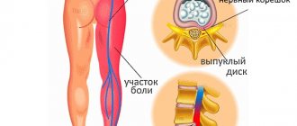
During attacks of hysteria, the patient also experiences double vision. Double vision often occurs with cervical osteochondrosis, when the nerve roots associated with the functions of the visual apparatus are pinched.
Note: diplopia almost always develops against the background of paresis of the extraocular muscles responsible for coordinating the movements of the eyeball. The cause of complete or partial paresis is ocular myasthenia gravis - muscle weakness, or damage to the nerves of the eye that control these muscles.
Binocular and monocular pathology
These are two types of diplopia. The first is characterized as follows: when viewing an object with both eyes, it forms a split. It disappears if you close one eye.
The second type manifests itself as follows: a split image is present in only one eye. And it does not disappear when the second one is closed.
The scheme of binocular diplopia is as follows:
And its reasons are:
- Tumors in the orbital area.
- Neurological pathologies.
- Vascular disorders.
- Malfunctions of the oculomotor muscles.
- Injuries and bruises.
- A sharp rise or decrease in intracranial pressure.
It also occurs when a person is sick:
- pathologies of the nervous system, for example, meningitis or multiple sclerosis;
- infectious diseases, examples - rubella, diphtheria, influenza;
- retinopathy due to diabetes;
- vasculitis.
- epilepsy, brain tumor.
When there is double vision in one eye, there may be abnormalities in the cornea, retinal tissue, or lens. Patients with dry eye often complain of this condition.
The monocular duality scheme is as follows:
Reasons for appearance
Monocular diplopia often acts as a congenital disease. Signs appear in early childhood and worsen as the child grows older.
Diplopia appears in a child due to astigmatism.
However, there are common reasons that in most cases provoke the development of the disease:
- Infectious diseases affecting different parts of the visual system. Usually the cornea is affected, and after healing, scars remain on it, which provoke diplopia. Experts note that in case of an infectious disease, the symptoms disappear when the previously affected eye is closed.
- Astigmatism often becomes the cause of the disease. In this case, objects in the patient’s eyes not only split into two, but also lose clear boundaries and become blurred, which only worsens the clinical picture.
- Cataracts more often provoke diplopia in elderly patients. In this case, double vision becomes a constant symptom, which gets worse over time.
- Diseases accompanied by damage to the thyroid gland. With such disorders, the elasticity of the muscles supporting the eyeball may decrease, which leads to double vision. Men rarely suffer from such disorders; in most cases, hypothyroidism or thyrotoxicosis affects women.
- A decrease in the amount of fluid responsible for moisturizing the eyeball often provokes severe eye fatigue. However, diplopia in this case is rarely permanent. Often the problem can be solved with the help of special drops that moisturize the eyes.
- Uncontrolled hypertension, stroke and heart disease can cause temporary diplopia. Usually the signs disappear after the attack ends. But for many patients with chronic diseases, visual impairment is an accompanying symptom.
- Diseases accompanied by damage to the lens. In this case, diplopia is permanent, the patient’s vision quickly deteriorates, and treatment is long and complex.
- Congenital or acquired strabismus can also cause diplopia. Typically, a violation leads to the occurrence of a cross-type disease.
- Injuries to the brain and cranium. Mild damage almost never leads to double vision, but severe injuries are always complicated by disturbances in the visual apparatus.
- Atherosclerotic changes in the blood vessels of the brain and visual apparatus, provoking poor circulation of the muscles supporting the eye.
- Diseases of the peripheral and central nervous system that lead to disruption of the nutrition of nerve fibers.
Any of the factors can provoke diplopia, but in most cases the disease is the result of several causes.
Vertical and horizontal
The localization of the pathology determines the directions of image ghosting. The two most common options here are horizontal or vertical. There is another type - diagonal. But it is diagnosed extremely rarely.
In this matter, the type of affected eye muscles is of decisive importance. So, if there is double vision in one eye horizontally, it means that the rectus muscles are damaged:
- Bottom.
- Upper.
- Lateral.
- Medial.
Items 1 and 2 are internal. Items 3 and 4 – external.
If there is double vision in one eye vertically, the pathology has affected the oblique muscles: lower or upper.
Both of them are internal.
The diagram of the muscle groups presented is as follows:
Problem with eyes vertically, horizontally
What disease causes double vision? Depending on the location of the pathological process, the image doubles either horizontally or vertically, and sometimes a diagonal split occurs. It all depends on which eye muscles are affected. When the oblique muscles are affected, the picture doubles vertically; when the straight muscles are affected, the image doubles horizontally.
For effective treatment, it is necessary to identify the cause that caused diplopia, and then select options to correct the situation. When the cause is eliminated, as a rule, the ghosting also disappears. If the reasons are not very serious and deep, then it is possible to preserve vision, avoiding various side effects.
To find out why the disease occurred and what led to it, the specialist conducts a comprehensive diagnosis. If it turns out that the pathology is not in the eyes themselves, then other studies of the body are prescribed, since various infectious diseases, intoxication, diabetes, inflammatory processes in the meninges, tumors, injuries, banal fatigue, overheating, and nervous tension lead to diplopia. .
It is impossible to make an unambiguous diagnosis from the patient’s words; a comprehensive examination is required.
Treatment follows the line of the underlying disease. For partial diplopia, eye exercises and wearing special glasses are effective.
Treatment. General provisions
For it to bring the desired effect, it is necessary to discover the real causes of diplopia.
If a connection with any illness is found, the patient is sent to the appropriate specialist. For example, an endocrinologist and a neurologist. And an established course of treatment almost always leads to elimination of the problem.
If the cause of diplopia is eye diseases, then the patient is treated by an ophthalmologist. The most serious pathologies can only be eliminated surgically.
If strabismus is detected, correction is carried out using professional optics. If these methods do not bring the desired effect, a surgeon is required, and the doctor changes the length of the eye muscle.
If you see double in one eye or both when looking at a long distance, then the accommodation of the lens is impaired.
When the cause of the pathology is impaired cerebral circulation, nootropics and drugs to lower blood pressure are used in treatment. Based on the doctor’s testimony, prismatic correction is organized. This procedure may have a similar effect associated with a decrease in vigilance. To solve the problem, special visual gymnastics are prescribed.
The selection of glasses is based on personal characteristics and disease. These optics help to combine a double image into a single one.
Treatment of diplopia
Treatment of diplopia begins with identifying the cause of double vision. To eliminate double vision, it is necessary to treat the underlying disease. Treatment of diplopia is therefore not only the responsibility of the ophthalmologist. In many cases, an examination by a neurologist is required.
In a significant number of cases, treatment is carried out surgically. In particular, surgical treatment is used for damage to the extraocular muscles.
To reduce discomfort during treatment, prismatic optical correction can be used - using specially selected prismatic lenses.
In case of partial diplopia, eye gymnastics can be useful: exercises to expand the field of vision, to restore binocular vision, etc., some of which should be performed using special devices.
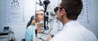
Basic diagnostic methods
During the examination, the specialist determines:
- The nature of diplopia is vertical or horizontal.
- Is there any rotation of objects?
- Quality of vision, light perception and refraction.
- Unsteadiness of gait.
- Deviation of the eyes, parameters of their section, position and movements of the eyelids.
The doctor also sets the contrast and brightness of the image.
The main diagnostic methods are:
- Cover testing. Allows you to identify hidden strabismus.
- Ophthalmoscopy. Special instruments are used to study the vessels in the fundus, the optic nerve and the macula zone. In this procedure, the patient is in a supine position.
- Coordimetry. For the procedure, an ophthalmocoordimeter device is used. In this way, the focus and nature of the disease, as well as the damaged muscle sector, are revealed. Haab procedure. Helps identify split points.
- Laboratory blood tests. With their help, the proportions of glucose are established. They are prescribed by a doctor when he suspects a patient has diabetes.
- Examination of the brain using an MRI procedure. Helps identify abnormalities in its structure or problems with blood circulation.
- Taking a proserine sample. The doctor prescribes it for suspected myasthenia gravis.
- Strabometry. Allows you to measure the angle of strabismus.
Diagnostics
The primary diagnosis of diplopia is made when the patient complains of a double image. The ophthalmologist determines visual acuity, refraction and color perception. Diagnosis of diplopia includes an ophthalmological examination, blood testing, computed tomography (CT) or magnetic resonance imaging (MRI), and a cover test.
If diplopia is the underlying disease, observation by a neurosurgeon or neurologist is necessary.
If diplopia is caused by injury, surgery is necessary. Diplopia can be corrected using prismatic glasses, which significantly improves the patient's vision. Effective correction in the treatment of diplopia is approximately six prism diopters per eye. Up
Treatment courses
The main task in the fight against diplopia lies in eliminating its causes. Typically, the following therapeutic methods are used:
1. Occlusion. It is used when several nerves are affected at once and three-dimensional perception (3D) is lost. During the procedure, one eye is “switched off”. To do this, use special lenses or a very thin tape that does not transmit light. It sticks to glasses. The duration of treatment is determined by the treating specialist and the form of pathology.
2. Prismatic correction. Manifests itself in wearing glasses equipped with prisms. They deflect light rays and shift the image. Fresnel prisms are also often used - special linings on glasses. They can be replaced with similar devices with a different refraction angle when the patient restores vision.
3. Botulinum injections. They are placed when monocular diplopia occurs suddenly to provide temporary relief. Botox is injected into the injured muscles. To carry out the procedure, local anesthesia is administered. This method eliminates the formation of contractures.
4. Operational method. Necessary in extremely severe cases. Allows you to restore the symmetry of the eye position. The operation is performed under general anesthesia. The result is completely restored vision.
If there is double vision in one eye, and this condition is a symptom of any disease, then therapeutic methods are aimed at combating it. So, for example, for infectious diseases antibiotics and immunomodulators are prescribed, for neuralgia - neuroprotectors and analgesics, for hematomas in the orbit - special creams and ointments, for example, "Troxevasin", "Fastum Gel", etc.
To achieve the desired effect faster, the doctor prescribes special exercises to correct vision. They usually stimulate the muscles of the neck and eyes. With their help, the perception of binocular vision is trained. Their selection is based on an individual basis based on the type and format of the pathology. They bring the greatest effect when fighting partial diplopia.
Treatment with folk remedies
Traditional recipes for the treatment of diplopia can also be used in the absence of serious diseases as a complement to therapy prescribed by a doctor. The most effective remedies are considered to be decoctions and infusions of medicinal plants with anti-inflammatory and strengthening effects.
- Eyebright. One of the best medicines for the treatment of ophthalmic diseases. Take 50 g of the crushed plant, pour half a liter of hot water, leave for several hours, then filter and take 0.5 cup three times a day.
Eyebright - Pollen . Flower pollen can be bought at a pharmacy or from beekeepers - it is a good remedy for boosting immunity and combating diseases of the visual system. The standard dosage is 3 g, the course of treatment is a month.
- Blueberry . The consumption of the berries and leaves of the plant is recommended for everyone who suffers from decreased visual acuity and other ophthalmological disorders. Take a heaped tablespoon of blueberry leaves, add water so that the liquid completely covers the raw material, bring to a boil and cool. Drink half a glass of tea several times a day instead of tea. In addition, eating fresh or dried blueberries is beneficial.
- Lavender. To prepare a lavender tincture, take 40 g of crushed leaves of the plant, the same amount of valerian rhizome, put it in a glass container and pour in a liter of good white wine. Leave for three days, then drink a teaspoon three times a day before meals.
Lavender - Vitamin infusion . Mix equal parts of viburnum and rose hips, pour in 0.5 liters of boiled water and simmer over low heat for 15 minutes. Cool the broth, filter and drink half a glass 30 minutes before meals. This remedy helps well not only with vision disorders, but also with colds.
Along with the use of the above remedies, it is necessary to include in the diet carrots, fatty fish, pumpkin, spinach and other foods that contain substances beneficial to eye health.
Spinach
Complications
If diplopia is not treated, then over time the functions of the eyes are suppressed and vision is impaired, and cataracts may occur. Therefore, you need to promptly contact specialists to find out the causes of double vision and eliminate harmful consequences.
What is a cataract? It is a disease that obscures the lens. It is one of the most important structural components of the eye. In its normal state it is transparent. Streams of light pass through it without obstacles and are displayed on the retina. From there, the picture travels along the nerve to the brain.
The simplest explanation of what a cataract is is a disease in which the lens becomes opaque and vision is seriously impaired. If complications occur, complete blindness may occur.
The main symptom of this disease is a condition when a person sees all objects as if through foggy glass. Other manifestations are:
- Poor visibility in the evening.
- Impaired color identification.
- Double vision.
- High sensitivity to bright light.
Cataracts can only be cured through surgery. The cloudy lens is removed. Instead, a special transparent lens is inserted.
How to treat diplopia
If you have double vision, treatment for dyslopia will depend on whether both eyes (binocular dyslopia) or one eye (monocular dyslopia) are affected, and what exactly is causing the problem.
Treatment of monocular diplopia
If you are diagnosed with monocular dyslopia, treatment will depend on the underlying condition causing the double vision.
Astigmatism
If monocular dyslopia is caused by astigmatism (an abnormally curved cornea), the patient may be prescribed corrective contact lenses or glasses. Most children over the age of 12 can wear contact lenses.
Laser surgery is another possible treatment option for astigmatism, but only your doctor can determine whether it is necessary.
Cataract
If monocular dyslopia is caused by cataracts (clouding of the lens), you may need cataract surgery to remove them. Cataract surgery is usually performed on the same day you are admitted to the hospital, which means you can return home soon after.
Treatment of binocular diplopia
Treatment for binocular dyslopia also depends on the underlying cause of the eye disease. Possible treatment options include: glasses
- special exercises for the eyes
- attaching prisms to your glasses
- eye patch for one eye
- intraocular lenses (IOLs)
- botulinum toxin (Botox) injection into the eye muscle
- eye muscle surgery
Sometimes children may ignore double vision because their brain is dealing with the problem by ignoring, or "suppressing", one of the two images. Most cases of double vision in children (when the eyes look at different angles) can be treated effectively if the condition is detected and treated carried out at an early stage.
Prisms on glasses
A prism is a wedge-shaped piece of glass or plastic that refracts light. Special Fresnel prisms can be attached to glasses and are an effective way to treat dyslopia.
You may need to wear the prisms for several months. The strength can be adjusted according to the condition of your eyes.
Botulinum toxin
Botulinum toxin (Botox) injections are sometimes used to treat eye movement disorders such as squint. Botulinum toxin is injected into one of the muscles that controls eye movement. It blocks chemical messages sent from nerves to cause muscles to relax. After Botox, the muscle is unable to move, so other muscles take over control of eye movement, which corrects double vision.
Eye muscle surgery
If double vision is caused by strabismus, eye muscle surgery can correct the position of your eyes. Any decision to proceed with an operation is made on the basis that the benefits offered by the operation are greater than any risks.
There are six muscles that control the position of each eye. Each muscle can be weakened, strengthened or moved. Depending on the specific case of strabismus, surgery may be performed to reposition the eyes and "reorient" them.
Intraocular lenses (IOLs)
For some cases of diplopia, one option to resolve the problem may be a surgical procedure to insert an intraocular lens (IOL). However, this procedure is only recommended when other treatments for diplopia have failed.
An intraocular lens may only be indicated for the treatment of people with binocular diplopia (both eyes). The procedure typically involves removing the lens and replacing it with an implant.
Although it is considered a safe procedure, there are risks associated with inserting intraocular lenses:
- deformation of the lens after installation
- damage to the natural lens of the eye (if it is not removed)
- inability to examine the back of the eye (retina)
- decreased visual acuity
Before inserting intraocular lenses (IOLs), it is necessary to discuss the consequences of the procedure with an ophthalmologist. The procedure is irreversible, and removal of the implant involves a significant risk of eye damage and visual impairment.
Author of the article: Andrey Selin, Moscow Medicine Portal
Diplopia in children
If a child sees the image in two ways, he also needs to undergo a full diagnosis of the whole body without delay. And doctors must determine the cause of this condition.
Most often, bifurcation in children occurs due to:
- Brain circulatory disorders.
- Strabismus.
- Metamorphosis in the cornea.
- Nervous disorders.
- Drooping eyelids.
- Pathologies of the lens.
- Disturbances in the eye muscles.
Is monocular diplopia or its binocular variation treated quickly and effectively in children? Here a lot depends on the efficiency of the parents. If they pay attention in time to the child’s complaints of pain in the eyes, dizziness and other symptoms, then they immediately contact a doctor. This is usually a pediatrician. And he already refers the patient for tests, to an ophthalmologist and a neurologist.
The necessary diagnostic and treatment procedures are carried out.
Types of diplopia
This disease is divided into binocular diplopia and monocular diplopia.
In most cases, binocular diplopia occurs. With this type of pathology, the parallelism of the visual axes of the eyes is disrupted. They move, and the patient sees a double image of objects. In this case, binocular diplopia comes in different forms. Most often, sensory, motor or mixed, temporary or permanent, orbital, and neuroparalytic forms are diagnosed.
In rare cases, the patient develops monocular diplopia. With this type of disease, image disturbances also appear in vision with one eye. This occurs as a result of the projection of images of objects onto two different parts of the retina of one eye simultaneously. Monocular diplopia is usually caused by partial clouding or subluxation of the lens of the eye. In addition, diseases such as polycoria (a congenital pathological structure of the iris with several holes) or iridodialysis (an eye injury in which the iris is torn from the ciliary body) can contribute to the appearance of this pathology.
The following forms of monocular diplopia exist:
- refractive – the most common and mildest form; well corrected with glasses and contact lenses;
- aberration – appears due to unevenness of the lens or cornea; its cause is eye diseases that cause coloboma (lack of certain eye tissue) and lens opacities, disturbances in the sphericity of the cornea;
- Pupillary - appears as a result of the presence of additional holes in the iris;
- retinal – a consequence of distortion of the relief and disruption of the sphericity of the retina in inflammatory, vascular and degenerative eye diseases;
- neurogenic is the least understood form of the disease, which is a functional disorder and occurs in hysteria, endocrine diseases, meningitis, and encephalitis.
Double vision after drinking alcohol
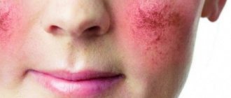
After drinking, alcohol moves through the blood vessels and narrows them. This occurs in the oculomotor muscles and optic nerves. As a result, blood flows worse to the eyes, there is a lack of oxygen, and the person begins to see the image darkened.
Due to the narrowing of blood vessels, blood pressure increases in them. Small capillaries burst. As a result, the eyes turn red, itching and burning appear in them, as well as double images.
Due to the toxic effects of alcohol, the eye muscles work inhibited and uncoordinated. And normalization of the condition often occurs after the alcohol has completely left the body. If the pathology persists for a long period, the help of doctors is required.
Disease prevention
It is impossible to give effective advice on preventive actions for diplopia, since the disease can develop unpredictably .
And even with careful observation by specialists, development forecasts may remain unclear for a long time.
However, you can reduce the likelihood of such a pathology if you follow the following general instructions:
- If possible, it is necessary to lead a healthy lifestyle . In particular, it is necessary to get rid of bad habits such as smoking and drinking alcohol. Since this can lead to a decrease in the tone of the eye muscles, as a result of which the risk of developing diplopia increases;
- in order to strengthen the immune system, it is necessary to take daily walks , during which you need to walk at least one and a half kilometers;
- Any ophthalmological diseases require timely and correct treatment. Since in the case of the organs of vision, any diseases can provoke the development of more serious ailments.
Gymnastics for the eyes
Attention! Special ophthalmological gymnastics according to
Dr. Bates' technique
can help get rid of the disease in the initial stages, as well as reduce the likelihood of its occurrence in a healthy person.
to perform such gymnastics before meals , as well as after heavy eye strain. You can perform these simple exercises up to five times a day.
It is optimal to do gymnastics at the first signs of eye fatigue (irritation of the visual organs, redness, itching, drowsiness).
the exercises while sitting on a chair ; first you should blink well, taking off your glasses or contact lenses before doing this.
Before proceeding directly to the set of exercises, you can do palming (that is, press your palms against your closed eyes for about one minute, but without exerting strong pressure).
The following describes a series of exercises, each of which should be performed, depending on your well-being and degree of fatigue, from five to twenty times:
- The gaze is alternately moved up and down , and in each position the organs of vision must be fixed in a stationary position for one or two seconds. Then the same exercise is repeated in lateral and diagonal directions.
- After resting for a minute, you need to make several circular movements with your eyes (first clockwise and then counterclockwise).
- The same continuous movements must be performed again , but this time describing with the eyes not a circle, but any other geometric figure (square or triangle).
Need to know! A remarkable feature of this set of exercises is that it can be performed with either closed or open eyes.
The effect will be approximately the same, but if you are very tired, moving your eyes open can cause discomfort and even pain.
Traditional methods
Traditional medicine is often an effective aid in the treatment of ophthalmic pathologies , but such methods should be used with caution so as not to worsen the condition of the visual organs.
For diplopia, such methods are far from the most effective compared to surgical intervention, but the development of the disease can be slightly slowed down using the following traditional methods:
- Flower pollen purchased at a pharmacy is taken twice a day, one teaspoon, for a month. Before use, you must read the instructions for possible contraindications.
- crushed valerian root and lavender leaves in equal proportions, then pour 200 milliliters of dry white wine into a tablespoon and leave for three days in a dark place. At the end of each day, the container with the product must be shaken so that sediment does not fall to the bottom. Take one tablespoon one hour before any meal.
- The fruits of viburnum and rose hips are mixed in equal proportions , after which a tablespoon of the mixture is poured into 0.5 liters of boiling water and boiled over the fire for another ten minutes. Then the product is allowed to cool and filtered. You can take it before meals twice a day, half a tea cup.
Conservative therapy
Before choosing treatment methods, the patient is prescribed a comprehensive examination. Depending on the causes and symptoms of the disease, treatment is carried out by a neurologist, ophthalmologist or neurosurgeon. An ophthalmologist provides assistance to injured patients, a neurosurgeon performs surgery, and a neurologist treats neurological diplopia.
Therapy methods:
- prismatic correction;
- functional therapy;
- drug treatment;
- surgical intervention.
Let's consider the methods in detail.
Prismatic correction
The patient wears glasses with a special lens that corrects the focus. The prism has the shape of a straight pyramid with a pointed apex. The refraction of light occurs so that the image of an object hits the retina at the correct angle. As a result, the patient sees a clear, undistorted image of the object.
Depending on the nature of the pathology, the prism is placed horizontally, vertically or diagonally in one/both lenses of medical glasses.
Prismatic correction is indicated for:
- pathologies of the eye muscles;
- concomitant strabismus;
- neurological pathologies (stroke, head injuries);
- diabetes mellitus;
- multiple sclerosis.
Double vision sometimes manifests itself partially: when viewing objects close or far away, when looking at an object from the side or in front of you. In severe forms of the pathology, the patient sees two different objects in front of him.
When corrected with prisms, a temporary deterioration in vision may occur, but the double vision will disappear.
Functional therapy
This treatment method is aimed at strengthening the eye muscles. The method is based on:
- accommodation training;
- orthopedic gymnastics;
- activation of fusion reserves.
For diplopia of a sensory nature, methods of hardware intervention are indicative. In cases of motor bifurcation, the method of developing single visualization fields is used. However, if the result is not achieved after six months of training, surgical correction is prescribed or other methods of influence are chosen.
Drug treatment and occlusion
The method involves injecting a drug (Botox) into the muscle of the diseased eye. Due to the long-term presence of botulinum toxin in tissues, it is possible to achieve lasting results. Drug treatment methods also include the use of eye drops and ointments as prescribed by a doctor.
The occlusion method helps to get rid of unpleasant symptoms, although it is not a therapeutic measure. The eyes are closed one by one, depriving vision of the panoramic image. This improves visual clarity.
Also, to normalize the visual system, the disease that caused diplopia should be cured. If it is high blood pressure, take the appropriate pills. In case of diabetes mellitus, the endocrine system is restored. If cerebral circulation is impaired, nootropics are prescribed.
For double vision, a variety of measures can be prescribed: plasmapheresis, steroids, hormones and antibiotics. The treatment regimen will depend on the concomitant disease, so it may not be the same as the treatment for diplopia in another person.
Surgery
Surgical correction is prescribed when conservative therapy fails, coupled with traditional methods of treatment to correct the situation and eliminate double vision. Indications for surgery are:
- irreversibility;
- critical risk of complications;
- high degree of deviation.
During the operation, the muscles of the visual apparatus are tightened or weakened. Surgical correction eliminates double vision and strabismus. The operation can be prescribed after injury, but not earlier than six months from the moment of deformation of the visual organs. After surgery, functional therapy, prismatic correction and the use of medications are carried out. Eye drops, ointments and eye exercises significantly speed up the healing process.
Possible treatment
Diplopia is curable, but the possibility of complete relief of the pathology depends on how correctly the treatment tactics for the underlying cause are chosen. In most cases, not just correction by an ophthalmologist is necessary, but also intervention by neurosurgeons and neuropathologists. Diplopia is treated by various means. Some causes that provoke the development of pathology require surgical intervention, while others can be treated through exercises and the use of instruments.
Exercises for diplopia
You can get rid of double vision by performing special exercises aimed at strengthening the muscles of the eyeball. Bates exercises are a complex technique that includes elements for the visual apparatus and the cervical spine. The former help strengthen the eye muscles, while the latter relax the nerve endings. The Bates gymnastics complex includes the following elements:
- Turns the head right and left.
- Tilts of the head with laying on the shoulder.
- Bend forward and backward.
- Circular movements clockwise and counterclockwise.
- Move up and down and then side to side.
- Circular movements of the shoulders with arms extended and elbows bent.
- Slow movements of the eyeball with fixation at a point.
- Movements in different directions.
- Moving eyeballs diagonally.
- Rotate your eyes clockwise and counterclockwise.
- Imagining a square before your eyes, move your eyes along its edges.
- Bring your outstretched hand closer and further away with a raised finger and focus your gaze.
- Close your eyes and open your eyes for a few seconds.
Eye exercises according to Kashchenko include the following elements, which are best performed in a medical facility. The execution algorithm is as follows:
- The patient sits in front of a lighted wall at a distance of 1 meter.
- A white sheet with a black stripe measuring 10 cm * 1 cm is placed at eye level.
- The patient positions the head so that the line is continuous.
- The patient performs head movements until bifurcation appears.
- The procedure is repeated three times a day for 5 minutes.
Medications
Medicines for double vision are prescribed to eliminate the root cause of the pathology. If the deviation develops due to damage to the central nervous system, the patient is admitted to a hospital. If there is an ophthalmological factor that provokes the development of diplopia, the decision on the necessary treatment is made by the ophthalmologist.
Operations for diplopia
If the deviation is caused by the presence of neoplasms, surgical intervention is necessary. The pathology will eliminate itself along with the root cause. If the wall of the orbit of the eye is damaged, it is replaced with a silicone implant. After the operation, double vision disappears and vision remains at the same level. However, this technique does not always 100% guarantee elimination of the deviation; sometimes additional surgical intervention is necessary.
Hardware treatment
A course of instrumental therapy is prescribed by an ophthalmologist based on individual indications and the desired result. Typically, laser exposure is used on the eyeball, as well as an effect produced by a photoflash. To strengthen the muscle tissue of the eye, magnetic therapy is used, which also has an anti-inflammatory effect and stimulates blood circulation.
Therapy with folk remedies
Elimination of diplopia using alternative medicine methods is aimed at strengthening the body and improving the effect of basic therapy. The most effective folk remedies are:
- lavender-valerian tincture with white wine;
- brewed flower pollen;
- vitamins from viburnum and rose hips;
- eyebright decoction;
- blueberries in any form;
- decoction of calendula;
- Mint tea.