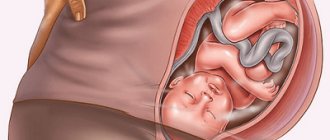
Interpretation of an ultrasound scan during pregnancy provides the doctor with a lot of valuable information that allows the doctor to monitor whether the growth of the embryo corresponds to the norms. An ultrasound done at the appropriate time shows many key moments in the life of the fetus, including the appearance of a heartbeat and the development of internal organs. In addition, using an ultrasound during pregnancy, the doctor can identify genetic abnormalities at an early stage, quite accurately calculate the date of birth, the number of embryos in the uterus, the position of the placenta (this is a very important indicator during childbirth), and the gender of the unborn child. This is why a woman should not ignore this examination method.
Description of the procedure
Ultrasound is absolutely safe for the health of the woman and the fetus. During this examination, you don’t have to tremble at the thought of needles and syringes: this procedure is painless. Ultrasound scanning during pregnancy is interpreted using special computer programs.
During this examination, experienced professionals use plastic probes that contain a transmitter that sends ultrasound waves through the uterus. These signals are reflected from the tissue and go back to a receiver located in the sensor, which transmits them to the machine, after which the signals are translated into an image on the screen.
The examination is usually carried out transabdominally (the probe is passed over the skin of the abdominal area). While the pregnant woman is comfortably positioned on the gynecological table, the doctor applies a special ultrasound gel to the skin of the abdomen. After that, he moves a plastic probe across the stomach from side to side to find the best angle.
The ultrasound test should not be accompanied by pain, although the gel may feel cold and unpleasant. To go for an ultrasound, it is advisable to wear two-piece clothing, trousers (skirt) and a blouse. This will provide the doctor with quick access to the abdominal area.
Interpretation of fetal ultrasound is carried out by selecting the options of the biometric program displayed on the monitor screen. That is why, having modern equipment at his disposal, the doctor can think not about how to decipher an ultrasound during pregnancy, but how to correctly position the sensor relative to the fetus in order to obtain the highest quality image. The results of an ultrasound scan during pregnancy are stored in the computer’s memory, and based on them the doctor makes a conclusion.
In the most advanced ultrasound centers, where 3-D ultrasound is used, the sonologist can take very high-quality photographs of the embryo in the smallest detail. The latest equipment with modern software allows you to better assess the growth and development of the child, determine anomalies in the structure of the face or neural tube, and also more accurately determine the date of birth.
One type of ultrasound is monitoring the baby using Doppler ultrasound, which allows you to determine the state of the embryo's blood supply. This test is usually performed in the last trimester of pregnancy in women with gestational diabetes. There are some differences between Doppler ultrasound and regular ultrasound. In a conventional exam, sound waves bounce off tissue to create images, while Doppler ultrasound receives echoes from red blood cells to measure blood flow speed and blood pressure.
What does ultrasound screening show?
Ultrasound scanning is a highly informative, non-invasive and safe way to obtain data on the health of the expectant mother and fetus. The method is based on the analysis of differences in the reflection of ultrasonic waves from structures of unequal density.
The purpose of an ultrasound examination is to assess the condition of a woman and her unborn child, as well as to identify possible deviations from the norm in order to take the necessary measures (ultrasound for fetal pathology).
Ultrasound diagnostics allows you to confirm the fact of pregnancy, obtain data on the formation of the fetus, the structure of the uterus and appendages, as well as the condition of the placenta, umbilical cord, and amniotic fluid. The results of ultrasound largely determine the tactics of pregnancy management and preparation for childbirth, as well as the choice of method of delivery.
After confirming the fact of pregnancy, all subsequent ultrasounds are performed according to the schedule drawn up by the attending gynecologist.
The following reasons may be the reason for an unscheduled ultrasound in early pregnancy:
- discrepancy between the size of the uterus and the norm for the current stage of pregnancy;
- the woman has bleeding (single or frequent);
- pain and discomfort in the lower abdomen;
- suspicion of a “frozen” pregnancy, ectopic pregnancy and other pathologies of early pregnancy;
- the age of one or both future parents exceeding 35 years.
1 fetal ultrasound
2 fetal ultrasound
3 Fetal ultrasound
First trimester
If a woman has a delay lasting a week, and some time before her expected period she had sexual intercourse, she needs to take a pregnancy test. To do this, you can go to the doctor and donate blood for an hCG test, or purchase a special test at the pharmacy (the first option is more reliable). If the assumption is confirmed, after some time it will be necessary to do an ultrasound.
The first ultrasound, also called a sonogram, must be performed from 6 to 8 weeks of pregnancy. Before the procedure, you should definitely ask whether you should come with a full bladder or an empty one. Sound waves travel much better through fluid, so a fluid-filled bladder can improve imaging. But if examination through the vagina is envisaged, the bladder must be empty. As the uterus and embryo increase in size, the amount of amniotic fluid also increases. Therefore, the fullness of the bladder for a longer period will no longer matter much.
In the first trimester, the embryo is still very small, and the uterus and fallopian tubes are located extremely close to the birth canal. Much closer than to the abdominal wall. Therefore, the gynecologist may prescribe a transvaginal ultrasound in order to get a clearer picture. During this test, the gynecologist places a thin plastic cylinder with a probe that emits high-frequency ultrasound into the vagina. In this case, ultrasonic waves are reflected from the fetal tissues. But at this stage the child is still invisible. A good and clear photograph of the fetus is obtained no earlier than the 13th week.
In the fifth week, the fetal sac (the so-called structure located in the uterus surrounding the embryo) begins to be visible, which by this time reaches 8-12 mm. This is the very first indicator that ultrasound can determine during pregnancy. The sac not only surrounds the embryo, but also contains amniotic fluid (amniotic fluid), which nourishes and protects the embryo. The size of the fetal sac is examined during an ultrasound to confirm the presence of pregnancy and the viability of the fetus. Based on the size of the fetal sac, the time of conception can be determined with an accuracy of up to 5 days.
When the gestational sac reaches 8 mm, there is a noticeable yolk sac (resembling the yolk in an egg) that surrounds the embryo. Its main purpose is to provide the embryo with nutrition before the circulatory system and placenta develop.
How to decipher ultrasound results in pregnant women
When interpreting the results obtained, the doctor compares the obtained ASI values with tabulated norms according to the gestational period. During pregnancy, the uterus and placenta communicate through terminal villi, which contain a large number of vessels. These formations deliver oxygen, nutrients and remove waste products.
With an insufficient number of villi, fetoplacental insufficiency develops. As a result, the value of IR and SDO will be high. Due to disruption of the uteroplacental blood flow, an increase in the vascular resistance of the uterine arteries is also noted. The IR norm is 0.62-0.82 depending on the period, and the SDO should vary between 2.19-4.67.
The nature of blood flow in the uterine arteries is of great importance during intrauterine development. A normal value is considered to be SDO in the range of 1.66-2.10, PI - 0.40-0.65, IR - 0.3-0.9. A decrease in blood flow velocity leads to exceeding the standard values of SDO and IR. If there is a significant disturbance in blood flow, the development of the fetus stops. As a result, the value of SDO and IR of the carotid and cerebral arteries of the unborn child increases.
Investigation for anomalies
When the ultrasound can clearly see the embryo (week 8), the doctor can measure it from the crown to the tailbone to determine the gestational age. The embryo at this time is still very small and grows only one millimeter per day. The doctor must decipher the information received in order to more accurately determine the date of birth, as well as key dates that are important for each pregnant woman.
In addition, in the first trimester, ultrasound detects multiple pregnancies, and also makes it possible to detect such markers of chromosomal pathology of the fetus as an increase in the nuchal region and a decrease in the nasal bones in Down syndrome. Ultrasound markers also detect other chromosomal abnormalities, including trisomy 18.
Pathological markers include TPT, prenasal tissue thickness. TPT is measured at the bridge of the nose. In the third trimester, TPT levels may be increased in cases of Down syndrome and other genetic abnormalities. Based on the results of an ultrasound scan of the first trimester, a gynecologist is able to exclude an ectopic pregnancy, which develops if the embryo develops not in the uterus, but in the fallopian tubes. But you should not worry about this pathology: it occurs in 1% of cases.
Each pregnant woman is asked to undergo an ultrasound to examine the nuchal region of the embryo. This should be done between 11 and 13 weeks (on the verge of the first and second trimester). A sonologist must determine the risk of having a child with Down syndrome, trisomy 18 (another chromosomal abnormality), or heart disease.
This examination consists of two parts: this includes a blood test for hormones and proteins. In addition, ultrasound determines the thickness of the newborn’s neck (exceeding the norm may indicate genetic abnormalities, Down syndrome and trisomy 18).
Why is a routine ultrasound performed from 32 to 34 weeks of pregnancy necessary?
32-34 weeks
- Determination of the position and presentation of the fetus. This is necessary for labor management tactics.
- The presence of the umbilical cord in the fetal neck and its entanglement.
- Localization of the placenta and its quality, in case of its dysfunction. If the placenta is located centrally or low, perform a planned cesarean section.
- Detection of late fetal malformations, such as heart defects and skeletal abnormalities, that appear late in pregnancy.
- Elimination of complications that prevent normal childbirth.
- Condition of the cervix.
Second and third trimester
In the second trimester (weeks 13-27), the embryo is already mature enough for ultrasound photos to be truly impressive. An ultrasound scan at this stage is usually performed between 18 and 20 weeks and is called an anatomical scan.
The gynecologist uses ultrasound to evaluate the child’s growth and make sure that their values can be considered normal. For assessment, you can use computer programs, or you can use fetal tables by week. At this time, all organs of the embryo are viewed in detailed detail, but for an untrained person it is quite difficult to distinguish the kidneys from the stomach. Therefore, you can ask the doctor to tell him what is shown on the screen and name each embryonic organ visible in the image.
In the third trimester, an ultrasound is usually done at 20 weeks, when an anatomical examination is performed. If the estimated date of birth has not arrived, an ultrasound is prescribed to monitor the fetal heartbeat and the level of amniotic fluid. Other reasons to perform an ultrasound in the third trimester are to check the healthy position of the placenta and to question the growth of the fetus.
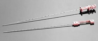
Between 14 and 20 weeks, amniocentesis may be done, where a sample of amniotic fluid is taken using a needle (puncture). This procedure is performed under local anesthesia and determines genetic abnormalities in the fetus, such as Down syndrome. This procedure is not prescribed to everyone, but to women who are at risk (women over 35 years of age, as well as those with a family history of genetic diseases), or pathological problems are observed in the fetus.
During amniocentesis, a needle is used to pierce the skin of the abdomen and tissue of the uterus, guiding the needle using ultrasound. After entering the uterus, a sample of amniotic fluid is taken. There is a small (0.5%) risk of miscarriage with amniocentesis.
Changes in the child's body
Already from the 29th week of pregnancy, the child begins active preparation for the upcoming birth. The baby’s immunity is fully formed, and thermoregulation is improving. The blood composition is stabilized. Almost all of the baby’s systems are actively functioning and he is almost ready for a future independent life.
Intensive growth of the baby is observed at the 29th week of its development. At this time, the growth of the uterus itself slows down slightly. At the 29th week of pregnancy, during an ultrasound, the type of presentation is established, which usually persists until the time of birth. Indeed, in such a cramped space with very large parameters it is very difficult to turn over. The norm is cephalic presentation.
In the case of a transverse, pelvic presentation, a cesarean section is recommended. This will minimize the risk of injury to the fetus during childbirth. To change malpresentation, an obstetrician-gynecologist may suggest performing special exercises that can increase the likelihood that the baby will independently change his position before labor begins. Turning the fetus in the womb with force is not performed. The effectiveness of the procedure is insignificant, and the risk of complications is high.
At the 29th week of development, the baby observes the production of red blood cells by the already developed bone marrow.
At this stage of pregnancy, the child experiences full development and functioning of most of the internal organs. The child rapidly increases his muscle mass, and fat storage is slightly inhibited. By the time of birth, the fetus will double its weight.
It is already difficult for the baby to perform large movements. He trains his muscles by pushing with his arms and legs, but somersaults and flips are no longer observed. These processes are clearly visible on ultrasound diagnostics, which are carried out unscheduled at the 29th week of pregnancy.
By the 29th week of development, the child already has 300 bones, the development of which has already been completed. The fetus is ready for birth. During the third trimester, some of the baby's bones will fuse, so at the time of birth he will have 206 bones.
During this period of development, the baby's accumulation of white fat increases. Until this time, the children's body was actively accumulating brown adipose tissue. Such adipose tissue is necessary for the process of thermoregulation (its action will become necessary after the birth of the baby). White fat deposits are necessary for the fetus to perform another function - energy reserve. After childbirth, it will be useful for the intensive growth of the child. This type of fat is an ideal source of energy for your baby.
Biometric parameters of the embryo
Ultrasound measurements of fetal and obstetric parameters reflect how developed the embryo is for its age and check whether these parameters are within acceptable limits. This allows timely detection of child abnormalities.
Ultrasound involves the study of the following indicators:
- the length of the fetus from the crown to the coccyx, in other words, the coccygeal-parietal size (CTR);
- the distance between the parietal tubercles of the embryo’s head, otherwise – the biparietal diameter of the head (BPR, BPD);
- length of the humerus or arm length (LA);
- head circumference (HC) is usually measured after 13 weeks;
- the distance between the back of the head and the forehead of the embryo or the fronto-occipital size (FOR);
- abdominal circumference (AC);
- length of the femur or femur (DB);
- estimated embryo weight (EFW).
The coccygeal-parietal size is the distance between the crown of the embryo and the end of its back, which is used to measure fetal growth. CTE is measured between 7 and 13 weeks. This indicator allows you to determine the exact time of conception. At this time, it is the most reliable and only it is taken into account, even if subsequent ultrasounds give reason to assume a different date (later indicators simply say that the embryo stops growing).
Biparietal head diameter (BCD) is the diameter between the two sides of the embryo's head. This indicator is measured by ultrasound after the 13th week of pregnancy. It increases from 2.4 cm at 13 weeks to 9.5 cm before birth. Different embryos of the same weight may have different head sizes. Therefore, it is not customary to set the date of conception using this parameter.
The BPR is located perpendicular to the fronto-occipital dimension (FOR). BPR, LZR and OG (head circumference) are placed in the same plane. Therefore, when deciphering, all three values, BPR, LZR and OG, are connected by one mathematical formula.
The femur is the longest bone in the human body. Its size reflects the growth of the fetus in length. The value of DB is the same as that of BPR. The length of the thigh at 14 weeks is 1.5 cm and increases to 7.8 cm before birth.
Abdominal circumference (AC) is the only indicator whose measurement is important only in late pregnancy. The coolant reflects more the size and weight of the child than his age.
The weight of the embryo at any time during gestation can be accurately determined using polynomial equations containing BPD (biparietal diameter), DB (femur length) and OB (abdominal circumference). To determine the weight of the fetus, many computer programs, online calculators and simple tables have been developed. For example, with a BPR of 9.0 cm and a coolant of 30.0 cm, the calculator will give a BWP value of 2.85 kg.
But we must keep in mind that even the best formulas calculate PVP with 15% accuracy in 85% of cases. In other words, approximately every seventh fruit has a non-standard weight, which may differ from the calculated one by more than 15%.
The following table shows the following ultrasound parameters and norms by week of average fetal size (data is given in millimeters by week of pregnancy). Using them, you can determine how the ultrasound result is deciphered. But we must remember that due to the fact that laboratories use different equipment, these data may differ. Therefore, it is better to entrust the conclusion to specialists.
Consultation
Hello, according to an ultrasound at 20 weeks, the shortening of the nasal bones was 5.0 mm and 3.8 mm. The first screening at 12 weeks is normal, the second was not prescribed. What does it mean? What kind of examination should I take? The placenta is also very low. Is this normal?
Guaranteed response within an hour
Doctors' answers
The length of the nasal bone at 20 weeks of pregnancy is normally 5.2 mm, but at the moment, according to many sources, a length of 5.0 mm is considered normal. In your case, this may be an individual characteristic, measurement error, etc., so TPT is measured to assess the risk of chromosomal abnormalities. TNT is the thickness of prenasal tissues and to exclude the risk of chromosomal abnormalities, the ratio of TPT to the length of the nasal bone is assessed: TPT/DNA. Normally, up to 26 weeks of pregnancy, this ratio should be less than 0.8; if this figure is higher, then there is a risk of chromosomal abnormalities. In your case, this figure is 0.69. Therefore, taking into account all the above factors, as well as the normal ultrasound results at 12 weeks, I see no reason for concern. The doctor will prescribe the necessary examination, as scheduled. Low placentation at 20 weeks of pregnancy is a very common occurrence, the placenta will most likely rise, follow all the doctor’s recommendations, this will help avoid possible complications.
Stomach, intestines, kidneys
The study of these abdominal structures is to determine the presence or absence of a lesion. If deviated, fluid may accumulate in the intestinal lumen.
Ultrasound of the stomach and intestines is carried out according to the doctor’s indications - it is not included in the standard examination of the obstructive system.
If necessary, an ultrasound examination of the kidneys is additionally included in the conclusion. Kidney diagnostics are normal:
- width: 5-6 cm;
- length: 11 cm;
- thickness: 4-5 cm;
- parenchyma: no more than 23 mm;
- pelvis: no changes;
- lumens of the pelvis and ureters without unnecessary inclusions.