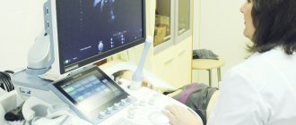
Ultrasound, or echography, is the study of internal organs using sound waves. The waves, reflected from the internal organs, are recorded using special instruments and create images of anatomical details. In this case, ionizing radiation (X-rays) is not used. The normal size of the uterus according to ultrasound in adults serves as an indicator of the health of the genitourinary system in women.
For women, this test is most often used to examine the uterus and ovaries before, after and during pregnancy to monitor the health of the organs and the development of the embryo or fetus. Ultrasound images are captured in real time so they can show the movement of internal tissues in organs, such as the flow of blood in arteries and veins. Standards for the size of the uterus according to ultrasound have been developed and calculated for any condition of a woman.
Types of uterine contractions
In different situations, contractions are different, they can range from weak, which the woman does not actually feel, to strong (during childbirth), they cause pain.
During menstruation, the uterus contracts intensively to remove the endometrium from the woman’s body. Since the uterus is surrounded by many nerve endings, when it contracts, pain appears in the lower abdomen. And if a woman produces a lot of oxytocin (a hormone that causes uterine contractions), then the pain does not go away immediately after the end of her period.
Also, the formation of pain during menstruation may be associated with a high level of prostaglandins in the blood, which are produced by the uterine tissue and cause its contractile effects. The higher the level of prostaglandins in a woman’s body, the more intense the uterine contraction occurs and, accordingly, the pain increases.
Before menstruation, the uterus opens slightly (closes when it ends), rises up, and is slightly enlarged. During constant contractions that cause mild pain, it reaches its normal size.
The cause of postmenstrual pain can be improper location of the organ, stress, or overexertion.
To reduce the pain threshold during uterine contractions, it is recommended to use No-shpa, Ibuprofen, Naproxen, and Diclofenac suppositories.
During orgasm, contractile activity of the uterus is also observed, as endorphin hormones are produced, which are often called the hormones of happiness. It is because of the production of endorphins that a woman’s mood significantly improves.
During orgasm, under the influence of the hormone oxytocin, the body of the uterus slightly enlarges and expands, and the length of the cervix decreases; in addition, the walls of the entire organ begin to shrink evenly over the course of several minutes.
Doctors say that uterine contractions during orgasm are not at all the same as during childbirth and they do not pose a threat to the further development of pregnancy. It can be considered an undesirable sign only in the very last stages of pregnancy. Having sex during early pregnancy does not have a bad effect on a woman’s well-being and allows her to carry and give birth to a healthy baby without complications.
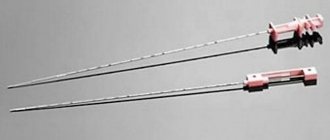
Curettage (cleaning) is a surgical intervention in gynecology, during which the top layer of the lining inside the uterus is removed. Very often, bleeding begins after curettage. The need for this procedure often causes cessation of fetal development and signs of its death.
If curettage is performed by an experienced doctor, then, as a rule, it will not cause complications. Typically, during the rehabilitation period, a woman feels slight pain in the lower abdomen, as during menstruation. This indicates that the uterus is restored to its previous size. This happens quite quickly, in about a month.
There are cases of severe bleeding after curettage (you need to change the pad several times an hour), in such a situation you should immediately call a doctor. Often the cause of curettage is polyps and prolonged periods.
Abnormalities of contractile activity
Anomalies of contractile activity of the uterus are divided into two types:
- Primary weak labor is poor dilatation of the uterine cervix before childbirth.
- Secondary weak labor - characterized by interruption of contractions during childbirth after their regular sequence.
Other types of anomalies are also identified. Some women have no uterine contractions at all, some have irregular contractions, and sometimes rapid labor. Let's take a closer look:
If the placenta and lochia are not completely released, curettage is performed, before which the woman is given general anesthesia.
The reasons for all this can be various diseases, insufficient production of contractile proteins and hormones by the body, and the anatomical structure of the birth canal.
The hormone progesterone plays a very important role, which prevents contractions of the uterus; it also affects the emotional state of a woman.
Normal uterus in women according to ultrasound
If a woman exhibits alarming symptoms, she is sent for examination. With normal functioning of the pelvic organs, all indicators should be normal. During an ultrasound, the ophthalmologist must examine:
- How is the uterus located in the pelvic area? Normally, it should be slightly tilted forward.
- What is the outline of the uterus? Normally, they should be smooth and clearly visible.
- What are the sizes of the uterus?
Parameters of the uterus during reproductive age
| Group | Neck length(cm) | Neck thickness (cm) | Neck width (cm) | Uterine body length (cm) | Thickness of the uterine body (cm) | Width of the uterine body (cm) |
| There were no pregnancies | 2,9+-0,5 | 2,6+-0,4 | 2,9+-0,5 | 4,4+-0,6 | 3,2+-0,5 | 4,3+-0,6 |
| Only abortions | 3,1+-0,5 | 2,7+-0,4 | 3,1+-0,5 | 4,9+-0,6 | 3,7+-0,5 | 4,6+-0,5 |
| Childbirth 1 | 3,4+-0,6 | 2,8+-0,4 | 3,3+-0,5 | 5,1+-0,6 | 3,9+-0,5 | 5,0+-0,5 |
| Childbirth >1 | 3,7+-0,6 | 3,0+-0,5 | 3,4+-0,5 | 5,6+-0,9 | 4,3+-0,6 | 5,5+-0,5 |
Medications to normalize contractile activity
For treatment at home, you can use both medications and herbs.
Medicines that can act on uterine contractions are divided into four groups:
All of these medications are very common in gynecology and many contain hormones:
After curettage and to stop heavy and protracted periods (with such a pathology, you should definitely consult a doctor without delay), hemostatic agents are used, which are prescribed by a specialist based on the specific situation.
Traditional medicine recipes and exercises for uterine contractions
It is better to use traditional medicine recipes at home before going to see a doctor, and not to replace it.
Although uterine contraction is a natural phenomenon, sometimes it needs help, especially during the period after childbirth and during prolonged periods. Herbal infusions can help here, for example:
After childbirth, you need to breastfeed the baby, as during this process the hormone prolactin is produced, which promotes contractile movements of the uterus.
In addition to medications and herbs, you can do special exercises to contract the uterus, and a huge advantage is that you can do them at home.
Exercises are good after childbirth, curettage and cesarean:
Exercises to contract the uterus need to be done for quite a long time (about three months). But after this you can maintain a good figure. The reasons for uterine contractions can be different, so to normalize this process you need to consult a specialist.
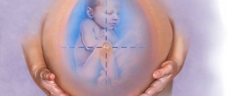
During pregnancy, metamorphoses occur throughout the body. The uterus, as one of the most important organs, is no exception. As the fetus grows, so does it.
This organ is distinguished by its unique properties, because during pregnancy it increases significantly, and after the baby leaves it, it gradually returns to its standard size.
It is difficult to say how long it takes to return to its previous size, because every woman’s body is unique. There are several factors that can speed up or, conversely, slow down this process.
How does the uterus contract after childbirth?
After the fetus leaves the womb, the uterus looks like a large wound. Particularly severe damage is observed in the area of the placenta, because there are many clogged vessels there. At this time, there are blood clots and parts of the fetal membrane in the cavity.
Cleansing occurs within 3 days after delivery. In this process, an important role is played by phagocytosis - the dissolution of bacteria by leukocytes, and extracellular proteolysis - the dissolution of bacteria by proteolytic enzymes.
These processes contribute to the release of wound secretions - those same lochia. On the first day they look more like blood, and on the 3rd-4th they become serous-hysterical with leukocytes. At the end of the third week, they are normally light and liquid, and by the sixth week they are completely gone.
The restoration of the epithelial layer occurs over about 20 days, and the placenta attachment site heals by the end of the postpartum period.
How long does it take for the uterus to contract after childbirth?
The average period is from 1.5 to 2.5 months. It is worth noting that the highest activity is observed in the first few days after delivery.
Immediately after the baby leaves the mother's womb, the cervix measures 12 cm in diameter. If necessary, the doctor can insert his hand there to clear the cavity of any remaining placenta.
But at the end of the first day, the cervix narrows so that only a couple of fingers can be inserted, on the third day - 1. The external pharynx closes completely in the third week.
As for the weight of the organ, immediately after the birth of the baby it averages 1 kg, after a week - 500 g, after two - 350 g, and at the end of the postpartum period, after 2-3 months - 50 g, that is, it reaches its prenatal weight .
The contraction process is always accompanied by minor cramping pain in the lower abdomen. It is worth noting that they are most pronounced after repeated births. For some women, this phenomenon causes severe discomfort, so the doctor may prescribe certain painkillers or antispasmodics, but it is recommended to refrain from them, especially if you are breastfeeding a newborn baby.
However, sometimes it happens that after childbirth atony is observed - the uterus does not contract, or the process progresses very slowly. Both phenomena pose a great danger because they can provoke postpartum hemorrhage and a number of other complications.
Why are there no cuts?
A slowdown in the process can cause:
- Multiple births;
- Low attachment of the placenta;
- Large fruit;
- Complications during childbirth (eg weak labor);
- The state of health of the woman herself, for example, the body can be severely depleted.
Their complete absence occurs in the case of uterine bending, trauma to the birth canal, organ underdevelopment, inflammatory processes in the uterine cavity itself or its appendages, including those in the anamnesis, fibroids (benign tumor), polyhydramnios or bleeding disorders.
Slow recovery of the uterus after childbirth
Even in the delivery room, a heating pad filled with cold water is placed on the mother's belly. This event helps stop bleeding and promote the contraction process. While the mother and child are in the maternity hospital, doctors regularly check the condition of the uterus and monitor the process of its recovery.
The gynecologist will be able to establish a slow recovery during a routine examination. In this case, the bottom of the organ will be soft. The woman is left within the walls of the maternity hospital until the doctor makes sure that the contractions are progressing at a normal pace.
If independent contractions are not observed, special drugs are prescribed that provoke the onset of this process - prostaglandins or oxytocin. The complex of therapy may include external massage of the fundus, which is carried out through the abdominal wall.
In addition, breastfeeding can serve as an impetus, so it is recommended to feed the baby as often as possible. A woman should move more, and it is recommended to rest and sleep on her stomach.
The contraction is affected by emptying the bladder, which should occur regularly. Very often this fact is missed, especially if stitches were placed that cause pain when urinating. But you should still visit the toilet more often.
If the methods described above do not produce results and the uterus does not contract, cleaning is performed. The need for such an event arises due to the fact that lochia or parts of the placenta may remain in the organ cavity. In addition, the cervix may be blocked by blood clots.
In the case when postpartum discharge or pathological clots remain, inflammation necessarily occurs, which affects not only the organ itself, but also adjacent tissues. Sometimes even cleaning does not bring the expected result, then surgical intervention is necessary, which may involve removing the uterus.
Delayed contraction is medically called subinvolution. Typically, this organ returns to its previous size approximately 5-7 weeks after delivery, and after 8 for those who do not breastfeed.
Normally, a rapid contraction is observed immediately after the birth of the baby. Only by the size of this organ can one judge the condition of the mother. When contractions occur normally, the process goes smoothly. Otherwise, the postpartum period is fraught with immune disorders and hormonal imbalance.
It is worth noting that in women who gave birth to a child by cesarean section, this process occurs much more slowly than in those who gave birth to a child naturally, but in such a situation this is considered the norm. But such mothers, despite the inconvenience, should get out of bed earlier and move more immediately after the side effects of anesthesia pass. Movement will promote contraction, and passivity will cause muscle flaccidity.
Norm of muscle contraction dynamics
After the placenta comes out, the fundus of the uterus should be located at the level of the navel. Prolapse of the uterus after childbirth without complications is approximately 2 cm per day. On the sixth day, when most women are discharged from the maternity hospital, it is normally located about 5 cm above the womb. A delay of at least a day is considered a pathology.
Causes of subinvolution:
- Lack of prolactin. The production of this hormone, which is responsible for the arrival of milk, provokes the release of oxytocin, which contracts muscles. Prolactin is produced when the nipples are irritated, that is, reflexively, so in women who are breastfeeding, recovery is faster. Accordingly, a lack of this hormone leads to disorders;
- The cervix, clogged after childbirth, and the remains of the placenta in the cavity prevent the body from recovering. If the placenta is not completely separated, the organ cannot function fully;
- Postpartum infection. Basically, this pathology is a continuation of a process that began during pregnancy. Postpartum endometritis is a complication after chorionitis - inflammation of the membranes. The inner surface of the organ is damaged and cannot respond to the production of oxytocin. The uterine tissue becomes flabby and activity decreases.
The causes of subinvolution are determined in the maternity ward. Treatment in all cases involves hospitalization.
After a cesarean section, the mother's uterus recovers somewhat more slowly than after a normal birth. This is due, first of all, to the fact that cesarean is still an abdominal operation, which is not natural for the body, but rather, on the contrary, traumatic. Thus, during surgery, blood vessels, muscle fibers, and nerve endings are damaged. After the procedure, the uterus has a scar, the healing of which requires some time and special care.
How long the uterus can contract after cesarean surgery depends on many factors. With a planned operation and the absence of any complications, in order for the woman’s body in general and the uterus, in particular, to reach a satisfactory state, it takes no less than 2 months, that is, almost the entire postpartum period. If the outcome is good, the reduction process occurs by itself. However, in some cases it is necessary to prescribe drugs that would stimulate this process to proceed somewhat more quickly. These drugs have a stimulating effect on contractile activity and also have a hemostatic effect on damaged vessels.
In general, the process of complete recovery of a woman’s body after a caesarean section takes about two years. Why so long, you ask? Did you know that during pregnancy, a woman’s uterus enlarges almost 500 times? So your body will have something to work on.
In the meantime, when the uterus after cesarean is still quite stretched and measures from 10 to 12 cm in diameter, when the scar is still very fresh, the woman feels a certain discomfort. It may be associated with pain in the incision area, fear and difficulty going to the toilet, coughing, turning over from side to side, etc.
All these nuances are discussed and decided with the doctor who sees the mother during the postpartum period.
As measures to help more comfortably survive the postpartum period, in addition to medications, the woman is prescribed a diet that is acceptable for both her and the baby, as well as wearing a special bandage that protects the tissue from further stretching at a time when effective physical exercise is not yet possible for the woman. not possible. At first, while the uterus has not yet contracted after the cesarean section procedure, and the suture has not healed properly, the woman should get an assistant who will relieve her of housework and heavy lifting, which can very significantly affect the quality and speed of the recovery process.
What can you eat after a caesarean section?
What pathologies are there?
Over the course of nine months, the uterus has stretched and increased in size, so it takes some time for it to return to normal. Immediately after the birth of the child, its weight is about one kilogram, and the upper limit is in the navel area. Under the influence of natural hormones secreted by the mother's body, its contraction occurs, and after 1-1.5 months the uterus becomes the size of a fist, and its weight is no more than 40-50 grams.
After childbirth, the cervix also undergoes changes. In the first hours after the birth process, the diameter of its lumen reaches 10-12 cm, which corresponds to full dilatation. On the second day after birth, the size of the cervix does not exceed 4 cm, but by the tenth day of the postpartum period the muscle ring should close, allowing only the tip of the finger to pass through. After three weeks, the cervix takes on a slit-like shape, which is a diagnostic criterion by which a gynecologist can identify a woman who has given birth.
Due to the fact that in the early recovery period the birth canal is open, there is a risk of penetration of pathogenic microflora, which can cause inflammation not only of the cervix after childbirth, but also of deeper organs.
For several weeks after childbirth, the inner surface of the uterus is a wound bleeding surface that has no protective barriers. The discharge is called lochia and is characterized by an alkaline environment.
It is important to note that the normal pH of the female reproductive system is acidic, which is detrimental to pathogens. An alkaline reaction, as well as the absence of a mucus plug in the cervical canal, increases the likelihood of inflammation of the endometrium of the uterus in the absence of proper hygienic care.
Therefore, young mothers are advised to carefully clean the external genitalia:
- use a low-concentrated solution of potassium permanganate for washing;
- use sterile pads and promptly replace them with clean ones (at least once every four hours);
- after defecation, wash the perineal area under running water in the direction from the vagina to the anus.
Involution of the uterus occurs slowly: it returns to its natural size no earlier than 6-8 weeks after birth. In some cases, the recovery period may extend over a longer period of time.
The color of postpartum discharge from the uterus gradually changes:
- Red lochia is the primary discharge, mainly consisting of blood. They are especially abundant in the first 3-4 days after birth.
- Gray lochia - appears to replace red discharge and has a brown color. In addition to blood, they contain a mucous secretion.
- White lochia - represent the final stage of the lochial period. The discharge is characterized by a yellowish or white color.
With slow contraction of the uterus, as well as in the presence of anatomical features such as bends, lochia can linger in the uterine cavity, which disrupts its involution.
Usually the recovery period ends well, however, there are situations when, after childbirth, prolapse of the internal genital organs occurs, and the cervix is visible in the vagina. Other pathologies of the female reproductive system may also develop. Therefore, if you have any suspicions that something is going wrong, it is better to consult a gynecologist for advice.
Sometimes a woman’s postpartum period is difficult, which is associated with the development of complications.
For these reasons, both external and internal ruptures often occur, or doctors prevent them and perform an episiotomy, which requires suturing the cervix after childbirth.
The consequences of injuries associated with damage to the entrance to the cervical canal are of several types:
- Cervical ruptures require careful examination and careful suturing to prevent subsequent pathological changes.
- Ectropion of the cervix - eversion of the soft tissue structure occurs due to inaccurate sutures or their absence after ruptures.
- Cicatricial deformities - occur in cases where birth injuries to the cervix were multiple and of great depth.
- Erosion is a frequent companion of women who have given birth. Their formation occurs due to the fact that injured tissues have a lower level of trophism. This leads to the fact that various mechanical or microbial damage has excessive force on the weakened mucous membrane of the cervix and cervical canal, which leads to erosion.
We suggest you read: When to go to the gynecologist for the first examination after childbirth
Separately, you should pay attention to such a postpartum complication as cervical prolapse. Cases where the cervix descends after childbirth are rare in gynecological practice. However, this pathology has serious consequences for a woman’s health. Therefore, it requires high-quality diagnostics and treatment methods.
Mostly women who decide to have a baby at a fairly mature age suffer from this pathology.
In addition to age-related predisposition, there are other reasons for uterine prolapse:
- severe natural childbirth - when a mother gives birth multiple times or her children are born large;
- excessive physical activity - athletes involved in weightlifting, as well as women forced to do hard male work, are most susceptible;
- congenital pathology - a violation of the anatomical structure of the vagina or uterus can lead to the cervix descending after childbirth;
- chronic pathology of the respiratory system - a strong regular cough leads to the fact that the pressure in the abdominal cavity often increases, and this, in turn, creates additional stress on the reproductive organs.
There are four degrees of cervical prolapse after childbirth:
- I degree - upon examination, the gynecologist determines a decrease in the tone of the perineal muscles. The prolapse of the vaginal walls and its incomplete closure are also determined.
- II degree - a woman experiences a sensation of a foreign body inside the vagina, and the discomfort intensifies with movement. Upon examination, the doctor notes prolapse of the uterus and weakened tone of the pelvic muscles. In this case, prolapse of the bladder and intestines often occurs.
- III degree - the cervix descends into the lower third of the vagina. Due to low prolapse, compression of the blood vessels occurs, as a result of which tissue nutrition is disrupted, and sometimes their ischemia develops.
- IV degree – incomplete prolapse, when the cervix is visible after childbirth.
- V degree – complete prolapse, in which all uterine structures are outside the woman’s body.
Treatment methods are prescribed depending on the degree of prolapse. At the initial stages, anti-inflammatory and hormonal therapy is recommended; if the cervix prolapses, doctors install a pessary. In severe cases, surgery is required.
The birth of a child, although a natural process, often brings a lot of trauma to a woman. For this reason, even during pregnancy, you should engage in strengthening the pelvic floor muscles and general strengthening exercises, and do not lift weights.
During the birth process, you should carefully listen to the advice of the midwife and follow her instructions when pushing. This will reduce the likelihood of cervical injury and avoid the development of further complications.
The uterus is a muscular organ, so stretching is a normal process for it. Its usual size does not exceed a woman’s fist, and during pregnancy it can increase 500 times. After the baby is born, this organ should return to its normal size within just a few weeks.
Contractions place a very large load on the uterus, and after the placenta is rejected, a damaged area and thrombosed vessels remain. Nature has designed it so that the process of cleansing and contraction of the uterus occurs quite quickly - after just 6 weeks it returns to its original size.
After childbirth, a special secretion is formed in the uterus - lochia, which looks like bloody discharge and contributes to the rapid healing of the wound. Within a couple of weeks they become thin and light, and by week 6 the discharge should completely disappear.
The uterus is a sterile organ, and after the birth of a child, the body does everything to ensure this indicator. Immediately after birth, the processes of phagocytosis and proteolysis begin, which kill all pathogenic microflora and promote the restoration of endometrial tissue.
The cervix immediately after the birth of the baby is quite dilated - its lumen reaches approximately 12 cm. However, after just a day, the cervix contracts and becomes dilated by only 1-2 fingers. Complete closure of the pharynx to its original state occurs 3 weeks after birth. At the same time, visually the cervix of a woman who has given birth differs from the cervix of a nulliparous woman, which is clearly visible to the gynecologist.
Before pregnancy, the cervix has a round shape and a round opening of the throat; after childbirth, it becomes cylindrical, and the shape of the throat resembles a slit.
Contractions of the uterus to normal sizes are also accompanied by cramping pains, but they are quite tolerable, unlike labor pains. In multiparous women, the uterus returns to its original state faster, but the pain is felt more strongly.
As soon as the placenta is released, the fundus of the uterus is located approximately at the level of the navel. Every day it drops 1-2 cm lower. The doctor observes the dynamics of the process every day, probing the localization of the organ in the woman in labor.
After childbirth the following changes occur:
- On the third day - a spherical shape of the uterus and bright red discharge from the genital tract. The length of the uterus is about 136 mm.
- Fifth day - the uterus takes on an oval shape, and the discharge becomes yellowish. The length of the uterus is about 110 mm.
- After the seventh day, the uterus becomes pear-shaped, and the discharge takes on a familiar character, the same as before pregnancy. The length of the uterus is about 90 mm.
Below are the dimensions of the uterine cavity, in accordance with the period that has passed since childbirth (weeks):
- 1 (week) – 11 cm;
- 2 – 10 cm;
- 3 – 8 cm;
- 5 – 7.5 cm;
- 6 – 7 cm;
- 7 – 6.9 cm;
- 9 – 6.5 cm.
Slow uterine contraction may occur for the following reasons:
- multiple pregnancy;
- birth by caesarean section;
- large fruit;
- complications or injuries during childbirth;
- individual characteristics of the physical development of a woman in labor;
- incorrect location of the placenta;
- infection;
- hormonal disbalance;
- lack of physical activity after childbirth;
- pathologies of the uterus (kink, underdevelopment, inflammation);
- bleeding disorders and others.
We invite you to read: Restoring bladder control after childbirth
While the uterus contracts...
After a cesarean section, as well as after a normal birth, the uterus is one continuous bleeding wound. The greatest damage to the organ is at the placenta attachment site and the incision area. There are still remnants of blood clots and membranes. On days 3-4 the bulk of the bloody discharge (lochia) comes out. Afterwards, the color of the discharge gradually becomes lighter, it takes on the character of ichor (about the third week after the operation) and after 6-7 weeks it should stop altogether. By this time, the process of epithelial regeneration ends.
How is everything going?
How long does it take for the uterus to contract after a cesarean section? We have already said that in 9 months it increases almost 500 times. Immediately after childbirth or cesarean section, the weight of the organ is 1 kg. A week later - already half as much, that is, half a kilogram. In another 7 days - about 350 grams, and by the end of the third month she should have returned to her prenatal size and weight.
Thus, we see that the uterus contracts most actively in the first days after childbirth. Then this process gradually slows down.
Sometimes the period of uterine contraction is accompanied by cramping, nagging pain in the lower abdomen. As a rule, they are not a cause for concern and are not permanent. However, sometimes, especially after repeated childbirth, these sensations can cause some inconvenience, and some women cannot lead a normal life due to the pain syndrome. In this case, you should consult a doctor. Most often, such situations are resolved by prescribing antispasmodic drugs.
Sometimes there are cases when the uterus does not contract or this process occurs too slowly. Such situations can become harbingers of bleeding or other complications, which means that you simply need to seek help from a specialist.
What if the contractile activity is too long?
What can affect the timing of uterine contractions after a caesarean section? First of all, it is worth mentioning about multiple pregnancies or large fetal weight. At the same time, the uterus stretches more and, accordingly, the mother’s body requires more strength and time for it to contract.
The contraction process can also be slowed down if the placenta was low, if the caesarean section was not planned, but was due to weak labor, and also if the woman after childbirth leads a very passive lifestyle and moves too little. Among other things, one cannot discount the woman’s general health, her readiness for cesarean section, concomitant diseases (hyper- or hypotension, nephropathy, etc.).
When does milk come in after a caesarean section?
Speaking about how long the uterus can contract after a cesarean section, one cannot help but draw your attention to inflammatory processes, physiological developmental features (such as bending or underdevelopment of the uterus), which, by the way, may cause the uterus not to contract at all. This is also possible in the case of an injured birth canal, the presence of fibrous formations in the walls of the uterus, inflammation of the appendages present or even in the past, with a blood clotting disorder or polyhydramnios that accompanied pregnancy. Then the time required for uterine contraction can only be affected by timely and correctly prescribed treatment.
The doctor must determine how normally the process of uterine contraction is proceeding before discharge.
If he deems it necessary, the mother will be prescribed drugs that stimulate contractility - oxytocin or prostaglandins.
Sometimes a massage of the fundus of the uterus is also prescribed, which is performed through the anterior wall of the peritoneum.
Possible deviations from the norm
When the uterus does not contract after childbirth, this is a significant complication for the mother in labor, since this condition is dangerous to life and health. Deviations from the norm in the intensity of contraction of the uterine body can be observed in women at risk:
- giving birth after 30 years;
- multiple pregnancy;
- early birth (before 35 weeks);
- anomaly of the anatomy of the uterus (sidoloid, horn-shaped);
- polyhydramnios;
- heavy weight of the child;
- injuries of the birth canal;
- the presence of fibroids in the woman in labor;
- poor blood clotting.
If the contractions go poorly, and the woman in labor feels worse, then a decision is made on additional drug stimulation. But the best preventive medicine is the natural hormone prolactin and oxytocin, which is produced every time a baby is applied to the breast. This is natural stimulation, which is provided by nature itself.
Also read our article: “Restoration of the female body after childbirth” https://prorozhdenie.ru/652-vosstanovlenie-posle-rodov.html
What else causes the uterus to contract?
An excellent stimulator of contraction is breastfeeding, during which oxytocin is also released. That is why, in order for the uterus to contract more actively, women who have given birth (here - caesareans) are recommended to start breastfeeding their babies as often as possible. In some maternity hospitals, mothers are allowed to rest in the first days, and antibiotics are also prescribed to prevent postoperative infection, and for these reasons, babies are bottle-fed for the first days. Therefore, being aware of these issues, you can discuss such nuances with your obstetrician-gynecologist in advance.
An active lifestyle, in this case – regular walks in the fresh air – walking.
Such physical activity not only helps to reduce the time of uterine contraction, but is also a preventive measure for starting the adhesive process after surgery.
In addition, in order for the uterus to contract better after childbirth by cesarean section, women are advised to lie on their stomachs more often, ideally (if their breasts allow) to sleep on it.
It is also worth taking hygiene procedures very seriously, treating the seam in a timely and correct manner, preventing the penetration and spread of infections.
Length of hospital stay after caesarean section
An important factor influencing the normal contraction of the uterine body after a cesarean section is the timely emptying of the bladder and bowel movements. Often these processes, which are completely natural for a healthy body, give the woman who gave birth (whether independently or by cesarean section) a lot of unpleasant sensations. However, these nuances are extremely important not only for the normal contraction of the uterus, but also for the functioning of other systems and organs, and the recovery of the body after surgery in general. Unfortunately, many women are embarrassed by such problems and delay contacting a doctor. This should under no circumstances be done in order to avoid problems in the future and ensure a good quality of life for yourself and your baby. After all, healthy children most often grow up with healthy mothers.
If the uterus does not contract at all...
There are cases when the time of uterine contraction has been too long and the woman experiences some discomfort and inconvenience. As a rule, this happens because lochia remains in the cavity, which should have gradually come out naturally. However, the os of the uterus can be blocked and this does not happen. Then doctors resort to cleaning (also called curettage), when the remains of labor are removed mechanically.
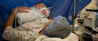
This article will discuss the changes that occur in the uterus after the birth of a child. The uterus undergoes massive changes after childbirth. Its volume and contents are sharply reduced. This entails significant changes among the organs surrounding it. It is the condition of the uterus and the speed of its contraction that is the most important criterion for the recovery of the body as a whole. Any illness or illness will affect the recovery of the uterus.
Let's take a closer look at these changes. This will help you understand why sports and weights are prohibited in the postpartum period, as well as why constipation occurs in the first days after childbirth.
Immediately after birth, the uterus is stretched, enlarged, and has a volume of about 5 liters. Its inner surface is bleeding. In order to return to normal, it needs to shrink.
Contractions of the uterus stop bleeding. The uterus contracts on its own and during stimulation of the nipples. The hormone of happiness - oxytocin, which is generously released into the blood during feeding of the baby, promotes uterine contractions. They are also called postpartum contractions. Such contractions can be quite painful.
Tell your doctor if you cannot bear the pain. He will help ease it.
It is difficult to answer the question of how long the reductions will continue. They can continue for 2-2.5 months - until the uterus is completely restored and the hormonal levels stabilize. This happens differently for every woman. Moreover, while breastfeeding, contractions may resume with the onset of a new pregnancy or before the return of menstruation.
It happens that after childbirth the uterus contracts poorly or does not contract at all. This happens extremely rarely and leads to massive bleeding. Uterine atony (lack of contraction) often ends in the death of a woman, especially if the birth takes place outside an obstetric hospital.
What diseases does ultrasound detect?
The transcript is made by the doctor conducting the study. In this case, an ultrasound alone may not be enough to make a diagnosis, then a specialist will prescribe tests. Based on the results of an additional examination, the initial conclusion is confirmed or refuted. Interpretation of an ultrasound of the pelvic organs in women can detect pregnancy, any developmental disorders of the observed fetus, and show various abnormalities (thickening of the tubes, adnexal inflammation, etc.).
Ovarian cyst in women
The pathology takes the form of a fluid-filled formation localized on the wall of the ovary. The cyst looks like a sac with a thin shell and has a diameter of up to 20 cm. To achieve the most accurate picture, ultrasound examinations are interpreted immediately after the completion of the menstrual cycle. Sometimes a doctor, along with this disease, diagnoses polycystic disease - an enlarged ovary, the formation of fibrosis zones, and thickening of the walls.
Cervical endometriosis
When performing an ultrasound of the uterus and appendages, the doctor may detect small bubbles in the muscle tissue of the tubes and cervix. With this research method, it is possible to detect foci of formations and endometrioid cysts in the ovaries. Endometriosis is characterized by the growth of the endometrium outside the uterine cavity. Using a pelvic ultrasound, the doctor can also detect internal adenomyosis - the spread of the endometrium into the uterine wall. Since endometriosis sometimes causes infertility, interpretation is carried out to predict the likelihood of pregnancy.
Myoma
The disease is a benign tumor. With this pathology, the body of the uterus is enlarged, its contours are heterogeneous, in addition, a node (or several) is observed in the myometrium. Interpretation of ultrasound helps determine the composition, size, and location of formations. In addition, this research method makes it possible to monitor the dynamics of nodule growth in order, if necessary, to carry out surgical treatment in a timely manner. Women who have been diagnosed with fibroids should have an ultrasound scan twice a year.
Abnormalities of the uterus
Uterine defects can also be diagnosed through examination. Interpretation of a pelvic ultrasound in women can show deformation, thickening of the walls, and an increase in the volume of the organ. In addition, the study provides the opportunity to observe pathological changes in the uterine cavity. Possible organ abnormalities:
- Doubling. It is extremely rare and is characterized by the presence of two vaginas and two uterine cervixes in a girl. At the same time, reproductive function is preserved.
- Bicornus. A woman with this anomaly has less space for the fetus, but this does not affect her ability to become pregnant.
- Arcing. There is a depression at the bottom of the organ, and its upper part is practically no different from the normal uterus.
- One-horned. It is half the normal size and has only 1 fallopian tube. If she and the ovary are healthy, then pregnancy is possible.
- Formation of the septum. The uterus has an extra wall inside, which consists of a fibrous or muscular wall. Sometimes this anomaly prevents pregnancy.
- Agenesia. It is extremely rare, characterized by the complete absence of the uterus or its small size, and underdevelopment of the vagina. This anomaly excludes conception.
Discharge after childbirth
Postpartum bleeding (lochia) is guaranteed by pregnancy itself. Over the previous nine months, maternal blood volume had increased significantly. After the birth of the baby, the need for additional blood disappears and part of it is successfully used for postpartum washing of the uterus. This protects it from infection.
Lochia continues for almost the entire postpartum period. At first they have the scarlet color of blood, then gradually they acquire the color and smell of normal menstrual flow. After a while they turn pale, discolored and turn into ichor, rich in leukocytes.
Such discharge continues for an average of 6-8 weeks. Each woman has her own characteristics and to say that lochia will end in 45 days is the same as saying that all women have 3-day menstruation.
The diagram below clearly shows how the color of the lochia changes during the first 28 days.
- The Y axis is the percentage of women experiencing one or another type of lochia
- X axis: days after birth
- Bright red - bright red
- Red/brown - red-brown
- Pink/red - red-pink
- Pale pink - pale pink
- Cream - creamy
- Variable - changeable
- None - absent
As you can see, the options for the norm are quite vague. However, if lochia ends before the fifth week or continues after the eighth, be sure to visit your doctor.
Also, contact your gynecologist if the lochia takes the form of meat slop (water in a bowl after washing the meat) and has an unpleasant (foul) odor.
The sudden cessation of red lochia is a reason to urgently seek medical help.
A small piece of placenta remaining in the uterine cavity can “confuse all the cards” and prolong the period of bleeding (scarlet blood). It can also interfere with the development of breastfeeding - milk simply will not come for 3-4 days, but there will still be colostrum. Cleaning may be necessary to eliminate it.
In the first ten days, the maca shrinks, returns to its pre-pregnancy size and hides behind the pubis. The uterine os is finally restored in the period from the 4th to the 6th week or later. The cervix will no longer have a round entrance - the “pupil”, but a slit-like one.
The uterine epithelium will recover unevenly. It will be the last place to recover at the placenta attachment site.
It is this uneven recovery that explains the possibility of menstruation occurring a month after childbirth, against the background of ongoing lochia. If after 4-5 weeks you notice an increase in lochia or the return of a bright color, this may indicate both pathology and the beginning of the menstrual cycle. This happens rarely, but it does happen.
To summarize this section, let's once again review the situations in which you need to see a doctor:
- The largest pad lasts less than 1 hour.
- Lochia appears scarlet after the 4th day.
- Increased body temperature.
- The discharge has an unpleasant (fetid, putrid) odor.
- Sudden cessation of discharge.
- Sudden increase in discharge or return of scarlet color.
- Several clots larger than a walnut in size came out.
- Discharge stops before the 5th week after birth.
- The discharge continues for more than 8 weeks.
Due to the presence of a wound surface (in the uterine cavity) and the natural postpartum stretching of the pelvic floor muscles and ligaments that support the internal organs, heavy lifting and vaginal sex are sharply limited until the discharge stops.
Normal cervical size
The cervix is a muscular organ that closes the entrance to the uterine body. The opening in the cervix is called the cervical canal and serves as a passage for sperm into the uterine cavity and an outlet for menstruation and the fetus.
Cervix dimensions: normal 4 cm, 2.6 cm, 3.5 cm (length, thickness, width). Deviations of about 0.5 -1 centimeter are allowed. After the first birth, the dimensions change upward by 0.5 - 1 centimeter. After the second, another 0.3 centimeters. Previous abortions affect the size in almost the same way as childbirth, because the uterus is artificially opened. Because of this, it also increases, but only by 0.1-0.2 cm.
Cervical size during pregnancy:
An important parameter for determining the possibility of bearing a child is the length of the cervix, the norm is 3.5 cm - 4 cm. If the cervix is less than 25 millimeters it is called shortened, and less than 20 is called short. In the case of a shortened cervix, pregnancy will pass without complications, simply under the strict supervision of a doctor. With a cervix shorter than 20 mm, pregnancy is more risky. It is she who holds the fetus in the uterus and the short cervix is weaker. Bearing a child will be a constant threat. This is not a contraindication, but a signal for more careful examinations and frequent visits to the doctor.
When a woman is in position, the length of the cervix is constantly monitored. This is necessary since it should not be shortened before 38 weeks. If the neck has reached a size of 20 mm, a woman needs to undergo a simple surgical procedure to carry out correction. In this case, sutures are placed on the cervical canal, which help keep the baby in the uterus.
Before birth, the length is already 10-15 mm, and the body of the cervix becomes soft.
Organs surrounding the uterus after childbirth
Now let's turn our attention to the structures surrounding the uterus.
During the process of growth, the uterus gradually raised and moved the surrounding organs to the sides. You can see this process very clearly in the video.
After childbirth, the uterus sharply decreases in volume, and the surrounding structures are released from its pressure. It would seem that everything should be fine - everything is back in place. But no! Not so simple. It takes time for the surrounding organs, like the uterus itself, to take their places.
Remember how hard it was to breathe while standing after giving birth. And all because the diaphragm suddenly lost support from below! Same with other organs.
The loops of the small intestine sharply fall down, the large intestine, like the diaphragm, loses its close neighbor (the usual tone disappears) and cannot immediately come to its senses - constipation occurs. Fortunately, breastfeeding (oxytocin helps the intestines contract), diet, and walking can help relieve constipation.
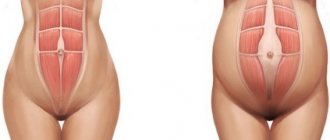
The pelvic floor muscles were under a lot of pressure for the entire 9 months. During childbirth they stretched even more. This can cause the bladder and rectum to become dislodged, making it difficult to control urination and bowel movements.
It is the stretching of the pelvic floor muscles and ligaments that support the internal organs that makes the use of a postpartum bandage undesirable. Tightening your stomach may create an outwardly neat figure, but it will lead to chaos inside. It will be difficult for the organs to return to their rightful places, and the “lower floor” - the uterus, vagina, bladder and rectum may descend.
Dimensions of the uterus after childbirth
Approximately 5-50 minutes after the birth of the child, the placenta and membranes (afterbirth) emerge from the genital tract of the woman in labor. Following this, the reverse contraction of the uterus occurs - it takes the shape of a ball.
If it were possible to weigh this important female organ immediately after delivery, one would have to agree that the size of the uterus after childbirth is quite large, because its weight is approximately 1 kg. After a week, the weight of the uterus is halved, and after two it is no more than 350 g.
In case of uterine contraction disorders, these indicators may differ slightly. If there is a discrepancy in these important parameters, an urgent consultation with a gynecologist is necessary.
After childbirth, the uterine muscles contract, some of the blood and lymphatic vessels dry out, and the muscle cells formed during 9 months of pregnancy dissolve. Upon completion of this process, the uterus returns to its normal parameters. The normal size of the uterus after childbirth (after a month and a half) is about 50 g.
Norms for the size of the uterus according to ultrasound
When the ovaries undergo involution, there is an associated decrease in estrogen production. This leads to gradual atrophy and involution of the endometrium. In postmenopausal women, the average endometrial thickness is reported to be 3.2 +/- 0.5 mm. Studies generally find an inverse relationship between uterine size and time after menopause: uterine size and volume gradually decrease. The greatest changes occur during the first ten years after menopause, and then gradually. In postmenopause, the normal size of the uterus according to ultrasound is: 8.0 +/- 1.3 cm in length, 5.0 +/- 0.8 cm in width and 3.2 +/- 0.6 cm in depth (antero-posterior size). If there is no menstrual cycle, subsequent changes in the blood supply to the uterus are usually undetectable. If the patient is on hormone replacement therapy, the size of the uterus, endometrium and cyclic changes may remain. Even the size of the uterus is approaching that of the premenopausal state.
In general, estrogen therapy affects the postmenopausal endometrium in a manner similar to estrogens during the normal cycle. Conjugated estrogens have a proliferative effect. Progestin therapy may cause the endometrium to react in a manner similar to normal secretory endometrium.
And when used together with exogenous estrogens, synthetic progestogens reproduce characteristic biochemical and morphological changes in the secretory phase of the normal menstrual cycle.
The blood flow to the uterus also changes while taking hormone replacement therapy. The thickness of the endometrium almost doubles. For example, before treatment, the average thickness was 0.37 +/- 0.08 cm. After treatment, the value became 0.68 +/- 0.13 cm.
In the study of postmenopausal women, one of the most important uses of ultrasound is the diagnosis and treatment of endometrial cancer. Such studies make it possible to determine the normal size of the uterus and ovaries using ultrasound. And in general, intravaginal ultrasound is superior to transabdominal ultrasound for visualizing the myometrium and endometrium.
Uterine contractions after childbirth
Contractions of the uterus are felt by a woman after childbirth as aching pain in the lower abdomen. During feeding, when the nipple is stimulated, the hormone oxytocin is released into the blood, which has a contractile effect. Therefore, in breastfeeding women, involution occurs by the end of the 6th week, and in non-breastfeeding women, involution occurs only at the 8th week.
After childbirth via cesarean section, the ability of the uterus to contract is much lower, so doctors recommend that women who have undergone such an operation move more and more actively to speed up the process of involution.
By how the process of uterine contraction occurs after childbirth, one can judge the woman’s condition. If the reverse development of the process occurs slowly, hormonal and immune disorders are possible in the body of the young mother.
Uterine prolapse after childbirth
Prolapse, or prolapse of the uterus after childbirth, is a fairly common consequence of injury to the pelvic floor muscles received during childbirth. The risk of this complication increases in women who have had a difficult birth or who have given birth more than once.
Normally, after the placenta has passed, the uterus is at the level of the navel. Further after childbirth, uterine prolapse occurs by approximately 1-2 cm per day. By the end of the first postpartum week, the normal height of the organ is 4-5 cm from the womb. Any deviation from this is considered a pathology and requires medical intervention.
When the uterus prolapses, the cervix is significantly lower than normal: it protrudes into the vagina or may even extend beyond the perineum. If the diagnosis reveals violations of its descent, the woman needs urgent surgical intervention. If treatment is left untreated, not only does sexual life become difficult, but there is also a high risk of developing urinary tract infections, prolapse of internal organs, and difficulties with the outflow of urine.
What complications may arise?
After the birth process, various complications can occur.
Postpartum hemorrhage
It can begin immediately after delivery. Bleeding is not accompanied by pain and can be very severe. Without surgical intervention, a woman's life may be at risk.
Causes of bleeding:
- disturbances occurred during the separation of the placenta and membranes;
- trauma during childbirth;
- uterine contraction is impaired.
Medicines and donated blood are used for treatment. Due to the risk of bleeding, the woman is always left in the delivery room for several hours.
Subinvolution of the uterus
As a result of delayed postpartum discharge, the organ contracts poorly. Often this disease appears on days 6–7: clots in the uterus after childbirth or part of the fetal membrane block the cervical canal.
Endometritis
Inflammation of the uterus is called endometritis. It appears due to infection of the cavity.
The occurrence may be due to the following factors:
- difficult labor process;
- the placenta separated incorrectly during childbirth;
- diseases of the genital organs during pregnancy;
- abortions;
- decreased immunity.
Symptoms of endometritis:
- heat;
- after childbirth the uterus hurts;
- rotten smell of postpartum discharge.
To make an accurate diagnosis, an ultrasound of the uterus is performed after childbirth. In case of endometritis, the contents of the cavity are removed, washed or scraped out. After surgery, antibiotics are prescribed. Read more: postpartum endometritis→
Prolapse
It may happen that after childbirth the uterus prolapses. This occurs as a result of injury to the muscle tissue of the pelvic floor. Women carrying a second child are often susceptible to this complication.
Normally, the uterus after childbirth is located in the navel area when the placenta comes out. The bottom drops 1–2 cm daily.
Any deviations from the norm are considered pathology. In severe forms, the uterus may prolapse into the vagina and come out after childbirth.
It is necessary to treat prolapse in a timely manner so as not to be at risk of infection or prolapse of the internal organs of the urinary system. When the uterus prolapses, sexual activity is prohibited.
To identify possible pathologies, it is recommended to visit a gynecologist 6–9 weeks after birth. Even if you feel good, there is no pain or discomfort.
If the uterus decreases in size slowly after childbirth, this indicates hypotension or atony.
In some cases, lochia or parts of the placenta may remain in the uterus. This condition cannot be ignored, so the woman undergoes cleaning - mechanical removal of pathological residues. The problem is usually detected by ultrasound.
A woman may notice increased uterine bleeding, which occurs for various reasons. The release of a large amount of blood is observed both with poor contraction of the uterus and with an infectious process.
One of the common complications is cervical erosion. If the process is not accompanied by infection, doctors recommend continuing dynamic monitoring, since the structure of the cervix can recover after some time. To exclude pathogenic microorganisms, a smear is taken from the cervix, and colposcopy is performed to confirm tissue eversion.
Erosion is often asymptomatic, so a few weeks after giving birth, a woman needs to come for a routine examination so that the doctor can identify the disorder.
In some cases, after childbirth, a complication such as uterine prolapse occurs. It is caused by weakness of the ligamentous-muscular system and is typical for women who have given birth many times.
If lochia continues to be discharged 6 weeks after giving birth, this is not considered normal and the woman should consult a doctor. Early disappearance of lochia can also be a pathology. This condition can occur when there is retention of secretions in the uterine cavity, uterine atony, or blockage of the cervical canal. If its secretion is released, an infection begins to develop in it.
With rapid labor or a large fetus, cervical ruptures are possible, which necessitates the need for sutures.
Endometritis after childbirth occurs when an infection enters the uterine cavity. Symptoms appear a few days after birth. Most often, a woman’s temperature rises, chills, nausea, and increased pulse and heart rate occur. At the same time, she feels severe pain in the lower abdomen, which radiates to the lumbar region. The color and smell of the discharge becomes pathological.
We invite you to read: Feelings before and after childbirth
Causes of uterine contraction disorders
The causes of uterine contraction disorders after childbirth may be different.
First of all, the involution process is affected by a lack of prolactin, which is produced reflexively when the nipples are irritated. With its deficiency, involution slows down.
Delayed uterine contractions can be caused by the presence of remnants of the placenta attached to the walls of the uterus.
In addition, an infection suffered by a woman can reduce the contractility of the uterus.
All these cases require consultation with a specialist; self-medication in such situations aggravates the situation of the young mother.
Clots in the uterus after childbirth
The uterus after childbirth is a big wound. From the inside, it is severely damaged in the place where the placenta was attached. On its inner membrane there are remnants of membranes and blood clots.
Normally, clots from the uterus are released only for 3-4 days. Thanks to wound healing processes in a woman’s body, wound secretion, lochia, begins to be released from the uterus.
In the first days, lochia is bloody, similar to menstrual discharge; on the 3rd day it becomes serous and bloody in nature, and by the end of the 20th day after birth it becomes liquid and light-colored. Lochia completely disappears by the end of the 6th postpartum week.
When involution slows down, lochia may be released longer. However, if after 2 weeks after birth there are still clots in the uterus, an urgent visit to the doctor is necessary. This can be guessed if the lochia does not change its color and the intensity of its secretion does not decrease. This can happen due to an infection or when the uterine pharynx is blocked by blood clots.
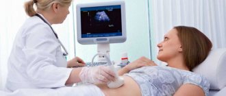
What does a pelvic ultrasound show?
Using the study, the specialist determines whether the structure and size of the organs of the reproductive system and the woman’s bladder are normal. This allows us to identify the presence of pathological changes - tumors and neoplasms. In addition, with the help of decoding pelvic ultrasound in women, you can establish:
- the presence or absence of congenital pathologies;
- pregnancy, approximate duration, any pathologies;
- presence of diseases (cancer, fibrosis, endometriosis, etc.);
- Are the volumes and locations of organs normal?
To avoid the development of serious gynecological diseases that affect reproductive abilities, experts recommend ultrasound and ECHO for girls for preventive purposes. So, women aged 18-40 years old should be examined at least once a year. After forty, in order to detect the development of pathological processes in time, you should visit the diagnostic room 1-2 times a year.
The advantages of ultrasound examination are:
- non-invasive method, painless;
- accessibility, ease of examination;
- absence of harmful radiation;
- obtaining clear, three-dimensional images;
- universality of the study (ultrasound helps to identify various diseases and anomalies, monitor the development of the fetus, etc.).
The size of the uterus is normal in women
The external contours of the uterine myometrium (muscle tissue), in the absence of pathological processes, should have clear, even lines. If during a pelvic ultrasound, some blurriness or unclear boundaries of the organ were detected, this indicates inflammation of the surrounding tissues and often indicates the presence of diseases. A woman’s uterus is pear-shaped; the following indicators are considered normal:
- length – 4.5-6.7 cm;
- width – 4.6-6.4 cm;
- thickness – 3-4 cm.
In women after menopause, the parameters of the uterus may change. Transformations of the organ in the postmenopausal period occur over a period of 20 years, after which it can significantly decrease. In this case, the normal sizes of the uterus are:
- length – 4.2 cm (maximum);
- width – 4.4 cm;
- thickness – 3 cm.
Normal ovarian size in women
Interpretation of pelvic ultrasound in women helps to monitor the condition of the ovaries - the organs responsible for the ability to conceive. Egg follicles are formed in them, which are subsequently fertilized by sperm. Due to the growth process of follicles, the walls of the ovaries are uneven and lumpy, but they should have clear outlines. The echostructure of organs in the absence of pathologies is homogeneous, with minor areas of fibrosis. Any changes in the echostructure of the ovaries indicate an inflammatory process. Normal ovarian sizes:
- volume – 2-8 cubic centimeters;
- length – 30 millimeters;
- width – 25 millimeters;
- thickness – 15 millimeters.
Articles on the topic
- Fibromyalgia – treatment with traditional and alternative methods, preventive measures, complications
- Libido in women - what is it, deviations, home correction methods
- Testosterone analysis in women - when to prescribe and how to take it, reasons for increased and decreased values
Normal cervical size
This is an organ consisting of muscle tissue that closes the entrance to the body of the uterus. The cervix has an opening for the passage of sperm and removal of secretions during menstruation, it is called the cervical canal. When interpreting an ultrasound, it is worth considering that the normal length of the organ is 3.5-4 cm. At the same time, the endocervix (canal) should remain within 3 mm; its increase indicates the presence of a serious pathology - endometriosis or cancer. After childbirth, the size of the cervix increases by a maximum of 1 cm, and at the birth of the second child by another 3 mm.