Why carry out diagnostics
Pregnancy is diagnosed at different stages. At the first external signs of pregnancy, a woman turns to various ways to determine the fact of conception.
This is necessary to change lifestyle, diet, discontinue medications in case of saving the fetus or timely termination of pregnancy without further consequences. An abortion is done if the woman wishes or according to indications when diagnostics showed an ectopic pregnancy in the early stages.
The information content of the fact of conception is individual at different stages: early diagnostic methods are not suitable for determining late pregnancy.
Why does this happen?
The main reasons for late pregnancy can be divided into two large groups: social and medical. An unsettled life, low wages, difficult family relationships and the desire to “live for oneself” lead to the fact that some women do not want to have children between the ages of 23 and 33. In addition, in our turbulent age, many strive to ensure a fairly high level of education, occupy a high social position, make a successful career and provide prosperity for their family, postponing the birth of a child to a later date. These and other social aspects are the reason that many women begin to think about procreation after 30 years. The same reasons can influence a woman’s desire to give birth to a second or third child at a “respectable” age.
Medical causes of late pregnancy are certain diseases that have prevented conception or pregnancy for a long time.
What methods are there?
Diagnosis of early and late pregnancy differs. The results of the first studies will be irrelevant in the last trimester.
Early dates
The early period for pregnant women is the first 3 months, in medicine it is called the first trimester. The period lasts up to 12 weeks. Usually a woman thinks about the magical position in the fourth week, when menstruation should occur. But my periods stop.
Early diagnosis of pregnancy involves:
- carrying out urine analysis using pharmacy and laboratory tests for the hCG hormone;
- examination of the genital organs by a gynecologist;
- Ultrasound with a transvaginal sensor, or other sensors, pregnancy is not visible until 7 weeks;
- the presence of certain signs.
External signs
External signs that a woman is pregnant are suggestive and probable. Nowadays, diagnosis of zygote attachment is carried out mainly by modern ultrasound and laboratory tests.
But presumptive symptoms help the woman herself understand that she is pregnant:
- Periodic attacks of nausea and vomiting usually occur in the morning. Appetite changes. Unusual food cravings appear.
- The breasts swell, the mammary glands tense.
- A disgusting feeling appears towards smells: tobacco, food, eau de toilette.
- Urination becomes more frequent due to pressure from the growing uterus.
- The belly gradually grows, but at first it is not noticeable.
- The functioning of the nervous system is disrupted. The woman suffers from poor health, a constant craving for sleep, excessive irritability, and frequent mood swings. Sometimes headaches and dizziness are added. If pain and dizziness are periodic, it is better to consult a doctor.
- Change in skin color on the face, around the nipples. Protruding areolas on the chest cause the nipples to darken. A white line forms on the abdomen towards the middle of pregnancy.
- Increased hair growth on the skin of the abdomen. Sometimes excessive hair appears on the chest and hips.
Possible symptoms and signs of conception include objective changes in the genital organs in the first trimester:
- Menstruation stops in a healthy woman of reproductive age.
- Release of colostrum when pressing on the nipples. The symptom appears at the beginning of the second trimester. In rare cases, the release of colostrum is observed in the first months of pregnancy.
Pharmacy testing
If a woman suspects pregnancy, she tests at home. Pharmacies sell special tests that react to a surge in the hCG hormone to diagnose conception.
The test shows a reliable result 1-4 days after the delay. Typically, performance is affected by the quality of the test.
When the test shows 2 stripes, it is necessary to confirm the presence of pregnancy with a gynecologist.
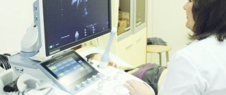
Carrying out an ultrasound procedure
The ultrasound method is always used to determine the attachment of the zygote to the wall of the uterus. Transvaginal echography diagnoses pregnancy already at 4-5 weeks, when menstruation stops. The ultrasound machine screen clearly displays the fertilized egg in the uterine cavity.
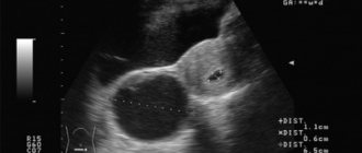
It is important to understand how the ultrasonic structure of the fetal egg works to eliminate diagnostic errors. When diagnosing, an indicator of echogenicity is used, the ability of internal organs to reflect ultrasonic waves. Sometimes a glandular polyp on the endometrium is perceived as pregnancy. During an ultrasound, the doctor relies on the following factors:
- The fertilized egg is visible on the screen as a black, anechoic spot.
- The surrounding tissue of the uterus, the endometrium, becomes thick up to 1.5 cm. The phenomenon is called the three-layer structure of Maho.
- A liquid formation is identified inside the egg, which means the chorion. The thickness of the chorion indicates the duration of pregnancy. Its contour is distinguished by a clear hyperechoic area.
From the 6th week, an embryo with a yolk sac is detected inside the amniotic cavity using an ultrasound sensor. The heartbeat of the embryo is visualized on the screen.
If there is a suspicion of a frozen pregnancy, a woman is recommended to undergo an ultrasound scan after 2 weeks. If the result has not changed, there is no heartbeat, the embryo is not detected, the woman is sent for cleaning.
Visit to the gynecologist
A positive reaction in the form of two stripes on a pharmacy test becomes a reason to visit a gynecologist. The doctor’s task is to assess the condition of the genital organs, in particular the cervix. It is cyanosis, cyanosis, of the cervix that indicates the development of pregnancy.
Afterwards, the gynecologist prescribes an ultrasound to determine the fact of intrauterine pregnancy. You can bring the research results in advance when registering.
Biochemical blood test for hCG
Human chorionic gonadotropin is produced in the growing villous chorion. With laboratory tests, diagnosis of this hormone is possible within a week from the moment of conception. The beta subunit of hCG is detected in the blood of a pregnant woman when it coincides with the attachment of the zygote to the endometrium of the uterus.
As pregnancy progresses, hCG constantly rises. The rate peaks at ten weeks. After this, the hCG level gradually decreases, and diagnosing pregnancy in this way becomes irrelevant. 2 weeks after birth, the hormone completely disappears from the woman’s blood.
HCG allows for early diagnosis of pregnancy, determining its normal or pathological development. During an ectopic pregnancy, the hormone is not produced, so pharmacy tests show a negative result.
Hormone levels are measured in a laboratory. To do this, a woman brings biomaterial: urine, or donates blood. In the latter case, the results are the most reliable. It should be remembered that the presence of hCG in the blood does not mean pregnancy. Additional diagnostic methods are always carried out.
Late dates
Diagnosis of pregnancy in late stages is usually not carried out. More often, women notice the first signs of successful conception in the first trimester. Only very young girls, recent women who gave birth and women on the eve of menopause may not suspect the presence of a small growing organism inside.
After childbirth, each female body recovers individually. Some people do not menstruate for a long time. Therefore, when the egg is fertilized and there is no menstruation, the woman who has just given birth does not think about a possible pregnancy.
Morning sickness, fatigue, and constant sleepiness after childbirth are associated with hormonal imbalances. A woman simply ignores obvious signs of pregnancy due to her constant busyness with the baby.
But then the belly begins to grow and colostrum appears. Diagnosis of pregnancy in this case involves an ultrasound scan and examination by a doctor.
Medical examination
Late periods mark the months of pregnancy from 5 to 9. The gynecologist accurately determines the development of the fetus inside a woman at the twentieth week:
- The child is palpated through the abdomen: large and small parts of the body are clearly recognized.
- The fetus moves in utero. The woman herself understands this.
- The heartbeat is listened to with a stethoscope, an obstetric instrument.
The size of the fetus affects the determination of the fact of pregnancy, clarification of its location and the presenting part inside.
Diagnosis of the baby's condition is often carried out before birth to ensure that the baby is present correctly in the uterus. A caesarean section is prescribed if the baby is positioned feet down towards the exit of the uterus or transversely.
A thorough examination of the woman by a gynecologist reveals the presence of a multiple pregnancy. Diagnosis in later stages is simplified by ultrasound.
Third screening
Laboratory and pharmacy tests for hCG in the third trimester are not informative. During this period, ultrasound examination is often performed. An ultrasound confirms the fact of conception with 100% accuracy.
But the main tasks of examining the fetus with an ultrasound sensor are to assess the degree of development of the child and its compliance with the gestational age. Ultrasound is designed to determine the number of fetuses.
From about 18 weeks, ultrasound screening shows future parents the sex of the child. The errors are minimal.
The long-awaited pregnancy!
Touching upon the topic of pregnancy in adulthood, one cannot help but say something about women for whom the opportunity to become pregnant only with the help of medicine has become the only chance to experience the happiness of motherhood.
After all, the chances of giving birth for those over 30, and even over 40, are two times less than for young people, but these are at least some chances in comparison with the diagnosis, which 20 years ago sounded like a merciless sentence. Today, doctors urge women not to delay infertility treatment and to resort to assisted reproductive technologies, because the younger the patient, the greater the chance of success.
Diagnosis of pregnancy and childbirth
The due date is calculated based on the date of the last menstruation and information about the first fetal movements. Conception occurs during ovulation; gestational age is often calculated based on the ovulatory day. This takes into account the duration of the menstrual cycle.
In obstetrics, the accepted norm is that the due date is calculated according to the 28-day menstrual cycle. Ovulation occurs 2 weeks after the last menstruation. Most often, pregnancy lasts 10 obstetric months or 40 weeks.
To diagnose the expected date of the onset of labor in a woman, 9 months plus 1 week are added to the date of the first day of her last menstruation. There is a simpler method: subtract 3 months from the date of the last menstruation and add a week.
When calculating the time of birth, the error in the onset of ovulation in the middle of the menstrual cycle is taken into account. The duration of pregnancy increases by one day if the menstrual cycle is not standard at 28 days.
The diagnosis of the duration of pregnancy is influenced by the period when the expectant mother begins to feel the first movements of the child. In first-time mothers, this happens at 20 weeks. Multiparous women notice the first movements from 16 weeks. But there are exceptional cases.
20 weeks are added to the period of the first fetal movements for primiparous women, 22 weeks for multiparous women. This gives the estimated time of onset of the labor process.
The obstetric calendar is considered convenient for calculating the gestational age based on menstruation and the first movements of the fetus. Diagnosis of the duration of gestation and date of birth is carried out through an objective study:
- size of the uterus;
- height of the uterine fundus;
- abdominal volumes;
- fetal length;
- head size.
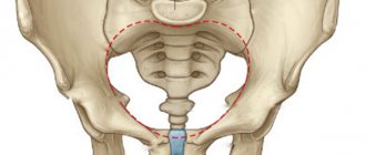
Growth of the uterine cavity by timing
When the first obstetric month ends, the size of the uterus corresponds to the size of a chicken egg. In the second month, the uterus enlarges to the size of a goose egg. By the end of the first trimester, the female reproductive organ matches the size of the newborn's head.
In this case, the asymmetry disappears. The uterus completely fills the upper part of the pelvis. The height of the uterine fundus approaches the upper edge of the pubic bone.
Starting from the fourth month of gestation, diagnosing the uterus becomes possible through the abdominal wall. The period is calculated based on the height of the uterine fundus. This indicator is affected by:
- child size;
- presence of multiple births;
- large volumes of amniotic fluid;
- location of the fetus;
- other features of bearing a child.
The position of the fetus inside
the height of the uterine fundus changes according to the timing:
- At the 4th obstetric month, diagnostics show a rise in the fundus of the uterus halfway between the pubis and the navel. By the middle of the second trimester, the indicator shifts: the fundus of the uterus is located 2 fingers below the navel. The abdominal wall begins to bulge.
- At the end of the second trimester, the fundus of the uterus is already at the level of the navel. After another obstetric month, the bottom moves 3 fingers above the navel. By the beginning of the third trimester, the indicator almost reaches the xiphoid process, the area where the ribs are attached in the middle of the chest. The navel is smoothed out.
- 2 weeks before birth, the uterine fundus is completely located at the level of the xiphoid process with the costal arches.
- At the tenth obstetric month, digital diagnostics show prolapse: the fundus of the uterus moves down to the level at which it was at the end of the eighth month. The navel protrudes outward. The baby's head drops and lies close to the entrance to the pelvis.
The size of the uterine fundus by period in weeks
Determination of gestational age by echography
Diagnosis of the duration of pregnancy is often performed through echography. Usually, at the first screening, the main indicator is the coccygeal-parietal size of the embryo, abbreviated CTE.
In subsequent trimesters, time is set based on other biometric indicators:
- abdominal circumference;
- biparietal size;
- average diameters of the abdomen, chest;
- femur length;
- head circumference.
As the time of gestation increases, the accuracy of determining the gestational period decreases due to the individual size.
Features of childbirth
The risk of complications during childbirth also increases slightly. Physiological changes in the structure of the body and cervix can cause weakness in labor. The likelihood of ruptures of the soft birth canal and bleeding increases.
Women often ask the question: is the first upcoming birth after the age of 30 an indication for a cesarean section? There is no clear answer to this. If, except for the patient’s age, all other figures (pelvic size, blood pressure, test data, number of heartbeats and respirations per minute, diopters for myopia - myopia, etc.) do not raise any doubts in the doctor, then the question of the method of delivery is resolved in favor of vaginal delivery. If, in addition to age, there are any indicators that indicate problems related to the health of the mother or baby, then, taking into account the age, a decision is made about a caesarean section. In each case, this issue is resolved individually. Since, as already mentioned, the number of diseases of both the organs of the reproductive system and organs not directly related to childbirth increases with age, the number of cesarean sections in women of the older age group also increases.
It should be noted that if the late birth is not the first and the older child can hardly be called a baby, then the mother’s body does not always retain the memory of the birth that took place 15-20 years ago and the process of dilation of the cervix can follow the “laws of the first birth,” i.e. There is a risk of complications during childbirth.
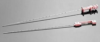
Invasive diagnostics
Invasive diagnostic methods are recognized as a set of studies in which biological material from the fetus is analyzed. Amniotic fluid, skin particles, fetal blood and placental villi are examined.
The purpose of the invasive test is to determine the baby’s developmental defects, diagnose chromosomal abnormalities, and inheritance of genetic diseases.
The diagnostic method is chosen by a geneticist with an obstetrician-gynecologist, guided by the pregnancy period and the expected pathology. The methods are considered dangerous for the unborn child. Such diagnostics are prescribed only in exceptional cases according to indications.
“Late” is a relative concept
Despite the fact that it is easiest for a woman to physically give birth at the age of 20–25, psychological readiness for this process comes to her much later. Here is what private family psychologist Evgenia NIKITINA says about late pregnancy and late childbirth:
— From a psychological point of view, having a child is a psychological and physiological norm for a woman at any childbearing age. However, both pregnancy and childbirth are serious stress for the body. Often women, especially young ones, are not ready for this. And a woman’s psychological unpreparedness for pregnancy and internal contradictions can lead not only to emotional anxiety, but also to very real physiological complications. And, on the contrary, if a woman is ready to accept herself in a new state, if she is not embarrassed by the presence of a belly, but on the contrary, she is happy about it, since a person lives in her who will soon be born and will become dearest to her, the chances of that everything will go well. The latter condition is more often typical for adult mothers.
For example, a friend of mine gave birth to her first child at 42 years old. She went to this for a long time. Not so much for medical reasons, but psychologically. In my opinion, the concepts of “late pregnancy”, “late childbirth”, “late child” exist only in a person’s head. Pregnancy in general is a harmonious state for a woman. Even if the expectant mother has physiological problems, she may well be able to bear and safely give birth to a healthy child, of course, under the supervision of obstetricians and gynecologists.
BY THE WAY:
Later offspring become increasingly popular among celebrities. Kim Basinger gave birth for the first time after 40 years. Madonna decided to do this at 36, but did not stop there and five years later gave her daughter a brother. Julia Roberts, America's leading runaway bride, gave birth to twins at the age of 37. Leading top models Claudia Schiffer and Cindy Crawford are a little behind, becoming mothers at 35 and 36.
As for our “stars”: they also do not lag behind the West. Elena Yakovleva, Tatyana Bulanova, Elena Proklova, Larisa Guzeeva, Lolita Milyavskaya decided on motherhood when they were well over 30.
Non-invasive examination methods
Ultrasound diagnostics is a non-invasive examination method. The method is considered accessible, informative and safe for determining the state of the child’s body in utero.
Dopplerography is used as an additional research measure. The procedure determines the nature of blood flow in the uterine, fetal-placental circulation. In this case, the doctor monitors the flow of blood in the arteries of the umbilical cord, uterus and the baby’s brain.
Fetus at 3rd screening
At the third screening, the pregnant woman undergoes CTG and cardiotocography. This study also applies to non-invasive diagnostic methods. The procedure involves simultaneous recording of fetal heart contractions and uterine tone. An image with a graph of physiological signals is displayed on the device screen.
The recording lasts for 30-50 minutes. The phases of activity and rest are recorded. The first phenomenon lasts 20 minutes, the second about 30 minutes. The image with the graph records only the activity phase of the child; the analysis is carried out on the basis of this data.
Diagnosis of pregnancy includes a set of procedures. It is important to promptly detect the fact of conception in order to prevent possible complications and other circumstances.
What types of diagnostic procedures did you have while you were pregnant? Share your opinion on the listed methods. Tell your friends about the article on social networks.
Risks of late pregnancy for the mother
It is worth mentioning the risks of late pregnancy. Remember that there is no need to be afraid - today millions of women around the world give birth to healthy children at 30 and 40 years old. However, potential problems exist both on the health side of the mother and on the part of the unborn child.
So, you should be aware of the threat of miscarriage (from 17 to 30%). This is due to age-related changes in the female body and a decrease in the quality of eggs.
It is also worth learning about problems with the formation of the placenta (chronic placental insufficiency, previa, premature placental abruption). These diagnoses can also affect the quality of your pregnancy and complicate both your 9 months of life and the birth process.
Don’t forget about chronic diseases, each of which can worsen during pregnancy. Pay attention to kidney and cardiovascular diseases. They can be dangerous to you and your baby's health. And, most importantly, inform your doctor - he can prevent possible risks and prescribe competent treatment.
Diabetes in pregnant women is another threat. Some expectant mothers are prescribed insulin injections and a special diet. Those with diabetes have an increased risk of complications during childbirth, late toxicosis, and stillbirth.
The next risk for you is a caesarean section in 40% of cases. Those women who plan to give birth to their first child after 35-40 years are especially susceptible to this factor.
Don't forget about other complications during childbirth and pregnancy. Most likely, your doctor will warn you about them, but you know the wonderful saying “forewarned is forearmed.”
Preparing for pregnancy after 35 years
When planning pregnancy, you need to evaluate the ovarian reserve - the supply of follicles in the ovaries. For this purpose the following is carried out:
- ultrasound examination of the ovaries;
- blood test for hormones (FSH, LH, estradiol, anti-Mullerian hormone).
If the ovarian reserve decreases, you should not delay conceiving a child or postpone IVF. For women over 40 years of age, in vitro fertilization with preimplantation genetic diagnosis is recommended. This will allow us to select only healthy embryos without chromosomal abnormalities.
Conceiving a child after the age of 45 practically does not happen. In this situation, IVF with a donor egg can be used. Preliminary cryopreservation (freezing) of eggs is also practiced. The procedure is performed at the age of 30-40 years. In the future, frozen eggs can be used in an IVF program.
Other examination methods in preparation for pregnancy:
- General clinical blood and urine tests. They allow you to assess the general condition of a woman and identify signs of pathological processes.
- Blood test for coagulation: coagulogram or hemostasiogram. According to indications, determination of D-dimer, antiphospholipid antibodies, etc. is prescribed.
- Blood test for thyroid hormones.
- Screening for sexually transmitted infections.
- Assessment of the condition of the cervix: smear for oncocytology and colposcopy, biopsy - according to indications.
- Hysteroscopy – for intrauterine pathology. If indicated, an endometrial biopsy is performed.
- Assessment of fallopian tube patency.
- Diagnostic laparoscopy. It is carried out for mass formations of the ovaries, uterus, adhesions, endometriosis, etc.
- ECG.
- Consultation with a gynecologist-reproductologist, therapist, ophthalmologist, ENT specialist and dentist; according to indications - other narrow specialists.
- Consultation with a geneticist if there are risk factors.
Before conceiving a child, it is recommended:
- Start taking folic acid 3 months before your expected pregnancy . This will reduce the risk of developing defects of the fetal nervous system and spontaneous miscarriage.
- Change your diet . The expectant mother's menu should contain more proteins and vitamins, less animal fats and carbohydrates.
- Move more . Adequate physical activity reduces the risk of exacerbations of chronic pathology, increases the overall tone of the body, and prolongs youth.
- Lose weight (for overweight and obesity) . Extra pounds provoke the development of gestosis, arterial hypertension, gestational diabetes and other complications.
- Get treatment . If a chronic pathology is detected, remission must be achieved before conceiving a child. It is worthwhile to undergo sanitation of foci of chronic infection - to cure teeth, get rid of rhinitis and otitis media.
It is also recommended to quit smoking, stop drinking alcohol, and avoid stress.
Risks of late pregnancy for the baby
As already noted, the possible risks of late pregnancy apply not only to you, but also to the unborn child.
First, preterm birth is not uncommon in late pregnancy, so be prepared for all the consequences. You can learn more about the risks in the article about premature birth.
Secondly, low birth weight. This can happen even if the birth was not premature. An underweight child will require certain conditions and maximum care from you. You will be interested in reading our article on this topic.
Thirdly, hypoxia, that is, lack of oxygen for the fetus. Hypoxia can lead to a number of unfavorable changes in the baby’s body. The main organs most susceptible to them are the liver, kidneys, cardiovascular system, and lungs.
Finally, chromosomal abnormalities. Unfortunately, science has proven that the risk of having children with Down syndrome increases with age. You can find out more about this terrible disease in the article.
All this should not scare you or stop you. Statistics show that a healthy child after 35 years of age is a pattern, not a rarity. Any risks can be prevented if you do certain examinations on time, take tests and listen to your doctor. So the health of both you and your baby directly depends on you.