Cordocentesis: the essence of the technique
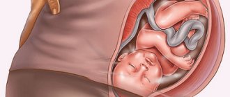
Cordocentesis is the removal of blood from the fetal umbilical cord for analysis. This method of prenatal diagnosis of fetal health began to be widely used in the second half of the last century, which is associated with the introduction of ultrasound research into medical practice. It is ultrasound that allows you to see the umbilical cord and insert a needle into it.
Many people consider cordocentesis and amniocentesis to be the same procedure, but this is not the case. The only thing they have in common is that these two examination methods relate to intrauterine examinations. Amniocentesis involves the collection of amniotic fluid, and cordocentesis specifically involves the umbilical cord blood of the fetus.
Under what conditions and how is cordocentesis performed?
Before 22 weeks of pregnancy, cordocentesis is usually performed in an outpatient facility or in a health care professional's office. After 23 weeks of pregnancy, cordocentesis is usually performed in a hospital if the baby develops complications that may require emergency delivery by caesarean section.
.
Before the procedure, a sample of your blood will be taken to compare with blood samples from the fetus
.
You may be given antibiotics approximately 30 to 60 minutes before the procedure to reduce the risk of uterine infection
. This is usually done through a venous catheter so that there is venous access if other drugs need to be administered during or after the procedure.
Your doctor or other specialist in the field will use ultrasound to locate the umbilical cord in your uterus. You will lie on your back and the attending physician will apply a special gel to your stomach
. He will then use a small device known as an ultrasound transducer to show your baby's position on the monitor.
Then your doctor will cleanse your stomach of gel and other possible contaminants with special antiseptics. Sometimes pain medication is prescribed to prevent discomfort during the procedure, but this is often not necessary
. If you read the opinions of patients who underwent cordocentesis during pregnancy, the reviews usually say that the procedure is quite unpleasant, but not too painful to require additional painkillers to be administered to the woman.
Under ultrasound guidance, the doctor will puncture the skin of the abdomen, then insert a thin, hollow needle through the abdominal wall and into the uterus. A small amount of blood from a vein in the umbilical cord will be drawn into a syringe and the needle will be removed
. You will need to lie still while the needle is inserted and blood is drawn. You may notice a burning sensation as the needle enters your skin, and you may feel cramping as the needle enters your uterus.
In addition, there are two techniques for performing cordocentesis. By the way, amniocentesis is usually performed at the same time. So, the single-needle method is when one needle is inserted, with the help of which amniotic fluid is collected, and then umbilical cord blood after puncture of the umbilical cord
.
Double-needle method - when a thicker needle is inserted into the amniotic sac, fluid is collected, and then a thinner one is inserted through the lumen of this needle, which pierces the umbilical cord vessel and blood is collected - that is, cordocentesis directly
.
The needles are removed in the reverse order - first a thin needle, which was used to draw blood, and then a needle with a wide lumen
. After removing the needle, the puncture site will be treated with an antiseptic.
What to do after the procedure?
After completing the puncture procedure and blood sampling, you may experience uterine tone or slight discomfort.
Your healthcare provider will use an ultrasound or external labor monitor to monitor your baby's heart rate after the procedure.
Once you have undergone cordocentesis, your doctor may suggest you rest for the rest of the day after the procedure, but this does not always mean staying in the hospital for the whole day. You will likely be able to resume normal activities the next day
.
At home on the day of cordocentesis, you will need to rest, lie down, and avoid excessive activity. If you have the slightest suspicion that something has gone wrong, you will need to immediately consult a doctor - call or call an ambulance
.
This could happen, for example, if you experience abdominal pain, a feeling of excessive uterine tone or cramping pain, the passage of even a small amount of amniotic fluid, or even slight bleeding from the genital tract
.
You will also need to pay attention to the baby’s activity - the nature of the movements should not change significantly from normal
. All these signs, as well as such uncomfortable sensations as severe dizziness, an attack of weakness, nausea, should be the reason for immediate contact with a specialist. This does not mean that you necessarily have a life-threatening condition or that your baby is suffering, but it is better to be on the safe side and make sure everything is okay.
Stages of performing the cordocentesis procedure:
- Treatment of the surgical field according to the standard method - the place on the woman’s anterior abdominal wall where the puncture needle will be inserted.
- Puncture of the anterior abdominal wall of a pregnant woman with a thin needle, which is controlled by an ultrasound sensor. Ultrasound allows you to control the advancement of the needle to the umbilical vein and makes it possible to avoid trauma to the fetus itself.
- Inserting a needle into the umbilical vein where it attaches to the placenta.
- Direct collection of cord blood in an amount of about 1 ml.
In general, no anesthesia is required to perform the cordocentesis procedure.
Algorithm and diagnostic methods
Technically, the manipulation, like other invasive procedures, is carried out either using the “free hand” method (more often) or using a puncture adapter (less often).
The puncture can be single-needle or double-needle.
- Single needle puncture. After the amniotic sac is punctured, a puncture of the umbilical cord vessel is made with the same needle, a syringe is attached to the needle and the amount of cord blood required for diagnosis is taken (1–4 ml), and then the needle is removed.
- Double needle puncture. In this case, a double needle is used: inside the large outer one there is a small one. The amniotic membrane is punctured first with a large needle. They bring it to a pre-selected point on the umbilical cord, and then they puncture the umbilical cord vessel with a small one and take cord blood for analysis. After which the needles are removed from the uterine cavity in the reverse order.
The operation is carried out under the continuous monitoring of an ultrasound machine, and in the third trimester, CTG (cardiotocography) is also used to monitor the condition of the fetus.
This is what puncture needles look like for invasive interventions and for ultrasound sensors
Sequencing
- The site of intervention is determined using an ultrasound sensor. The puncture should not affect the placenta. But there are situations when it is technically impossible to meet this condition. Then they try to mark the thinnest area on it.
- To obtain only fetal material (without any admixture of maternal material), the puncture is performed on the area of the free loop of the umbilical cord. It is, of course, more mobile, but the results of the analysis will be more reliable.
- If necessary, the fetus is immobilized with medication.
- The duration of the manipulation is about 30 minutes.
- After cordocentesis, the expectant mother remains in the hospital for 1–2 days under the supervision of doctors.
Cord blood collection is considered a painless procedure.
Cordocentesis during pregnancy: purpose of analysis
A fetal cord blood test can identify the following conditions:
- Down syndrome, Patau syndrome, Edwards syndrome, etc. (change in the number of autosomes in the chromosome set);
- Klinefelter and Shereshevsky-Turner syndrome, etc. (change in the number of sex chromosomes);
- Cystic fibrosis;
- Duchenne's disease;
- Thalassemia, hemophilia and other genetic diseases of the fetus;
- Intrauterine infection;
- Identification of the causes of intrauterine growth retardation.
Cordocentesis during pregnancy allows, in addition to diagnostic tests, intrauterine treatment of the fetus (blood transfusion, administration of various medications).
Most specialized clinics can perform the cordocentesis procedure. The price will be announced before the event.
When is cordocentesis analysis prescribed?
There are a number of strict indications
to carry out this type of diagnosis of the fetal condition, because the procedure is unsafe. Cordocentesis is performed only in cases where its risk does not exceed the probable risk to the future health of the child.
- High risk of having a child with developmental defects.
- Situations when the age of a primigravida exceeds 35 years, when the risk of chromosomal abnormalities in the fetus increases significantly.
- Cases in which screening tests have negative results and indicate health problems in the unborn child.
- Pathology of previous pregnancies (miscarriage, frozen pregnancy, etc.) and previously born children.
- The presence of any hereditary diseases in the mother or father, as well as in the family history (in relatives).
- Severe infections suffered by the mother during pregnancy, which often cause the development of defects.
- A number of complications during pregnancy.
- A sign of intrauterine infection of the fetus.
- Conditions of the fetus that require treatment, the introduction of special medications in the prenatal period (fetotherapy).
This is not the entire list of pathologies for which cordocentesis is prescribed. The timing of the procedure will also be selected strictly individually, depending on the expected diagnosis.
Indications
The main purpose of the procedure is the presence of questionable or unfavorable results during screening studies.
If the blood test shows deviations from the standard and ultrasound markers reveal chromosomal abnormalities, for example, a short nasal bone, the presence of an excessively thick nuchal space in the fetus.
When the doctor receives such results, the woman undergoes a consultation with a geneticist, and then the results are assessed by an expert commission. The decision to perform cordocentesis will depend on the doctor’s opinion.
The analysis is also prescribed if the child has a high risk of developing genetically determined pathologies. Additional prerequisites for this may include heredity and the presence of other children with pathological abnormalities in the family.
Specialists take into account the woman’s medical history (obstetric direction):
- the presence of other pregnancies that resulted in stillbirths;
- miscarriage;
- newborns with defects, dysmetabolic diseases;
- frozen pregnancy.
Indications for cordocentesis are the presence of Rh conflict, suspicion of hemophilia, infection of the fetus.
Cordocentesis: timing
It is not recommended to perform cordocentesis in all periods (trimesters) of pregnancy. Pregnancy should exceed 17 weeks, because before this time the umbilical vessels are not strong enough and the risk of complications increases.
There are several other reasons why they try to carry out cordocentesis analysis in later stages of pregnancy. Until the 17th week, the volume of circulating blood in the fetus is insignificant and the diameter of the vessels is small, which makes the procedure difficult.
How to prepare for research
Cordocentesis is an invasive procedure that does not require special preparation. On the eve of the study, an ultrasound scan of the pregnant woman is performed, which makes it possible to determine the number of fetuses, the exact duration of pregnancy, the amount of water and the location of the placenta. The study allows us to determine the presence of anatomical features of the woman and child that may affect the course of the procedure.
Cordocentesis analysis: contraindications
Even in situations where other tests and examinations of a pregnant woman indicate the likelihood of developmental defects and genetic diseases in the fetus, it is not always possible to perform cordocentesis. The consequences can be more dire for both the mother and the unborn child.
Cordocentesis is contraindicated in the following conditions of a pregnant woman:
- Hyperthermia (increased body temperature of a woman).
- The presence of symptoms that threaten termination of pregnancy (spontaneous miscarriage, fetal death, etc.).
- Exacerbation of chronic diseases of various organs and systems.
- Acute infectious disease of a pregnant woman.
- Uterine fibroids (significant sizes of one or more myomatous nodes).
- Chronic serious diseases in a pregnant woman who are in the stage of decompensation.
- Cervical incompetence (isthmic-cervical insufficiency), etc.
It is impossible to carry out cordocentesis analysis in the above conditions, which is associated with the high mine that the cordocentesis procedure creates. After this, a number of serious problems may develop.
The difference between cordocentesis and amniocentesis
If we talk about which invasive diagnostic method is the most common, then amniocentesis can confidently take the lead.
Amniocentesis involves puncturing the amniotic membrane and collecting amniotic fluid for analysis.
Amniocentesis is a puncture of the amniotic membrane, the purpose of which is to obtain amniotic fluid for analysis. This type of diagnosis is carried out at 14–20 weeks of pregnancy and is considered not so difficult, and also less associated with various risks.
Video “What is amniocentesis?”
When issuing a referral for diagnosis, the geneticist takes into account a number of factors that influence which method will be more preferable in each specific case. There are situations when cordocentesis becomes a priority for all indicators of pregnancy. But there are not many such circumstances.
Table “Cordocentesis or amniocentesis?”
| Factor influencing the choice of diagnostic method | Cordocentesis | Amniocentesis |
| Dates | Not earlier than 20 weeks of pregnancy | At any time (preferably after 16 weeks) |
| Difficulty of manipulation | High | Lower than with cordocentesis |
| Possibility of complications | Until 3% | Up to 0.5–1% |
| Deadlines for receiving results | Up to 10 days is the maximum period, but, as a rule, results come much faster | Up to 28 days (less often - up to 14 days) |
| Credibility | Up to 99.9% | Up to 99.5% |
For example, to confirm or refute the presence of fetal malformations that are incompatible with life, a pregnant woman will, of course, be sent for cordocentesis, since not only accuracy is important here, but also the speed of obtaining the result.
After all, if the diagnosis is confirmed, a decision will be made to terminate the pregnancy for medical reasons. And such an operation cannot be postponed in order to avoid complications in the woman.
If the fetal karyotype is determined by other indications, then amniocentesis is often preferred.
Cordocentesis analysis: preparation for the procedure
The doctor who prescribes the cordocentesis test and the specialist who will conduct it are obliged to explain to the pregnant woman what preparation should be done before the procedure. Most often, a woman is not required to follow any clear, strict recommendations. Meals should remain normal, and medications (if prescribed) should not be stopped. The last point is due to the fact that most often they do not affect the composition of umbilical cord blood, since the hematoplacental barrier operates, preventing drugs from passing from the maternal bloodstream into the fetal bloodstream.
Listed are those points that relate to the actions of a pregnant woman on the eve of the cordocentesis procedure, but there are a number of examinations that must be carried out in advance. These include:
- General clinical blood and urine tests of a pregnant woman, which were taken no more than 14 days ago.
- Vaginal smears for cleanliness are no more than three months old.
- Tests of a pregnant woman's venous blood for the presence of the virus itself or antibodies to HIV infection, hepatitis C and B, which were donated no more than three months ago.
- Conclusion of ultrasound examination.
- A referral from a geneticist specifically for cordocentesis analysis and other examinations if necessary.
Cordocentesis: reliability of the results obtained
Cordocentesis is an analysis after which there is a very high reliability of the results, which reaches values of more than 99%.
The only problem may be the presence of an admixture of maternal cells with fetal blood cells. The latest analyzers have been developed and are used that cope well with this problem, since they determine the “purity” of the taken material. In addition, errors are possible during the procedure for collecting fetal cord blood, which will result in a small amount of material for research. This factor directly depends on the qualifications of the medical worker who performs the cordocentesis procedure.
Interpretation of results
Performing cordocentesis allows you to diagnose fetal diseases associated with the number of chromosomes (the karyotype of the unborn baby is studied from its venous blood), such as Down syndrome, Klinefelter syndrome, Patau syndrome, Edwards syndrome, Shereshevsky-Turner syndrome and others. Also, this diagnostic method helps to identify severe hereditary diseases in a child that are not related to the structure and number of chromosomes (cystic fibrosis, chronic granulomatosis, phenylketonuria, hemophilia, thalassemia, and others).
note
Medicine today knows about 6 thousand hereditary diseases, a thousand of which can be diagnosed by timely cordocentesis.
An important role is played by umbilical vein puncture in identifying hemolytic disease of the fetus and its severity. A biochemical analysis of fetal blood makes it possible to determine the level of hemoglobin, hematocrit, platelet count, bilirubin, and the group/Rh factor of the unborn baby. The severity of fetal hemolytic disease is determined by the hematocrit number: severe is indicated by a decrease in hematocrit by 10–15%.
The results of the study become known on days 3–12, depending on them, the patient is offered to terminate the gestation if a severe hereditary or chromosomal pathology is detected, or further tactics for managing the pregnant woman are selected if she refuses to terminate (a specialized maternity hospital or perinatal center for delivery is determined).
In addition, the cordocentesis procedure involves fetotherapy (intravenous administration of antibiotics when an intrauterine infection is detected) or blood transfusion to the fetus (in case of hemolytic disease).
The reliability of the research results reaches 99%.
Cordocentesis: consequences
Like any other invasive examination method, cordocentesis is not a completely safe procedure. After it is carried out, a number of undesirable consequences may occur both for the pregnant woman herself and for her unborn child. Such complications include:
- The occurrence of fetal bradycardia is less than 100 beats per minute. This is manifested by a decrease in the frequency of his heartbeats, and in rare cases, even cardiac arrest is possible.
- The occurrence of bleeding from the blood vessels of the umbilical cord in the place where the puncture needle was punctured. In most cases, such bleeding lasts no more than one minute and does not in any way affect the health of the fetus and its mother. But in certain situations, when the duration of bleeding exceeds the specified limits, the situation can become dangerous for the fetus. To avoid such problems, it is recommended to puncture the venous rather than the arterial vessel of the umbilical cord, and also use a needle of the smallest possible diameter.
- The occurrence of a hematoma at the site of fetal umbilical cord puncture. This consequence of cordocentesis often does not affect the condition and further development of the fetus.
- Chorioamnionitis is an inflammatory disease that is prevented by prescribing a certain course of antibiotic therapy to all women after cordocentesis.
- Spontaneous miscarriage. According to all statistical data, such a complication of the cordocentesis procedure after its implementation is very rare. It is necessary to fear and prevent spontaneous miscarriage for two weeks after taking blood from the umbilical cord.
In cases where the full life and health of the unborn child is at stake, it is necessary to perform cordocentesis. The consequences mentioned above occur quite rarely.