A kidney cyst is a benign neoplasm, which is a thin-walled cavity with fluid inside. Typically, this disease affects men over 50 years of age, but a form such as a sinus cyst is most often found in women over 45 years of age. Treatment of kidney cysts in women can be either conservative or surgical.
A cyst in the renal sinus is a capsule located in the renal hilum, near the large calyces and renal pelvis. In medicine, such a capsular formation is called parapelvic.
general information
The term “cyst” primarily implies a pathological formation, or rather, a small cavity filled with fluid and framed by connective tissue. During growth, it can increase significantly.
A kidney cyst in women is usually a congenital pathology. However, in medical practice there are cases where the disease was formed as a result of a long-term inflammatory process in this organ.
The mechanism of development of this disease is quite simple. It is formed due to disruption of the normal outflow of urine through the so-called renal tubules. The result is dilation of the nephron due to accumulated urine. The immune system reacts to such changes and confines the accumulated fluid to a connective tissue capsule. The tumor cannot resolve or disappear on its own.
Diagnostics
Symptoms of cyst formation are often confused with other diseases of the genitourinary system, so cysts are often discovered by chance. Among diagnostic measures, clinical studies are mandatory. A general blood test allows you to assess the patient’s condition and the likelihood of infection. A general clinical urine test allows you to assess the condition of the kidneys and other organs of the urinary system. During the examination of urine, the presence of proteinuria, uremia, and leukocyturia is determined.
Among instrumental diagnostic methods, ultrasound examination of the kidneys is widely used. In the ultrasound image you can clearly see the size of the cyst, its appearance and location. X-ray diagnostics allows you to obtain more accurate data when ultrasound images are insufficient. An X-ray with a contrast agent helps determine the exact size.
Computed tomography and magnetic resonance therapy make it possible to study the physical properties of the formation, as well as the general condition of the organ tissues and the size of the pyelocaliceal system. In order to exclude oncology, a cytological examination of the fibrous contents is necessary.
Causes of kidney cysts in women
This pathology can be congenital, and in some cases acquired. A congenital cyst can be caused by two groups of reasons:
- Hereditary predisposition. The pathology is characterized by the occurrence of a mutation in the genetic material, which results in the gradual fusion of the renal tubules with the direct formation of a cyst.
- Congenital pathologies. In this case, no genetic changes are observed. However, due to the action of various factors on the fetus (alcohol, toxins, intrauterine infection), improper formation of the renal tubules occurs.
As for the main reasons for the formation of the acquired disease, experts identify the following:
- Chronic kidney infections (for example, pyelonephritis).
- Hypertonic disease.
- Age (in people over 60 years of age, the disease is diagnosed several times more often than in young people).
- Kidney tuberculosis.
- Urolithiasis disease.
Causes of cysts
The exact causes of cystic formations localized on the kidneys are unknown. Among the factors provoking their growth and education:
- congenital pathologies;
- kidney and lower back injuries;
- ecology that negatively affects kidney tissue;
- Unhealthy Lifestyle;
- hormonal imbalance;
- frequent hypothermia of the body;
- concomitant diseases of the urinary system.
Very often, parapelvic cystosis can have an unclear etiology, especially in cases where a person has a history of other chronic diseases.
Clinical picture
For a long time, a woman may not notice any obvious signs of the disease. This is explained by the small size of the tumor. The first symptoms appear only when the cyst begins to increase in size, squeezing nearby organs. Below we list only the most common of them.
- Painful discomfort in the lumbar area, which only intensifies with sudden body movements or after lifting heavy objects.
- Presence of bloody discharge in the urine.
- Arterial hypertension.
- Temperature increase.
- An increase in the size of the organ, which is easily determined by palpation.
If a woman’s immunity is weakened, an infection can join the disease and cause inflammation. In this kind of situation, patients, as a rule, note frequent urination, regular pain in the lumbar area, and general weakness throughout the body.
Treatment of kidney cysts in women should begin immediately after the appearance of primary symptoms and diagnostic examination. In the absence of adequate therapy, chronic renal failure may develop. This pathology is mainly manifested by frequent urge to urinate, thirst, and increased blood pressure. If the cyst has reached quite an impressive size by this time, it can compress not only nearby organs, but also important vessels. This situation, in turn, over time entails atrophy of the affected organ.
Symptoms of pathology
Simple kidney cysts are quite dangerous; their invisibility in the early stages of development plays a big role in this: they practically do not manifest themselves until they have already significantly increased in size. But the pathology has common symptoms that can be accidentally confused with other diseases:
- dull pain in the lumbar region
- tumor palpable through the abdomen
- the appearance of protein in the urine (in a healthy body there is no protein in the urine)
- onset of renal failure and its symptoms (drowsiness, fatigue, swelling of the skin on the face, insomnia at night)
To accurately confirm or refute the presence of pathology, a full diagnosis should be carried out.
Classification of the disease
Depending on the causes and mechanism of development, the following types of pathology are distinguished:
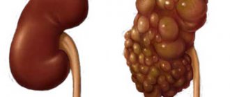
- Polycystic kidney disease. This is a disease of hereditary nature, characterized by the formation of numerous small tumors.
- Solitary (simple) cyst. The pathology is a single cavity formation. The disease develops predominantly unilaterally. A cyst of the left kidney is more often diagnosed. In women, the disease may not show clinical signs for a long time, but once it reaches a large size, the likelihood of developing complications increases.
- Parenchymal cyst. The neoplasm is localized in the thickness of the kidney tissue. Symptoms may not appear for a long time. If the tumor size exceeds 5 cm, surgical treatment is required.
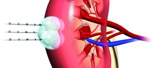
- Sinus cyst. It is a cavity formation localized in the sinus of the organ.
- Complex cyst. In this case, under one connecting capsule there is a small multi-chamber cavity with liquid inside. Treatment is exclusively surgical.
- Subcapsular cyst. The size of the formation is usually small. Complications occur extremely rarely and are treated by puncture using an ultrasound machine.
- Parapelvic cyst. The most common cyst is the right kidney. In women, this disease is diagnosed very rarely, mainly after the age of 50 years.
Kinds
As already noted, a kidney cyst can be acquired or congenital. There are other types of classification of neoplasms:
- By localization - one- or two-sided.
- According to location in the kidney - multilocular, intraparenchymal, cortical, peripelvic, sinus or parapelvic, subcapsular cyst.
- By filling – serous, infected, hemorrhagic.
- By quantity – multiple or single.
There is also a scheme according to Bosniak, implying the following division into types:
- simple cyst;
- education with a minimally complicated structure;
- inclusion resembling a benign tumor;
- pathological capsules that need to be removed only surgically;
- malignant tumors.
The last fifth group, after additional diagnostics of the third category, includes about 80% of identified cavities. In addition to cancerous structures, the cortical formation formed at the hilum is considered difficult and unpleasant in terms of development and treatment. Lack of therapy is fraught with complete urinary retention, acute pyelonephritis, and a serious condition of the entire body.
A multilocular or multilocular cyst with several internal septa does not require a radical approach. Its bulk is fibrous tissue, the key reason being improper formation in the embryonic period. You need to know that the pathology is extremely dangerous for newborns with bilateral damage.
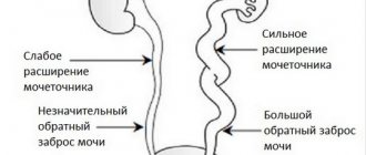
Sinus cysts are mostly filled with serous fluid with the presence of blood impurities. As in the previous case, such structures are often the result of abnormal intrauterine development. However, an acquired character cannot be excluded against the background of impaired evacuation of urine from the body. If a vessel does not communicate with the main circulatory system appears near the kidney, it begins to actively fill with fluid. The result is a formed parapelvic cyst.
In this video, Elena Malysheva in the program Live Great talks about cysts on the kidneys:
Establishing diagnosis
The disease is detected and subsequently confirmed exclusively by instrumental and laboratory examination. The latest diagnostic option includes:
- Analysis of urine.
- Blood test (increased erythrocyte sedimentation rate is a clear sign of the onset of an inflammatory process in the body).
- Biochemical blood test (changes in creatinine levels indicate the development of renal failure).
Instrumental diagnosis of kidney cysts in women implies:
- Ultrasound (allows you to detect the presence of a cavity formation).
- CT and MRI (the study helps to accurately recognize the location of the cyst and its size).
- Excretory urography (x-ray examination using a contrast agent).
What is the Bosniak classification of renal cysts?
Signs of malignancy of renal tumors are established by diagnosing magnetic resonance imaging (MRI) or computed tomography (CT). Classification of suspicious formations according to Bosniak gives the doctor the opportunity to choose the appropriate treatment tactics or method of eliminating the tumor.
Regarding the determination of tactics for further treatment of cysts, the Bosniak classification is as follows:
- Neoplasms of types 1, 2 (in relation to treatment, they are to be ignored)
- Cysts category 2F (require additional examination and medical supervision)
- Type 3 cysts (need to be removed urgently, as they have a high risk of degenerating into cancer).
The most informative diagnostic method for differentiating kidney tumors is histological laboratory examination.
Conservative treatment
Drug therapy is prescribed only to reduce the unpleasant symptoms of kidney cysts in women (pain, high blood pressure). Drugs are recommended to destroy the infection and normalize the salt balance in the body. Angiotensin-converting enzyme inhibitors are used to lower blood pressure. Long-term courses of antibiotics are prescribed (Ciprofloxacin, Tetracycline, Levomycetin).
Prevention
There are no specific preventive measures to prevent peripelvic renal cysts, but general recommendations will help reduce the risk of its formation:
- Once a year, undergo an ultrasound of the kidneys.
- Avoid hypothermia.
- Increase immunity.
- Treat all concomitant diseases in a timely manner.
- Stop smoking and drinking alcohol.
- Avoid lower back injuries.
- Eat properly and nutritiously.
Source: TvoyaPochka.ru
Surgery
For simple cysts, that is, of an uncomplicated nature, experts most often recommend drainage with subsequent emptying of the contents of the formation. The procedure is carried out under constant monitoring of an ultrasound machine. A needle is very slowly inserted into the cyst itself, through which all the fluid is pumped out of the formation. Then the cavity is treated with a special sclerosing substance, with the help of which its walls gradually stick together.
In some cases, when the cyst is large or oncological in nature, nephrectomy (removal of the organ) is performed.
Treatment through laparoscopic surgery is currently the most minimally traumatic way to remove a tumor. Initially, the surgeon introduces a gas substance into the operated field to expand it and increase the space for subsequent manipulations. Then the laparoscope and trocars are connected to the work. After removing the cystic formation, the doctor removes all the instruments and applies sutures.
Traditional medicine and kidney cysts in women
Treatment using the recipes of our grandmothers cannot be perceived as an alternative to conservative therapy or surgery. Herbal medicine should be used only as a supplement to the main course of treatment, and you must first consult with your doctor. Below we list the most common advice from traditional medicine.
- Pine nuts. You will need half a glass of nut shells and 0.5 liters of water. The shells must be boiled for an hour. The resulting infusion should be taken 70 ml three times a day before meals. The course of treatment is approximately 4 weeks.
- Burdock leaves. The leaves of the plant must be ground in a meat grinder, transferred to a jar and refrigerated. It is recommended to take this medicine one teaspoon twice a day for a month, then take a break.
- Yeast. In a three-liter vessel you need to put a tablespoon of the most common yeast, 30 g of grated elecampane root and two tablespoons of granulated sugar. This mixture should be left in a warm place for two days, and then consumed three times a day, 100 ml. The course of treatment is limited to one month.
Diet and its role in the treatment of the disease
For a pathology such as a kidney cyst in women, in addition to conservative therapy and surgical intervention, a special diet is also recommended. It implies special nutritional principles, including:
- Limiting the amount of salt consumed. This principle is recommended for those patients whose disease provokes renal dysfunction.
- Control the amount of fluid you drink. This rule applies to those representatives of the fair sex whose pathology is accompanied by the appearance of edema, symptoms of heart failure, and high blood pressure. If the neoplasm is not supported by such signs, the amount of fluid should not be limited.
- Refusal of junk food. This category includes fried and fatty foods, smoked foods, baked goods, and alcoholic drinks.
- Limiting protein intake. If these substances enter the body in large quantities along with food, the likelihood of releasing nitrogen metabolic products is very high. They are very toxic and have a negative effect on a weakened body.
Kidney cysts in women require adherence to the diet described above. However, dietary restriction is not the only sure way to combat pathology. Comprehensive health care and compliance with all recommendations from the attending physician is the key to a quick recovery.
Complications and consequences
In the absence of timely treatment, a kidney cyst in women can lead to the development of very unpleasant consequences, the main one of which is its rupture. In this case, its contents begin to pour into the abdominal cavity, which inevitably leads to its inflammation.
Suppuration is diagnosed much less frequently. In this kind of situation, patients note weakness throughout the body, increased pain in the lumbar area and a sudden increase in temperature. This condition absolutely always requires immediate surgical intervention.
Another complication of the disease is the degeneration of the neoplasm into a malignant tumor.