Classification
They are divided into several types according to location:
- Anarchidal . They are located between the meninges.
- Intracerebral . Located in the left or right hemispheres of the brain.
Also classified into:
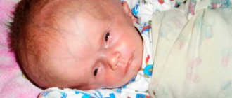
- Congenital . Diagnosed in infants in the first days of life.
- Purchased . Formed as a result of injuries and various inflammatory processes.
Features of the cyst
An epiphysis cyst is a neoplasm of a non-tumor nature, located on one of the lobes of the organ.
As a rule, its size is small, and there is no tendency to rapidly increase. Morphologically, such a formation is a small cavity that is filled with a specific liquid.
This formation of a benign nature usually does not affect the functioning of the pineal gland and extremely rarely affects the functioning of neighboring organs of the brain.
Important! In the initial stages, the disease is asymptomatic and most often can only manifest itself as a feeling of pressure in the affected area, which is accompanied by mild pain. Timely diagnosis allows you to avoid an increase in formation and further complications and manifestations
Timely diagnosis allows you to avoid an increase in formation and further complications and manifestations.
Cyst sizes
Small neoplasms, which are more common than others, do not exceed 5 mm in size.
They are not accompanied by special symptoms and do not provoke complications.
These cysts are mostly detected incidentally on MRI and respond well to treatment with medications.
As a rule, cystic neoplasms do not require special treatment. With this diagnosis, in most cases, only systematic monitoring is necessary.
However, some people with such a cyst may experience rapid growth and, as a result, various somatic and neurological disorders.
Symptoms
- Frequent headaches, migraines.
- Daily dizziness.
- Feeling of fullness in the head.
- Deterioration of vision. A person sees blurry objects around him.
- Multiple sclerosis.
- Hearing loss. Hearing is getting worse. A person constantly experiences tinnitus.
- Periodic loss of consciousness. The patient may faint.
- Nausea and vomiting.
- Epileptic seizures.
- Trembling hands or feet. Very common among all patients.
Diagnostics
The first diagnostic method is taking an anamnesis. The doctor collects information about previous illnesses and injuries. The patient must describe in detail the disturbing symptoms.
The danger of pathology lies in the difficulty of diagnosing it in the early stages. The person and the doctor may mistakenly attribute the symptoms that appear to related diseases. To make an accurate diagnosis, the doctor prescribes additional diagnostic methods.
- Magnetic resonance imaging and ultrasound examination. The research results will allow you to see the location of the formation, its size, and structure. It is also determined how strong the compression of surrounding tissues is or its absence. Based on the available results, the nature of education is determined. To monitor the formation, MRI is performed several times. The doctor notes the growth dynamics and possible structural changes.
- The potential for convulsive readiness of the brain can be detected by an electroencephalogram. Based on the results of the study, the specialist makes a forecast of the further development of the pathology and selects treatment.
- Neurosonography is used to examine children under 2 years of age. It is as informative as possible and safe for the child’s health.
In order to see the most complete clinical picture of the course of the disease, cerebrospinal fluid sampling is prescribed. Its results make it possible to determine the presence or absence of an inflammatory process or a clearing infection.
Histological examination is necessary to determine the nature of the cystic formation. Depending on whether it is malignant or benign, treatment is selected. The patient also undergoes a blood test when there is a suspicion of the development of multiple sclerosis and other infectious brain diseases.
Causes
Congenital neoplasms occur as a result of the following reasons:
- Penetration of infections into the fetus through the blood . The expectant mother may suffer from various sexually transmitted infections. To avoid this, it is necessary to check the blood of all pregnant women for hidden infectious diseases.
- If the pregnant woman took various antibacterial medications . Taking certain medications can trigger this disease. It is especially dangerous to get sick at the beginning of pregnancy. At this time, all the most important organs of the future human being are formed.
- Rh factor . Rhesus conflict may occur in both mother and fetus. When the mother is Rh negative and the child is Rh positive. This can lead to a congenital brain cyst. To avoid this situation, expectant mothers must donate blood for antibodies.
- Fetal hypoxia . Oxygen starvation of an unborn child is very dangerous. Expectant mothers need to walk in the fresh air for two hours every day.
- Birth injury . The fruit is often large in size. As it travels down the birth canal, injury can occur.
- took alcohol, drugs or smoked during pregnancy . As a result, the child will almost 100% be born sick. A young woman should not drink strong alcoholic drinks or smoke cigarettes. Drugs should be completely excluded from the life of any person. Otherwise the child will be a freak.
An acquired cyst occurs as a result of the following reasons:
- Trauma . Receiving a traumatic brain injury leads to the development of a tumor. Trauma can happen at any age.
- Transport accidents are especially dangerous . Car accidents lead to brain injury. After some time, it may turn out that a person has a brain cyst.
- Sports injuries . Hitting the head with a ball in football, volleyball, or basketball can lead to the disease.
- Inflammatory diseases . Exposure to various inflammatory processes can provoke this disease. Encephalitis, meningitis, abscess - all these are dangerous diseases that can cause the formation of a cyst.
- Post-stroke complication . A stroke in many cases will lead to the growth of a tumor. Vascular disorders lead to this disease.
- Subanarchoidal cerebral hemorrhage.
- Brain infarction . It is often observed in older people.
- Subepidermal ischemia . Violation of venous outflow is very dangerous.
- Hydrocephalus.
Prognosis and prevention
The prognosis for this disease, despite its seriousness, is very optimistic and life after the operation is quite normal. With timely treatment, the patient recovers completely. Naturally, if there is no treatment or it is of poor quality, certain consequences may occur, and in especially advanced cases, even death.
Normally, the patient recovers within several weeks.
As for preventive measures, there are no measures aimed at reducing the risk of cyst development. However, there are general recommendations that will help not only reduce the risk of arachnoid cyst formation, but also other tumors, including:
- all bad habits (especially smoking) are contraindicated for expectant mothers, as they can provoke oxygen starvation of the fetus, and this is not good;
- healthy eating;
- compliance with the work and rest regime;
- avoidance of stressful situations;
- maintain normal cholesterol levels;
- blood pressure control (upper and lower);
- timely and high-quality treatment of any inflammatory processes in the body;
- regular preventive examination by a doctor.
As for the consequences for later life, they may occur in the case of an uncontrolled course of the disease, including:
- Neurological problems (loss of sensitivity, problems with internal organs at the level of the nervous system).
- Epilepsy and seizures.
So, an arachnoid cyst is a serious and unpredictable disease, treatment or observation, which should only be carried out under the supervision of a specialist. Don’t aggravate your situation, get proper treatment from doctors!
With timely diagnosis and treatment of arachnoid cysts, the prognosis is very favorable. The main risks associated with the development of an arachnoid cyst are the increased compressive effect of the cyst body on the brain centers, resulting in disturbances in body functions, as well as rupture of the cyst.
If a brain cyst is detected early, the prognosis is favorable. The main risks associated with arachnoid formation are compression of the brain centers, after which disturbances in the functioning of the body occur. After cyst removal, speech, hearing, or vision problems are sometimes observed. If diagnosis is delayed, cyst rupture, hydrocephalus, and death are possible.
Tags: intrasellar, cyst, such
About the author: admin4ik
« Previous entry
Consequences
- The cyst is growing . Over time, if left untreated, the cyst will increase in size. Urgent surgery is required. Complete removal of the cyst is indicated in this case.
- Squeezing . The cyst will put pressure on neighboring parts of the brain. The person will feel a throbbing pain in the temples.
- Vessel rupture . A vessel may rupture and bleeding into the brain may occur.
- Epileptic seizures . A large cyst can cause epilepsy.
- Impaired mental function . A child with a congenital cyst will experience developmental delays.
- Oligophrenia . Children, if left untreated, will become oligophrenic. There will be complete degradation of personality. The child will not develop. He will have to study in a special educational institution.
- Increased intracranial pressure . Often people do not understand why they have intracranial hypertension. Unfortunately, only through MRI can a correct diagnosis be made.
- Stroke . A cyst can cause a stroke. Especially in people suffering from hypertension.
- Paralysis . The compressed areas of the brain will not be able to function, and they will die. The person is paralyzed. Can immobilize the right or left hand. The entire half of the body may be lost.
Why is a brain cyst dangerous?
Classification of brain cysts is made by specialists. Doctors divide liquid neoplasms:
- Depending on the location: intracerebral and arachnoid, located in the intercerebral membranes.
- By origin: congenital and acquired.
- According to the characteristics of the damaged tissues: with the content of cerebrospinal fluid - arachnoid, resulting from scarring - colloidal, from the germ cells of the face - dermoid, from cells intended for the formation of hair, nails - epidermoid, on the tissues of the pineal gland - peneal.
Each type has its own differences: in symptoms, location, nature of growth. Based on the type of pathology identified, the neurologist plans treatment or complete elimination of the tumor.
Arachnoid
A benign formation in the form of a bubble filled with fluid, located between the brain tissue and the arachnoid membrane, is called an arachnoid cyst.
Most often diagnosed in males, it may not manifest itself for a long time, not grow, or cause discomfort. A congenital arachnoid cyst consists of tissue from the arachnoid membrane of the brain. Formed as a result of an illness - from scar tissue.
Diagnosis is carried out using MRI. Surgery is used to treat dynamic formation. If it does not grow, does not pose a health threat, does not damage the membranes of the brain, the person is simply under medical supervision and takes medications that help improve cerebral circulation.
Retrocerebellar
When fluid accumulates in places where brain cells have died in the cerebellum area, a retrocerebellar cyst of the brain can form. There are several factors that provoke the development of education:
- Stroke, post-ischemic condition.
- Circulatory disorders.
- Trauma to the skull and brain.
- Encephalitis, meningitis.
Based on what caused the damage to the gray matter, treatment is selected. If the brain cyst grows, shunting or craniotomy is recommended. If the patient refuses surgical intervention, then such consequences as asthenic syndrome, seizures, epilepsy are possible.
Subarachnoid
Retrocerebellar cyst of the brain has a subtype - subarachnoid formation. It looks like a bubble with liquid and is located between the structures of the brain. To diagnose the disease, the patient undergoes an MRI.
The usual treatment is taking medications that normalize vascular tone, vitamin therapy. Surgical intervention is performed only if the subarachnoid formation grows, puts pressure on the gray matter, hemorrhage is observed in it, and the functioning of the blood vessels is disrupted.
Pineal
The epiphysis cyst of the brain is located on the pineal (pineal) gland, which is responsible for endocrine function. A fluid-filled formation grows if the outflow of secretion synthesized by the pineal gland is disrupted.
Experts name several reasons for the development of the disease:
- The excretory channel becomes clogged due to viscous secretion. May occur due to genetic predisposition. The sac of liquid rarely reaches a large size and does not manifest itself symptomatically.
- The pineal gland is affected by echinococcus. Infection can occur due to human contact with farm animals and dogs. The tumor grows, puts pressure on the brain tissue, causes discomfort, headache, vomiting, and blurred vision.
A cyst that occurs due to a hereditary predisposition is usually not treated. A person simply undergoes an examination once a year. If the neoplasm provokes an infection with echinococcus, the cause of the disease is first eliminated. Small formations are treated with medications, large ones are removed surgically.
Liquor
The brain is protected from contact with the skull by cerebrospinal fluid (CSF). A cyst in the brain filled with cerebrospinal fluid is called a cerebrospinal fluid cyst. It can be an arachnoid, penial, or retrocerebellar cyst - it depends on the location of the tumor.
Neurologists offer surgical removal of formations that cause significant discomfort to the patient. An asymptomatic cerebrospinal fluid lacunar cyst that is not growing is simply observed with periodic MRI scans of the brain.
Porencephalic
Porencephalic cyst of the brain is quite rare, most often in children. Intrauterine infections can cause the development of the disease:
- Cytomegalovirus.
- Herpes.
- Colds.
Or diseases that the newborn suffered after being born. Several formations may appear. The symptoms are severe, manifested in convulsive seizures, hydrocephalus, mental retardation, and loss of vision.
Treatment usually involves relieving symptoms. Surgeries are not performed due to low efficiency. Most often, children suffering from porencephalic pathology do not live to see one year of age.
A choroid plexus cyst of the brain may be detected in a fetus at 6-7 months of intrauterine development. On an ultrasound, the doctor can see fluid-filled bubbles in the network of small blood vessels in the brain.
Such a neoplasm is not pathological and resolves without treatment by the birth of the child. Doctors pay attention to it, since the appearance of bubbles on the choroid plexuses may indicate the development of Down syndrome in a newborn.
The doctor does not always recommend removing a brain cyst. If the liquid formation does not grow, does not cause any discomfort to the person, or does not affect health, then therapy consists only of observation.
Medication is prescribed if the cyst found on the cerebral cortex is of average size and does not develop dynamically. Some patients get scared: “I have been diagnosed with a cyst, and the doctor suggests taking pills for atherosclerosis.” First of all, the specialist prescribes drugs that eliminate the cause of the disease:
- For atherosclerosis - drugs to lower cholesterol levels.
- If vasoconstriction is detected, antispasmodics and vasodilators are prescribed.
- If hypertension is detected, it is recommended to take medications to normalize blood pressure.
In addition, medications are prescribed to help resolve vascular adhesions, improve blood circulation, and restore vascular tone. If your doctor has prescribed complex treatment for you, you strictly follow the recommendations, regularly take medications in sufficient dosage, the fluid formation in the brain tissue may simply resolve.
Why might it be necessary to completely remove a brain cyst? If the disease occurs with significant painful symptoms, the amount of fluid in the cyst increases, and the neoplasm occupies a large area, surgery is necessary to eliminate the discomfort. Surgeons use several methods to eliminate cyst formations:
- Endoscopy. Fluid is removed from the cyst through a puncture. A highly effective method, but not suitable for all types of cysts.
- Shunting. This method involves a puncture in the skull and installation of a drainage tube. Fluid from the cyst drains through the tube. The risk of infection during such an operation is high.
- Radical method. Trepanation of the cranial cavity is performed, the cyst along with its contents is removed. The method is traumatic, but you can be sure that the tumor is completely eliminated.
The postoperative period can take 4 days or more; the length of hospital stay depends on the patient’s condition, the type of operation performed, and the type of cyst.
We suggest you read: Frontal scalp cyst treatment
The cyst may require immediate treatment. In most cases, this is not required, especially if there are no complaints, clinical symptoms and the cyst does not increase in size. If it actively grows and compresses the cerebral cortex, this is an indication for medication or even surgical intervention.
Treatment is mainly aimed at treating the underlying disease that provoked its formation. If this is arachnoiditis, then treatment necessarily includes the use of absorbable drugs such as caripain or longidase.
If there is a circulatory disorder, treatment includes reducing blood clotting by administering anticoagulants, lowering blood cholesterol levels and normalizing blood pressure (treatment with antihypertensive drugs).
Treatment of pathology will require the prescription of nootropics, which help saturate brain cells with oxygen and glucose, which is very important. Treatment including antioxidants is recommended. The latter increase the resistance of brain cells to high pressure.
For dropsy and impaired outflow of intracerebral fluid, it is advisable to include furosemide in treatment. If the cause of the disease is infection or autoimmune pathology, then treatment includes the prescription of immunomodulators and anti-inflammatory drugs. Surgical treatment is possible. In this case, endoscopic operations are better suited.
As mentioned above, this disease can also occur in children. But in this population group, the disease appears as a result of an infectious disease suffered by the mother during pregnancy. Often the cause may be a birth injury.
There is no need to exclude an intracerebral cyst (cerebral). Its causes are trauma during childbirth as a result of unqualified assistance, a narrow maternal pelvis, exostoses, and so on. They are also possible in teenagers.
The location can be different: in the cerebellum, corpus callosum, trunk, nuclei, hemispheres. The fluid replaces the lost (dead) part of the brain. Adults have other reasons. Treatment of cysts in children is similar to adults.
Treatment for a cyst is chosen depending on the cause of its formation. A non-dynamic brain cyst does not require treatment. If a dynamic formation is detected, treatment may be as follows:
- Conservative treatment. It involves the use of medications, the action of which is aimed at eliminating the cause of the cyst. Medicines that restore blood supply and resolve adhesions can be used. Antibacterial, immunomodulatory, and antiviral drugs are also sometimes used - the need for them arises in the case of infections and autoimmune diseases.
- Radical treatment. It involves removing the cyst through surgery. For this purpose, craniotomy, endoscopy, and cyst shunting are used. However, in the latter case, the risk of developing an infection increases when the shunt remains in the skull for a long time.
A brain cyst is a benign formation
Only a doctor can prescribe correct and effective treatment!
Recommendations
Currently, diagnostics are carried out using CT and MRI . The resulting images provide a complete picture of the disease . If the above symptoms occur, you should consult a neurologist. Only a specialist will prescribe the necessary examination and make a diagnosis. It is necessary to follow the doctor's recommendations and take prescribed medications. If conservative treatment is unsuccessful, the neurosurgeon will prescribe surgery. But this happens only in extreme cases.
Treatment
If the disease is asymptomatic and there are no signs of cyst growth, treatment is not required, but dynamic observation by a neurologist and drug treatment of the disease that led to the formation of the cyst are prescribed. For example, the prescription of antibacterial, antiviral agents, immunomodulators, drugs that resolve adhesions, and restore blood supply.
If there are signs of enlargement of the cystic cavity, with pronounced symptoms, they resort to surgical methods of treatment, which can be divided into 3 groups.
- Radical operations, for example, craniotomy followed by removal of the cyst and its walls. They are characterized by good efficiency, but increased trauma.
- Bypassing the cyst cavity using a drainage tube; after evacuation of the contents, the walls of the cavity collapse. The disadvantage of this method is the increased risk of infection.
- Endoscopic methods, when punctures are used to remove the contents of the cyst. This is the least traumatic method, but it cannot be used for all types of formations.
Diagnostic features
Standard methods (blood tests, etc.) and even functional diagnostic methods (audiometry, viziometry, perimetry, ophthalmoscopy) are not reliable here.
- Ultrasound may show altered echogenicity, but this is not enough.
- Ultrasound is also used to identify certain types of cysts during the prenatal period.
- Encephalography can detect high blood pressure.
On MRI, a cystic formation can be visually localized with high accuracy, and the type of cyst can also be determined. However, it is better to conduct a study as part of differential diagnosis with contrast. Because only tumors are prone to accumulation of contrast agent.
Additionally, Doppler ultrasound can be performed to assess the condition of the vessels of the central nervous system. In newborn infants, the cyst is detected using neurosonography.