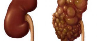
Angiomyolipoma of the left or right kidney - what is it? Angiomyolipoma is a benign kidney tumor, which is most often encountered in the practice of urologists at the Yusupov Hospital. Histologically, it is represented by thick-walled blood vessels, smooth muscle fibers and mature adipose tissue in various quantitative ratios. Angiomyolipoma of the kidney has a code in ICD-10 D30.
The Yusupov Hospital has created all the conditions for the treatment of patients with angiomyolipoma of the kidney:
- The wards are equipped with forced-air ventilation and air conditioning;
- The surgery clinic is equipped with the latest diagnostic equipment from leading American and European companies;
- Nephrologists and urologists use innovative methods for treating renal angiomyolipoma;
- The medical staff is attentive to the wishes of patients.
Severe cases of the disease are discussed at a meeting of the Expert Council. Candidates and doctors of medical sciences and doctors of the highest category take part in its work. Leading nephrologists collectively make decisions regarding the management of patients with renal angiomyolipoma.
Forms of pathology
There are two forms of this pathology. The name of the form indicates its feature:
- Congenital (hereditary). It affects two kidneys at once. The pathology consists of multiple formations resulting from tuberous sclerosis.
- Acquired sporadic (isolated). Accounts for 80-90% of cases of diagnosing angiomyolipoma. It affects one kidney.
If an angiomyolipoma of the kidney is detected, you must strictly follow all the doctor’s instructions. Neglecting health or self-medication can lead to dire consequences.
Is it possible to prevent the appearance of a tumor?
Prevention of angiomyolipoma consists of regular medical and genetic consultations, timely treatment of all concomitant diseases, as well as immediate consultation with doctors when the first signs of the disease appear.
Angiomyolipoma is a severe and progressive disease. If not properly treated, it can cause serious consequences, including disability and death. Persons suffering from pathologies of the urinary system should know what it is - renal angiomyolipoma and how the disease can harm health.
Factors of occurrence
The nature of the occurrence of kidney AML is still not fully understood. The reasons that provoke the occurrence of neoplasms are different. Often the disease develops under the influence of factors such as:
- Chronic or acute renal pathologies.
- Pregnancy. It is considered the most common cause. During pregnancy, a change occurs in a woman's hormonal background; female hormones - estrogen and progesterone - are actively produced, which provoke the development of a tumor. It is because of the action of these hormones that women are 4 times more likely to suffer from this pathology than men.
- The presence of similar tumors in other organs.
- Genetic predisposition.
Causes of AML
In most cases, the pathology is acquired (80-90%). Less commonly, the cause is a hereditary disease - tuberous sclerosis, which provokes the appearance of benign tumors in human organs. A gene with a defect is passed on to a child from the father or mother with a probability of 50%. Tuberous sclerosis is characterized by damage to small angiomyolipomas of the kidneys.
Angiomyolipoma is a hormone-dependent tumor, so it is detected in young women during pregnancy, due to an increase in estrogen levels. And it can be detected in them during an ultrasound scan. The tumor is not dangerous for the expectant mother and fetus, but during the period of pregnancy the tumor grows more intensively. In men, AML is diagnosed at an older age. The disease can provoke inflammatory diseases, other tumors, and kidney failure.
In rare cases, angiomyolipoma is found in the pancreas, on the skin or in the adrenal glands, although this pathology is characteristic of the kidneys. Angioma is a benign tumor formed by intertwined blood vessels (hemangioma) or (rarely) lymphatic vessels (lymphangioma). Kidney angioma, although a benign neoplasm, can cause unpleasant complications and there is a danger of its degeneration into malignant.
Main symptoms
Angiomyolipoma of the kidney forms and develops asymptomatically. The tumor grows quickly, but the vessels feeding the angiomyolipoma develop more slowly than the muscle tissue and, as a result, rupture. The onset of bleeding is accompanied by the following symptoms:
- There is constant pain in the lower back;
- there are sudden changes in blood pressure;
- loss of strength, dizziness, fainting;
- pale skin;
- blood in urine.
If these signs occur, it is necessary to immediately take the person to the hospital for diagnosis and treatment. The degree of danger depends on the size of angiomyolipoma, since a large tumor can rupture an organ. As a result, internal bleeding occurs, and the tumor grows into the nearest lymph nodes. This leads to the occurrence of multiple metastases.
Clinical picture of the disease
Symptoms of angiomyolipoma appear the same for the left and right kidneys and depend on concomitant pathologies and mutations in the affected organ.
With a single isolated neoplasm, the patient feels the following:
- Pain on the side of the abdomen. Depends on which kidney is affected by the disease.
- On palpation of the peritoneum, swelling is detected.
- When urinating, blood is detected.
Vascular-muscular lipoma takes a long time to develop and does not cause health problems. When the tumor size exceeds 4 cm, secondary changes occur in the parenchyma. Kidney functions are impaired. The patient experiences certain symptoms:
- Pulling pain in the peritoneum;
- Loss of body weight;
- Arterial hypertension;
- Increased fatigue;
- Lethargy.
Symptoms do not appear suddenly. Unpleasant sensations continue to grow for a long time. From the abdominal area the pain spreads to the side and lower back. The patient notes pressure surges. Hypertension is life-threatening because the tonometer readings reach high numbers. Back pain is often confused with osteochondrosis, so the patient does not see a doctor until his health suddenly deteriorates. The first trip to the hospital among most patients occurs after the appearance of blood in the urine.
If nothing is done about the tumor, the symptoms increase and complications develop. There are hemorrhages in the organ and in the surrounding space. When a tumor ruptures, the patient experiences:
- Acute pain;
- Feeling of fear;
- Nausea accompanied by vomiting;
- Paleness of the skin;
- Coldness in the extremities;
- A sharp decrease in temperature;
- The pressure drops sharply and rapidly decreases;
- There is weakness;
- Fainting;
- Urine filtering stops;
- Liver and heart failure develops;
- Brain functions are impaired, consciousness becomes confused.
There is a danger of developing peritonitis; a burst organ requires emergency surgery. With total damage, sometimes it is necessary to remove the kidney.
Is it dangerous for the patient's life?
The main danger to life of this disease is the rupture of angiomyolipoma. The cause of the rupture is a difference in the development of blood vessels and tumor tissue. In rare cases, the rupture occurs at the initial stage of development. Internal bleeding begins and urgent hospitalization is required. If the tumor grows significantly, it can cause rupture of the kidney parenchyma. Over the past 10 years of studying the disease, it has been found that this phenomenon can change and become a malignant tumor. In this case, the danger to life is comparable to any oncology. If treatment is not started in time, the pathology can cause liver dysfunction.
Possible complications
The growth of AML to a significant size or advanced cases of pathology can lead to:
- To the rupture of the hamartoma shell along with the vessels. The blood partially spills into the abdominal cavity, which is accompanied by pain and a drop in blood pressure. With massive bleeding, a state of shock develops, which is indicated by severe pain, a sharp decrease in blood pressure, tachycardia, nausea, and pallor of the skin;
- To kidney failure. It occurs due to the pressure of focal formation on the kidney parenchyma, which impairs the functioning of the organ;
- Towards malignancy. Malignancy of angiomyolipoma is rare and large neoplasms are more susceptible to this process.
Diagnostics
An ultrasound examination determines pathology by identifying compactions against the background of healthy renal parenchyma.
The earlier the pathology is diagnosed, the greater the chances of a full recovery. Since the pathology most often affects one organ, the result of the diagnosis is angiomyolipoma of the right kidney or the left. The following methods are used to detect tumors:
- Ultrasound. Determines the presence of seals.
- MRI and CT. Identifies areas of low density tissue (adipose tissue).
- Laboratory blood tests indicate the general condition of the kidneys.
- Ultrasound angiography. Detection of pathologies of renal blood vessels.
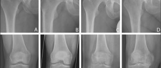
- X-ray diagnostics. Shows the condition of the organs and ureters, the presence of changes in structure and functioning.
- Biopsy. To exclude the possibility of developing a malignant tumor, a particle of the tumor is taken to study its nature and characteristics.
How is renal angiomyolipoma diagnosed?
It is important to note that most tumors are sporadic, while a minority of cases are associated with tuberous sclerosis. Diagnosis of renal angiomyolipoma includes the following:
Complete physical examination with medical and family history assessment.- Native radiography of the abdominal cavity.
- or CT with contrast of the abdominal cavity. These x-rays create detailed three-dimensional images of structures inside the body.
- MRI scan of the abdomen: Magnetic resonance imaging (MRI) uses a magnetic field to create high-quality images of specific parts of the body, such as tissue, muscles, nerves and bones. Tomograms in 100% of cases show signs of a tumor.
- Ultrasound scan of the abdominal cavity.
- General urine analysis.
- Blood biochemistry: urea and creatinine.
- Intravenous urography: after the administration of a contrast agent, a series of photographs are taken to obtain an image of the structure of the kidneys. The method allows you to assess the functional ability of the urinary organs.
- Angiographic studies of the tumor.
Invasive diagnostic procedures:
- Diagnostic (exploratory) laparoscopy: a special device with optics is inserted through a small hole in the abdomen.
- Diagnostic (exploratory) laparotomy: an incision is made and the abdominal cavity is examined.
Please note
The above methods can be used for initial diagnosis, but verification requires a morphological examination of the tumor tissue, which makes it possible to establish a final diagnosis and determine treatment tactics. It is preferable to obtain tissue by open biopsy.
Sometimes a pathologist may perform special tests that include immunohistochemical analysis, molecular testing, and electron microscopic examination to make a final diagnosis.
Differential diagnosis is carried out to exclude other types of tumors:
- carcinoma;
- oncocytoma;
- leiomyoma.
Important
Many clinical conditions have similar signs and symptoms, so additional testing may be performed as indicated.
Treatment and prognosis
Angiomyolipoma is a benign neoplasm located in a connective tissue capsule. Therefore, timely therapy has a favorable prognosis. After surgery, the patient recovers completely. However, surgery is a last resort; drug therapy in combination with diet is immediately used.
Observational tactics
The therapy program is developed by the doctor individually for each patient based on the diagnostic results. The number of tumors, their size and location are taken into account. If the patient is diagnosed with “Angiomyolipoma of the left kidney” and the tumor is less than 4 cm in diameter, then surgical intervention is not required, since small tumors develop slowly, without complications. Observation is prescribed, the patient periodically visits the doctor, and an ultrasound or computed tomography is performed once a year.
Surgery
If the diagnosis revealed a unilateral angiomyolipoma, the diameter of which is more than 5 cm, and the second kidney is functioning normally, surgery is prescribed. If the tumor develops quickly, the likelihood of complications increases. At any time, the disease can result in bleeding, blood poisoning and death. The tumor is removed to prevent this.
Kidney resection
Kidney resection involves removing only part of the organ along with the tumor. There are 2 types of this operation:
- Classical. A large incision is made in the lumbar region to access the organ.
- Laparoscopic. Several small incisions are made.
Enucleation
During the operation, the tumor is “husked” from the organ. Enucleation allows the tumor to be removed relatively easily if it is encapsulated, with little blood loss. This is a new way to remove AML from the kidney, as a result of which the kidney itself does not undergo any changes. This method is applicable only in the presence of a benign tumor.
Embolization
Embolization involves the injection of a special drug into the blood vessels feeding the tumor, which causes their blockage. The procedure is carried out under x-ray control. As a result, surgery is much easier. In some cases, embolization eliminates the need for surgery.
Cryoablation
This method is used to remove small angiomyolipoma by applying heat to it. The effect of the procedure is comparable to surgery with fewer contraindications and complications. In addition, the advantage of cryoablation is the minimal degree of intervention in the patient’s body, a short rehabilitation period and the possibility of a repeat procedure.
Nephrotomy
If the tumor increases significantly (more than 7 cm), the doctor is forced to perform a nephrotomy - complete removal of the affected kidney. This method is used if it is impossible to save the organ due to irreversible changes or a high risk of severe complications. It is important that the second kidney functions fully. The operation is performed under general anesthesia. The open (classical) method or laparoscopy is used.
Surgical methods
- Resection. The tumor and adjacent affected areas are removed while preserving the organ itself.
- Nephrectomy. Complete removal of the kidney and lymph nodes in case of malignant degeneration of tumor cells. Nephrectomy is followed by a course of chemotherapy.
- Enucleation. A benign neoplasm, enclosed in a dense capsule, is “husked” (removed) with minimal trauma to the organ during surgery.
- Cryoablation is a minimally invasive method of freezing tumors up to 4 cm in size with argon and helium.
Diet and nutrition
If renal angiomyolipoma is diagnosed, you should strictly adhere to a special diet that inhibits the development of the tumor and prevents exacerbation of the disease. In this case, it is necessary to minimize salt intake. Dietary rules for angiomyolipoma boil down to complete abstinence from alcoholic beverages and coffee, eating small portions 6 times a day, and consuming at least 1.5 liters of fluid daily. It is allowed to consume low-fat dairy products, vegetable broths, lean soups/borscht, low-fat meat, cereals, pasta, eggs, vegetables, and steamed cutlets. Weak tea is allowed. For sweets, dried fruits, baked apples, honey, and jam are allowed.
If you have angiomyolipoma, you should avoid the following products:
- broths (meat, fish);
- fatty meat/fish;
- smoked, salted products;
- legumes;
- spices, herbs, marinades, sauces;
- horseradish, garlic, onion, radish;
- parsley, spinach, sorrel.
What to do to avoid illness
Prevention of AML of the kidney comes down to eliminating factors from life that may contribute to its occurrence.
You just need to adhere to the well-known rules:
- during pregnancy, strictly follow the recommendations of the doctor who is seeing the woman, follow a diet and exercise regimen;
- monitor personal hygiene;
- prevent hypothermia of the body;
- avoid gaining excess weight.
To prevent extra pounds from causing kidney problems, it is recommended to follow a certain diet. There is no special diet for angiomyolipoma; it is enough just to limit the consumption of fatty, salty foods, foods containing various food additives and carbonated drinks.
If the patient has already been diagnosed with a small tumor, he should regularly visit a medical facility to monitor its condition and undergo all necessary tests.
Treatment with folk remedies
It is believed that the use of folk remedies for kidney angiomyolipoma is ineffective and can provoke serious complications, and wasting time on self-medication further aggravates the situation. There are a number of techniques that can be used in parallel with conservative therapy, but first you need to consult a doctor.
The following folk remedies are used:
- decoction or alcohol infusion of nut shells;
- decoction of calendula flowers, wormwood infusion;
- pollen;
- decoction of pine cones with honey.
The occurrence of renal angiomyolipoma cannot be predicted. Thanks to routine medical examinations, it is possible to diagnose pathology at the initial stage of development, prevent its progression and quickly eliminate it. Refusal of necessary therapy, or treatment with folk remedies without a doctor’s prescription leads to serious complications and death.