How and when are they formed?
The human kidney is formed as early as 1 month of pregnancy.
During the formation process, the following types of buds are distinguished:
- pronephros;
- mesonephros;
- metanephros.
The initial stage begins at the 3-4th week of pregnancy. At this time, it does not function: there are no glomeruli, and the tubules are not connected to the vessels. A renal capsule is formed, the shape of which is similar to a ball. The pronephros is quickly reduced and moves to the 2nd stage. Then the kidney becomes the only excretory organ in the child. It already performs functions, has gates, glomeruli and tubules. The vessels connect to two ducts: Wolffian and Müllerian, which will turn into the genital organs. The final stage of formation begins at the 4th-5th month. The function of the organ is similar to that of an adult.
Stabilizing duty
Automatic self-regulation of the body occurs on the basis of coordination of dynamic reactions, and is aimed at maintaining optimal balance. In medical reference books, homeostasis has the Latin definition homoios statis (a similar state).
This is a moving “living” balance, maintained by balancing and stabilizing the systemic norm under the influence of external and internal factors.
The homeostatic function of the kidneys is to adapt and maintain internal constancy to the changing influences of the environment. The excretory sphere is triggered automatically when the body is dehydrated (dehydrated):
- urinary excretion decreases;
- the concentration of decay elements in the urine increases;
- metabolism changes.
The paired urinary organs stimulate diarrhea (urinary excretion) in the process of increasing the volume of circulating blood, reducing the level of blood pressure blood flow. In this way, the kidney glands directly influence the normalization of blood circulation and the general condition of the body.
In the tubules - ducts, the acid balance in the bloodstream of the general circulation is stabilized.
A person’s daily diet significantly affects the percentage of acidification of urine produced. Protein nutrition causes an increase in uric acidity, as does physical muscle activity.
A plant-based diet has the opposite effect on metabolism and produces an alkaline reaction in secondary urine samples during laboratory testing.
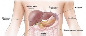
Location and anatomy of the human kidneys
The paired organ is located in the lumbar region behind the peritoneum.
The kidney is a paired organ that looks like a bean. Their anatomy is complex. Skeletotopy: the organs are located behind the peritoneal cavity in the lumbar area on the sides of the last 2 thoracic and first 2 lumbar vertebrae. Normally, the body of the left organ is located higher than the right one, which is explained by the placement of the liver. The height corresponds to the size of 3 lumbar vertebrae, width - 45-70 mm, thickness - 40-50. Both organs are joined by the renal vein and artery. Already purified blood moves through the veins, feeding them with oxygen and everything necessary. The vascular bed is well developed, there are straight and convoluted tubules.
Kidney membranes
The fibrous capsule protects organs from mechanical damage. Its structure is strong. The membranes of the kidney are easily separated from the organ. The presence of a fat capsule and fiber is normal. The layer of connective tissue fascia is formed by two membranes: the outer ball is connected by fibers to the fibrous capsule, and under the membrane is the renal cortex containing nephrons. The crust borders the pyramids. The parenchyma includes the medulla.
Protective stock
To prevent displacement of organs, kinks of blood vessels and ureters, there is a fixing device. The kidneys are located on a protective bed, the basis of which is adipose tissue. Intra-abdominal pressure is of great importance for the strengthening of organs. The kidney bed is formed by the quadratus, psoas minor and lateral transverse muscles, as well as the diaphragm.
Internal structure
The renal pelvis performs the function of transporting urine through the ureter to the bladder.
The medulla of the kidney forms 7 pyramids inside the organ. Each pyramid is attached to the pelvis with the help of a papilla. Urine passes through the ducts into small and large cups, where each cup passes urine through itself, ensuring the efficient functioning of the excretory apparatus. The renal pelvis is where the calyces deliver urine. The main gland of homeostasis, the pituitary gland, controls the functioning of the kidneys. When cut open, the structure of the human kidney is divided into 2 parts:
- shells;
- Malpighian bodies.
Renal nephrons
Taurus is a functional unit. The cortex contains more than 1 million nephrons, but a third of the total mass is active. The glomeruli are located in the medulla, and the main part of the organ consists of them. The corpuscles are arranged as clusters of vessels that filter the blood. The basement membrane does not allow large molecules and electrolytes to pass through. The size of the nephron is so small that it is impossible to see with the naked eye.
The number of nephrons depends on the age of the person: up to 40 years of age, 1% of the Malpighian bodies die off in a person every year, then the process slows down.
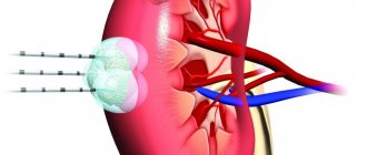
Blood flow system
When blood flow in the kidneys is impaired, the functions of many body systems suffer.
The organ filters fluids in the human body. The renal artery transports blood. It branches off from the aorta, and then divides at the hilum into interlobar vessels, arcuate arteries, and forms nephrons with a system of tubules. Kidney function depends on the pressure in the renal artery, which should be at least 70 mmHg. Art. When an organ is damaged, internal bleeding and hematomas occur.
Lymph movement
The lymphatic system cleanses the body of waste products of fungi, parasites and microorganisms. A network of vessels is located throughout the body and extends from each organ. The initial capillaries entwine the nephron capsules and tubules. Their lumen is larger than that of blood vessels. Further, the capillaries merge into interlobular, and then arcuate arteries and veins. Lymph from the organ enters the common thoracic duct. The lymphatic system of the kidney is considered a secondary link in reabsorption.
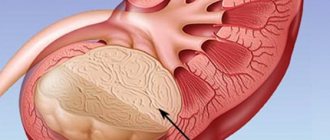
Common problems
The cortex is the outer part of the kidneys where urine is produced. With a long-term illness (chronic renal failure), if the organs work at less than 20% of their capacity, atrophy is detected.
Many diseases can affect the structure and function of all parts of the renal cortex.
The glomeruli are usually very susceptible to infections and autoimmune disorders (glomerulonephritis, SLE), and radioactive substances and some drugs can harm the tubules. When problems of this kind occur, the cortex can be damaged and ceases to fully cope with the purification or stops the filtration process altogether. These cases lead to a number of serious medical problems.
Main functions
The physiology of the kidneys is complex. The main task of the filtering organs is to cleanse the blood. The kidneys excrete water and water-soluble waste products. The reverse absorption and secretion system is responsible for the formation of urine and the support of mineral metabolism. Organs work continuously. The renal pelvis stores and removes urine. Other tasks include:
Organs are directly involved in the synthesis of calcitriol.
- homeostasis support;
- maintaining water-salt balance;
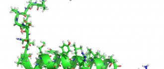
- synthesis of erythropoietin and calcitriol;
- nitrogen excretory, hydrouretic and osmoregulatory functions;
- urine formation;
- exchange of electrolytes: sodium, calcium and others.
The mechanism for reverse reabsorption of elements and water is a rotary-countercurrent system. It consists of the loop of Henle and collecting ducts. The proximal tubule contains a large number of mitochondria, which are responsible for energy production. The multiplying countercurrent system, due to the close contact of the loop legs, transports water, microelements and biologically active substances that affect the body back into the systemic bloodstream.
Diseases
There are a large number of kidney pathologies and among them:
- congenital;
- acquired.
Congenital
An unhealthy lifestyle during pregnancy can cause the development of kidney pathologies.
During the process of kidney formation, the anatomy may be disrupted, causing various types of changes to occur. The following pathologies are distinguished:
- violation of location and/or orientation;
- shape changes;
- fusion of organs - the upper segment unites;
- absence of an organ;
- the presence of an additional structure;
- improper tissue development;
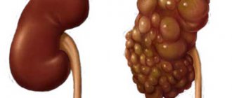
- polycystic disease
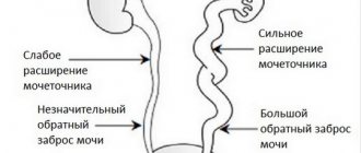
Congenital anomalies include narrowing and dilation of the ureters. Accumulating in the cups, urine cannot pass normally. When the ureteral valve does not function properly, urine from the bladder backs up into the ducts. Then pyelonephritis develops. The cause of congenital changes is the mother’s poor lifestyle during pregnancy or a hereditary predisposition.
Acquired diseases
The kidneys often get disrupted during pregnancy.
There are many kidney diseases. The table shows the most common ones:
| Pathology | Cause | Signs |
| Pyelonephritis | Inflammation of the parenchyma due to the proliferation of microorganisms | Lower back pain |
| Intoxication | ||
| General malaise | ||
| Fever | ||
| Changes in blood and urine tests | ||
| Glomerulonephritis | Autoimmune destruction of the kidney membrane | Similar to pyelonephritis |
| Diabetes insipidus | Lack of production or insensitivity to the action of antidiuretic hormone | Constant unquenchable thirst |
| Frequent and copious urination | ||
| Trophic skin changes | ||
| Urolithiasis disease | Mineral metabolism disorders | Renal colic |
| The appearance of blood and sand in the urine | ||
| Hydronephrosis | Extra valves | Lower back pain |
| Narrowing of ducts and structures | Increased frequency of urination | |
| Compression of the ureter | ||
| Blocking stones | Hematuria | |
| Chronic fatigue | ||
| Chronic form of renal failure | Infectious diseases | Lethargy |
| Muscle twitching | ||
| Metabolic disorders | Tendency to bleed | |
| Congenital pathologies | Frequent acute respiratory infections | |
| Decreased concentration function |
Excess weight contributes to organ hypoactivity.
The treatment regimen and main directions of treatment depend on the type and causes of the pathology. If a person is overweight, the cortex of the kidney becomes covered with fat, which reduces the functionality of the organ. A few facts about diseases:
- Diabetes mellitus provokes vascular changes. Glycogen accumulates in them.
- Rare infectious pathologies include kidney tuberculosis.
- Purulent lesions can provoke the formation of carbuncles and abscesses.
- Kidney injuries are accompanied by hematomas, which are removed surgically.
- Tumors occur 2 times more often in men.
Renal dysfunction
Unlike other organs, this internal organ is damaged almost imperceptibly. But some mild symptoms may hint at changes occurring. What are these “hints”? Let's look at examples:
- Puffiness under the eyes appears out of nowhere and disappears imperceptibly, and the skin also turns pale.
- Pain is extremely rare, only in case of inflammation or kidney stones. It is not the organ itself that hurts, but the ureters—the paths along which the stone moves.
- High blood pressure is not only a sign of hypertension. Nephritis or secondary lesions in other diseases (diabetes, atherosclerosis) increase blood pressure.
- Assessing the color of urine. When a reddish tint appears, urolithiasis or injury are possible. Colorless urine indicates that the concentration function is not working properly.
- Frequent urination or, conversely, insufficient production.
- Kidneys in children also do not show dysfunction until the last moment; disorders can be determined using laboratory tests for the amount and composition of urine.
Without kidneys, our body would not be able to function properly, and it is difficult to estimate the amount of work. If there is the slightest deviation in the functional state of the kidneys, you should immediately contact a specialist. In chronic disease, it is important to stop progress and work to preserve residual function. Residual function is the ability of the organ to remove toxins from the blood, as well as excess fluid from the body. Other vital processes of the body depend on the proper functioning of these organs, so restoring these functions should be an important undertaking.