Examination of internal organs using ultrasound is considered one of the main diagnostic methods in various fields of medicine. In cardiology, ultrasound of the heart, better known as echocardiography, which allows to identify morphological and functional changes in the functioning of the heart, anomalies and disorders in the valve apparatus.
Echocardiography (Echo CG) is a non-invasive diagnostic method that is highly informative, safe and is performed for people of different age groups, including newborns and pregnant women . This examination method does not require special preparation and can be carried out at any convenient time.
Unlike an X-ray examination, (Echo CG) can be performed several times. It is completely safe and allows the attending physician to monitor the patient’s health and the dynamics of cardiac pathologies. During the examination, a special gel is used, which allows ultrasound to better penetrate the heart muscles and other structures.
general description
Echocardiography (EchoCG) is a method for studying morphological and functional changes in the heart and its valvular apparatus using ultrasound.
The echocardiographic research method allows:
- Quantitatively and qualitatively assess the functional state of the LV and RV.
- Assess regional LV contractility (for example, in patients with coronary artery disease).
- Assess LVMM and identify ultrasound signs of symmetric and asymmetric hypertrophy and dilatation of the ventricles and atria.
- Assess the condition of the valve apparatus (stenosis, insufficiency, valve prolapse, presence of vegetations on the valve leaflets, etc.).
- Assess the level of pressure in the PA and identify signs of pulmonary hypertension.
- Identify morphological changes in the pericardium and the presence of fluid in the pericardial cavity.
- Identify intracardiac formations (thrombi, tumors, additional chords, etc.).
- Assess morphological and functional changes in main and peripheral arteries and veins.
Indications for echocardiography:
- suspicion of acquired or congenital heart defects;
- auscultation of heart murmurs;
- febrile states of unknown cause;
- ECG changes;
- previous myocardial infarction;
- increased blood pressure;
- regular sports training;
- suspicion of a heart tumor;
- suspected thoracic aortic aneurysm.
The main causes of local disturbances in LV myocardial contractility:
The most common causes of impaired RV systolic function:
- Tricuspid valve insufficiency.
- Pulmonary heart.
- Stenosis of the left atrioventricular orifice (mitral stenosis).
- Atrial septal defects.
- Congenital heart defects accompanied by severe pulmonary arterial hydrangea (for example, VSD).
- PA valve insufficiency.
- Primary pulmonary hypertension.
- Acute right ventricular myocardial infarction.
- Arrhythmogenic pancreatic dysplasia, etc.
An increase in normal values is observed, for example, with some heart defects.
Only the value of the volumetric volume at rest is determined. A value of less than 20 ml indicates a decrease in EDV, a value of more than 100 ml indicates its increase, and an EDV of more than 300 ml occurs with a very significant increase in the right atrium.
Echocardiographic examination of the valve apparatus reveals:
- fusion of valve leaflets;
- insufficiency of one or another valve (including signs of regurgitation);
- dysfunction of the valve apparatus, in particular the papillary muscles, leading to the development of prolapse of the valves;
- the presence of vegetation on the valve flaps and other signs of damage.
The presence of 100 ml of fluid in the pericardial cavity indicates a small accumulation, and over 500 - a significant accumulation of fluid, which can lead to compression of the heart.
Left ventricular parameters:
- Left ventricular myocardial mass: men - 135-182 g, women - 95-141 g.
- Left ventricular myocardial mass index (often referred to as LVMI on the form): men 71-94 g/m2, women 71-89 g/m2.
- End-diastolic volume (EDV) of the left ventricle (the volume of the ventricle that it has at rest): men - 112±27 (65-193) ml, women 89±20 (59-136) ml.
- End-diastolic dimension (EDD) of the left ventricle (the size of the ventricle in centimeters that it has at rest): 4.6-5.7 cm.
- End systolic dimension (ESD) of the left ventricle (the size of the ventricle it has during contraction): 3.1-4.3 cm.
- Wall thickness in diastole (outside of heart contractions): 1.1 cm. With hypertrophy - an increase in the thickness of the ventricular wall due to too much load on the heart - this figure increases. Figures of 1.2-1.4 cm indicate slight hypertrophy, 1.4-1.6 indicate moderate hypertrophy, 1.6-2.0 indicate significant hypertrophy, and a value of more than 2 cm indicates high degree hypertrophy.
- Ejection fraction (EF): 55-60%. The ejection fraction shows how much blood relative to the total amount the heart ejects with each contraction; normally it is slightly more than half. When the ejection fraction decreases, heart failure is indicated.
- Stroke volume (SV) is the amount of blood that is ejected by the left ventricle in one contraction: 60-100 ml.
Right ventricle parameters:
- Wall thickness: 5 ml.
- Size index 0.75-1.25 cm/m2.
- Diastolic size (size at rest) 0.95-2.05 cm.
Parameters of the interventricular septum:
- Resting thickness (diastolic thickness): 0.75-1.1 cm. Excursion (moving from side to side during heart contractions): 0.5-0.95 cm.
Left atrium parameters:
- Size: 1.85-3.3 cm.
- Size index: 1.45-2.9 cm/m2.
Standards for heart valves:
Norms for the pericardium:
- The pericardial cavity normally contains no more than 10-30 ml of fluid.
Indications for use
Echo CG is performed on all patients with suspected heart disease.
Such people usually come to the cardiologist with complaints of palpitations, chest discomfort or pain, fatigue, swelling, and shortness of breath. These are typical symptoms of chronic heart failure.
Also, a person who has an ECG abnormality may be referred for an ultrasound scan, even if there are no unpleasant symptoms.
It is advisable to undergo echo CG for preventive purposes:
- once every 1–2 years for healthy children and young adults;
- once a year - for athletes;
- once every six months to a year for people over 60 years old;
- once every 6 months for those who suffer from chronic cardiovascular diseases and those who have small congenital heart defects that are asymptomatic;
- all pregnant women (ultrasound of the mother’s heart is mandatory and, if indicated, ultrasound of the fetus’s heart).
Diseases for which a doctor may prescribe an echocardiogram
Hydropericardium and pathology of the valve apparatus are detected.
Echocardiography is performed to identify complications.
EchoCG reveals exudative pericarditis and valve pathology.
Dilatation of the heart chambers and valve defects are detected.
Ultrasound examination is one of the safest and most informative types of diagnostics.
This technique uses ultrasound to produce precise images of the organs and structures of the body. Using special sensors, reflected ultrasonic waves are recorded, and based on this, an image is formed on the screen. But in order to read the information received and understand what is happening to the heart, it is necessary to decipher the ultrasound of the heart
When conducting an echocardiogram, to obtain a complete picture of the condition of the heart, electrocardiogram readings are usually taken in parallel. As a result, the study gives a fairly accurate idea of the presence of functional and morphological changes in the heart muscle and valve apparatus.
For symptoms such as rapid heartbeat, chest pain, weakness, swelling of the extremities, a cardiac ultrasound may be prescribed to diagnose the condition. Decoding the data obtained will show what condition the heart is in and whether it is the cause of the above symptoms.
If a heart attack is suspected or there are symptoms of an aortic aneurysm, interpretation of an ultrasound of the heart can provide information that will help to quickly respond to the emerging pathology, select the optimal treatment and save the patient’s life.
What indicators does echocardiography contain?
Main indicators of cardiac ultrasound:
- ventricle mass (left and right)
- sizes of ventricles and atria
- thickness of the septum located between the atria
- thickness of the septum located between the ventricles
- wall thickness of the left and right ventricles
- left ventricular volume in systole and diastole
- ejection fraction - shows the volume of blood that was ejected when the heart compressed
- stroke volume - shows the amount of blood pushed out
- blood flow velocity in the pulmonary artery
- the rate at which the heart beats
- the condition of all valves, including the condition of the leaflets, the presence of reverse blood flow, fibrosis, calcification
If, after an ultrasound of the heart, the normal indicators coincide with yours, it means that your heart is working well and there is no reason to worry.
However, if, after decoding, the results obtained differ from the norm, there are anomalies in the functioning of the heart and cardiovascular system.
Normal indicators for ultrasound of the heart in adults
Heart ultrasound standards for adults:
- left ventricular size - 21-39
- left ventricular mass - 135-182 for men, 95-140 for women
- thickness of the wall of the left ventricle during compression - 10-16
- wall thickness of the left ventricle at the moment of relaxation - 8-11
- fractional emission percentage greater than 55
- stroke volume of blood 45 -100
- amount of blood expelled - from 60
- aorta size (ring diameter) 20-40
- left atrium wall thickness 19-40
As for the functioning of the valves, which is noted on an ultrasound of the heart, there should normally be no valvular insufficiency.
There are 4 degrees of disruption of valve closure:
- 1st degree - divergence of the valves by 2-3 mm
- 2nd degree - non-closure by 3-6 mm
- 3rd degree - gap 6-9 mm
- Grade 4 - more than 9 mm
Another type of valve pathology is stenosis, or excessive fusion of the valves, which interferes with normal blood flow. Heart ultrasound standards regarding valves also show this type of pathology.
Preparation for the event
No special complex preparation is required.
There are only a few recommendations:
- The day before the procedure, it is better not to drink alcohol, coffee, or strong tea.
- On the day you have your ultrasound (or at least 2-3 hours before), don't smoke.
- If you are taking any medications, notify the cardiologist who referred you for a heart ultrasound. He may advise you to skip medications on the day of the procedure.
- For the last 10 minutes before the test, sit and relax.
You will need to have a sheet with you to put on the couch and a towel to wipe off any remaining gel from your body. You can take one large towel, it will replace the sheet.
What are normal echocardiography indicators in children?
EchoCG standards in children depend on the age category.
Normal EchoCG indicators for infants:
- the size of the left ventricle in diastole is 18-24 mm for girls and 19-25 for boys
- the size of the left ventricle at the time of contraction is 12-17 for both girls and boys
- left atrium diameter 12-17 girls, 13-18 boys
- left ventricular diameter 15-13 girls, 6-14 boys
- thickness of the septum between the atria 3-5 mm
- thickness of the septum between the ventricles 2-3 mm
However, these are approximate figures, since in newborns, when performing an ultrasound of the heart, normal indicators are calculated depending on the child’s body weight.
Each age has its own normal echocardiography indicators. There are special tables showing the norms of echocardiography in children.
Decoding research results
Through the ultrasound procedure of the heart, it is possible to analyze in detail the entire cardiac cycle - a period that consists of one contraction (systole) and one relaxation (diastole). Assuming that the normal heart rate is about 75 beats per minute, the duration of the cardiac cycle should be 0.8 seconds.
Decoding of echocardiography indicators is carried out sequentially. Each unit of the cardiac structure is described by the diagnostician in the study protocol. This protocol is not a document with a final conclusion. The diagnosis is made by a cardiologist after a detailed analysis and comparison of protocol data. Therefore, when comparing your ultrasound results and standards, you should not engage in self-diagnosis.
Normal ultrasound readings are an average value. The results are influenced by the gender and age category of the patient. Men and women have different indicators of the mass of the myocardium (muscle tissue of the heart) of the left ventricle, the index coefficient of this mass, and the volume of the ventricle.
https://www.youtube.com/watch?v=DrI-_tTZHQI
For children, there are separate standards for the size, weight, volume, and functionality of the heart parts. However, they are different for boys and girls, for newborns and infants. For adolescents from the age of 14, indicators are compared against adult male and female standards.
Designation of the parts of the heart on an ultrasound image, the condition and size of which are assessed by an ophthalmologist
In the final protocol, the evaluation parameters are conventionally designated by the initial letters of their full names.
What do deviations from the norm on echocardiography indicate?
Exceeding the normal range of echocardiography may indicate a number of heart and vascular diseases.
Thus, a deviation from the norms of echocardiography is observed when:
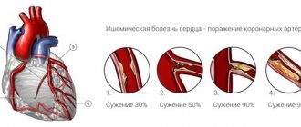
- coronary disease
- heart failure
- heart defects
- myocarditis, endocarditis
- tachycardia and bradycardia
- pre-infarction state
A discrepancy between your results and EchoCG norms can also occur in a number of other diseases. This test can also detect blood clots and aneurysms, heart tumors, and scar formation.
If you need to identify abnormal phenomena in the functioning of the heart, then one of the most suitable methods for this is echocardiography. The norms of the main indicators will help you navigate the results obtained. But it’s still better to leave the interpretation of the results to cardiologists. They will be able to get as much information as possible from this data and will explain to you whether there are any violations.
As a rule, an ultrasound of the heart is enough to make an accurate diagnosis and develop tactics for further action. However, in some cases, the doctor may prescribe additional examinations.
Regardless of heart disease, there are two main methods of instrumental diagnostics, which are quite informative and accessible to the population. An ECG allows you to assess the presence of pathologies in the conduction of impulses and create a general idea of the state of the organ. Using an ultrasound of the heart, you can evaluate its structure, the size of its components (walls, valves, septa), track the movement of blood through the departments and detect any space-occupying formations (tumors, abscesses, fibrinous deposits, and so on).
The quality of ultrasound depends not only on the technique, but also on the interpretation of the results. If the indicators are interpreted incorrectly, it is possible to make an incorrect diagnosis and choose inadequate treatment tactics. Despite the fact that if anyone knows the norms, they can determine the presence of abnormalities, only a specialist can predict a certain disease based on these data. Therefore, it is important that only a qualified doctor interprets the diagnostic results.