A general urine test is a laboratory test, the results of which are the main diagnostic indicator for most diseases, since urine contains more than 150 chemical compounds.
The entire process of formation and excretion of urine is called diuresis. The daily diuresis of a healthy person averages from 1.5 to 1.8 liters, depending on the air temperature, food and liquid consumed, and the time of day.
Also, the volume of daily urine changes under the influence of pathological processes in the body:
- the presence of diabetes mellitus or diabetes insipidus and some renal pathologies leads to increased fluid secretion (so-called polyuria);
- stones in the urinary system lead to a decrease in the volume of diuresis (oliguria);
- advanced forms of renal failure and blockage of the urinary tract can cause a lack of urination (anuria).
Why is this test prescribed?
Urine is a biological fluid in which the final waste products of the body are released from the human body.
It is conventionally divided into primary (formed by filtration in the glomeruli from blood plasma) and secondary (formed by reabsorption of water, necessary metabolites and other solutes in the renal tubules).
Disruption of this system entails characteristic changes in normal TAM indicators. Thus, the analysis can show:
- 1 Metabolic abnormalities;
- 2 Signs of urinary tract infection;
- 3Effectiveness of treatment and diet;
- 4Dynamics of recovery.
A person can contact a laboratory for a urine test on his own initiative if he notices sudden changes in his physical characteristics. But more often the patient receives a referral from a specialist at the clinic, who then deciphers the results obtained.
OAM is included in the list of basic studies during preventive examinations of the population, clinical examination, it is prescribed when seeking medical help from a specialist, during pregnancy, during hospitalization and in some other cases.
A general urine test consists of a sequential study of:
- 1Physical characteristics of the sample;
- 2Chemical composition;
- 3Microscopic examination of sediment.
Indications for urine testing
A general urine test is prescribed to healthy people undergoing annual medical examinations and to patients who have various complaints about deteriorating health.
Urine examination is carried out when:
- diagnosing pathological processes in the kidneys and urinary tract;
- detection of prostate diseases;
- mandatory examination before surgical interventions;
- recent illnesses in which kidney complications are possible;
- suspicion of diabetes mellitus, hepatitis, pancreatitis;
- assessing the toxic state of the patient’s body;
- monitoring the course of the disease and the effectiveness of treatment.
For children, the study is carried out before a scheduled visit to the pediatrician and each vaccination, as well as for various ailments. This makes it possible to promptly identify developing pathological processes in the child’s body.
Expectant mothers need to regularly undergo a urine test - in addition to monitoring the general condition of the body and urinary system, the study will allow for the timely detection of such a serious pathological condition as gestosis (late toxicosis)
Parameters by which urine decoding (UAM) occurs
The table shows the indicators of a general urine test that are normal in adults; if there are any deviations, it is necessary to decipher them.
| Indicator name | Normal values |
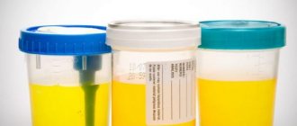
| Color | Straw color – dark yellow |
| Smell | Blurred, non-specific |
| Relative density | 1,010 – 1,025 |
| pH | 5-8 |
| Glucose (sugar in urine) | Up to 1 |
| Protein | Up to 0.14 |
| Ketone bodies | Up to 0.5 |
| Bilirubin | Up to 8.5 |
| Nitrites (bacteria) | No |
| Hemoglobin | No |
| Red blood cells | Up to 2 in sight |
| Leukocytes | Up to 5 |
| Epithelium (flat) | Up to 5 |
| Crystals | There is |
| Cylinders | Not detected (exception may be hyaline casts) |
Any urine test result should be discussed with your doctor to make decisions about further treatment. Only a specialist can make final conclusions about the state of health.
In adults, urine is examined according to several parameters. We will consider the norm and pathology below.
When hemoglobin breaks down after several transformations, the substance urochrome (a derivative of bile pigments) is obtained, which gives color to urine. This pigment normally gives the liquid a yellow color with different shades.
Most often, this indicator is normal in women and men. But sometimes a precipitate forms when salts, phosphates, and oxalates are detected.
If the urine is light, it means that the person does not have a lack of fluid. In the case of a dark yellow tint, a sharp loss of fluid from the body is possible, as a result
The color of urine changes as a result of consuming coloring foods and medications.
The norm in adult patients is completely clear urine. If it is cloudy, this happens for several reasons:
- various microorganisms and cells that normally should not be in it accumulate in the urine: leukocytes, pus, red blood cells, epithelial particles, bacteria;
- penetration of large amounts of salts into biological fluid;
- if the urine becomes cloudy after a certain time, when it sits for a while, this indicates that there are actively reproducing bacteria in it.
Specific gravity, also called urine density, depends on how much liquid a person drinks during the day. If there are chronic diseases of the excretory system, the density will decrease significantly.
This indicator depends on the release of electrolytes (calcium, sodium and potassium), as well as processed substances. They show whether all organs of the excretory system are working correctly.
Normal urine density in adults: 1003 – 1035 g/l. If the results of the TAM differ from the norm, this indicates the following pathologies:
- High values are observed with nephrotic syndrome or glomerulonephritis (then protein will be in the field of view), sudden losses of fluid in the body, intravenous injections of certain medications, increased blood sugar without the use of glucose-lowering drugs, toxicosis during pregnancy, minimal fluid intake during pregnancy. days.
- Low values are detected with the development of diabetes mellitus, pathological processes in the kidney tubules, drinking plenty of water during the day or taking diuretic medications, and chronic kidney failure.
Urine pH
The transcript of the urine test contains the pH indicator. What does it mean for an adult? This is the reaction or acidity of a biological fluid. It fluctuates significantly throughout the day, depending on the food consumed.
Normally, this indicator has the following range of values: from 4.5 to 8. The ideal option is values 5-6.
pH numbers increase when:
- constant consumption of vegetables and fruits (containing acids and fiber);
- with constant vomiting;
- infections that break down urea;
- neoplasms on the organs of the genitourinary system;
- decreased functionality of the parathyroid gland;
- renal failure.
The values of this indicator decrease when:
- frequent loose bowel movements;
- hypoglycemia;
- deliberate long-term refusal of food;
- protein diet;
- lack of fluid in the body;
- elevated body temperature;
- diabetes mellitus and pulmonary tuberculosis;
- use in the treatment of ascorbic acid and methionine.
A urine test provides useful information for a doctor about the capabilities of the kidneys; the norm for this indicator is up to 0.14 g/l.
Detection of protein in the urine of healthy people is possible if the person experienced severe stress the day before or was frozen. Traces of protein are noticed during testing if a person has not washed the dishes enough to collect the analysis or due to poor personal hygiene.
This value shows how much glucose is released in biological fluid. Moderate glucose in the urine is detected by excessive consumption of sweet foods before taking the test.
This indicator is important in determining diabetes mellitus, inflammation of the pancreas, various types of tumors, diseases of the thyroid gland, and adrenal glands.
The glucose norm in the study results is from 0.1 to 0.8 mmol/l.
If the value is high, additional diagnostics are carried out to determine diseases such as dumping syndrome, acute pancreatitis, chemical poisoning, diabetes mellitus, pheochromocytoma and some other diseases.
Medicines can also increase glucose levels in the body.
Urinalysis also helps to determine liver problems; the norm of the liver indicator - bilirubin - is its absence in the urine.
If it is detected, this indicates problems with the outflow of bile or damage to liver tissue. In addition, bilirubin in biological fluid can be detected when:
- hemolytic anemia;
- resorption of bruises (hematomas);
- exposure to toxins in the form of alcohol on the body;
- infectious liver pathologies.
As a result of the breakdown of fatty acids entering the body, acetone and acids are released. This indicator shows the functioning of the endocrine system and the performance of functions by the gastrointestinal tract. Norm in adult patients: 0-0.5 mmol/l.
The amount of ketone bodies is of decisive importance in therapy prescribed for diabetes mellitus. This value shows the degree of effect of sugar on internal organs.
In extreme cases, an increased concentration of ketone bodies leads to disruption of the central nervous system and coma due to a sharp increase in sugar in the body.
These elements may appear in urine due to:
- alcohol intoxication;
- impaired perception of insulin by the body or its overdose;
- long-term refusal of any food;
- lack of carbohydrates;
- severe fever and eclampsia.
Presence of hemoglobin
Deciphering a urine test is important for examining biological fluid for the presence of hemoglobin. Normally this substance is absent. The analysis results should say: “not detected.”
If hemoglobin or myoglobin is nevertheless detected in biological fluid, the following diseases are suspected in a person:
- burn;
- sepsis;
- severe hemolytic anemia;
- chemical poisoning;
- myocardial infarction;
- myopathy;
- damage to muscle tissue;
- excessive physical activity and training (the last 4 occur when myoglobin is detected).
The norm for this indicator is the complete absence of this substance. Due to the presence of urine in the bladder for a period of 4 hours, bacteria act on the fluid.
They are contained in the bladder as a result of the progression of a genitourinary tract infection. If nitrites are detected in the test results, this indicates bacteriuria.
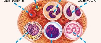
To examine biological fluid for the content of body cells, the method of analyzing urine sediment under a microscope is used. Ten ml of urine is passed through a centrifuge and the content of the following components is determined.
- Red blood cells. These are the cells that color the blood. Normally there should be up to 2 cells per µl. To exclude the mechanical entry of blood cells into the urine, it is better for women to refrain from this analysis during menstruation.
- Leukocytes. At elevated values, an inflammatory pathological process is observed in the organs of the excretory system. When collecting biological fluid for analysis, leukocytes can enter the external genitalia if they are inflamed. The norm for men is less than 3 in the field of view, for women: less than 5.
- Epithelium (squamous) - a large number indicates the presence of infection. Its normal content is less than 5 in women and less than 3 in men.
- Casts are casts of the kidney tubules and show poor functioning of the kidneys due to certain disease conditions. Normally they are not detected.
- Crystals are not normally detected. These are salt compounds that are formed due to the increased content of ampicillin and sulfonamides.
Main indicators of general urine analysis
There are three main groups by which doctors will determine the presence of diseases in the patient’s body. These are the physical properties of urine, the presence of organic matter and salts, and sediment. Moreover, these indicators are influenced by everything: a person’s diet, taking certain medications, and even lifestyle.
- In particular, the color of urine should ideally be yellow. Green, brown and black indicate medication use. Antipyrine, amidopyrine, and iron color urine pink or brown, and methylene preparations make it blue. Some food products also have coloring properties: rhubarb, bay leaves, beets or carrots.
- Products also affect the smell of urine , especially those that contain a lot of essential oils. The color or smell of urine caused by diet is accepted by doctors as normal, but complicates the research process.
- Doctors judge kidney function by density It is worth noting that fruits and vegetables reduce the density of urine, but the abundance of meat and fats in the diet, on the contrary, increases it.
Patient preparation
Before submitting the material for general (clinical) analysis, consult your doctor about the possible temporary cessation of taking certain pharmaceutical drugs. For example, diuretics should be stopped 48 hours before sample collection.
The day before collecting biomaterial, avoid foods with a high content of pigments, alcohol, fatty, smoked foods, sex, and excessive physical and psycho-emotional stress. All this can distort the OAM results.
For analysis, a morning urine sample is collected, optimally its middle part. Before collection, the patient must toilet the external genitalia (bath, shower, wet wipes).
After the start of urination, it is better to flush the first portion into the toilet, collect the middle portion in a clean, sterile container (optimally in a sterile pharmaceutical container). The minimum volume of urine required for testing is 50 ml. There is a mark on the medicine cup to the level at which it is advisable to fill the container.
In young children, it is often difficult to collect urine for analysis. Therefore, when collecting, you can use small tricks:
- 1Buy special soft polyethylene containers with a sticky edge at the pharmacy. Not all children like this procedure, but for some it is acceptable.
- 2Before picking up, take the baby to the bathroom and turn on the water. A child up to one year old can be breastfed beforehand, and an older baby can be given water to drink. Urination in babies is tied to feeding, so the task can be made easier.
- 3Some children pee several times with intervals between peeings of 10-15 minutes. To collect material from such babies, it is better to prepare several containers so that you can collect the droplets in different dishes without staining them during manipulation.
- 4Before the procedure, you can do a soft, stroking massage in the lower abdomen, in the bladder area.
What should you not do when collecting urine?
When collecting material for clinical urine analysis, it is not recommended:
- 1Use untreated dishes, the contents of a potty, a diaper, a diaper, a plastic bag. This analysis is called “dirty”; it is not suitable for assessing the condition of the urinary system.
- 2Use for analysis stale urine that has stood for more than 3 hours or has been in the refrigerator without a special preservative.
- 3Collect material for OAM after defecation, during menstruation or after sexual intercourse.
- 4Collect material for research during acute inflammatory diseases of the reproductive system, skin around the urethra and vagina (you must warn the doctor about this in advance). It will not be possible to collect such an analysis purely.
- 5Do not use a urinary catheter unless there is an urgent need for it (prostate cancer, prostate adenoma, a bedridden seriously ill patient and other situations that are discussed by the attending physician). When placing a catheter at home, there is a high risk of secondary infection.
The table below presents the main indicators, their standards and interpretation. Clinical urine analysis in women is practically no different from that in men, with the exception of some parameters. These small nuances are noted in the table.
| Index | Decoding | Norm |
| BLd | Red blood cells | 2-3 in the field of view in women (abbreviated as p/z) / Single in men |
| LEU | Leukocytes | 3-6 in p/z for women / Up to 3 - for men |
| Hb | Hemoglobin | Absent (sometimes they write the abbreviation neg - negative) |
| BIL | Bilirubin | Absent (neg) |
| UBG | Urobilinogen | 5-10 mg/l |
| PRO | Protein | Absent or up to 0.03 g/l |
| NIT | Nitrites | Absent |
| G.L.U. | Glucose | Absent |
| KET | Ketone bodies | Absent |
| pH | Acidity | 5-6 |
| S.G. | Density | 1012-1025 |
| COLOR | Color | Light yellow |
Physicochemical characteristics of urine
Relative density is an important indicator that can provide information about impairments in the ability of the kidneys to concentrate and dilute salts and various substances.
To measure the density of urine, a special device is used - a urometer.
Normally, the specific gravity of urine ranges from 1006 to 1026 g/l. Its increase is typical for:
- dehydration of the body;
- nephrotic syndrome;
- toxicosis in pregnant women;
- heart failure;
- diabetes mellitus;
- decreased production of urine in the body – oliguria;
- liver diseases.
A decrease in the indicator is observed with damage to the kidney tubules, diabetes insipidus, and renal failure.
The reaction of the urine environment is normally neutral, slightly acidic or slightly alkaline, the pH ranges from 5.0 to 7.0. A decrease in parameters is observed with a predominance of meat foods in the diet, an increase is observed with a plant diet.
Chemical properties
4.1. Quantity
When assessing the total amount of urine excreted, it is necessary to take into account the possible dietary characteristics of each patient. In an adult who follows a normal diet, daily diuresis ranges from 800 to 1500 ml.
Diuresis directly depends on the volume of fluid drunk. Typically, 60-80% of what you consume per day is eliminated from the body. The normal ratio of daytime to nighttime diuresis is 3:1 or 4:1.
A condition characterized by increased urine output (more than 2000 ml per day) is called polyuria.
A similar phenomenon is observed normally:
- 1If you have drunk a lot over the past day;
- 2In case of nervous excitement or overexertion.
Polyuria can be observed in the following pathological conditions:
- 1Kidney diseases (CKD, stage of resolution of acute renal failure);
- 2 Relief of edema, for example, against the background of diuretics;
- 3Diabetes insipidus and diabetes mellitus;
- 4Nephropathies (amyloidosis, myeloma, sarcoidosis);
- 5Taking certain medications.
The reverse condition is called oliguria. With oliguria, less than 500 ml of urine is excreted per day.
Physiologically it can occur with:
- 1Reduce fluid intake;
- 2Loss of fluid through sweat in the heat;
- 3 Significant physical activity.
It is noted in the following pathologies:
- 1Cardiac decompensation;
- 2 Poisoning;
- 3Excessive loss of water by the body (for example, during profuse diarrhea, vomiting);
- 4Burns;
- 5States of shock;
- 6 Fever of any origin;
- 7 Kidney damage of infectious, autoimmune and toxic origin.
Anuria is a condition in which urine production stops completely. Anuria is typical for:
- 1Initial stage of acute renal failure;
- 2Acute blood loss;
- 3Uncontrollable vomiting;
- 4 Stones in the urinary tract with obstruction of the lumen;
- 5 Oncological diseases accompanied by obstruction and compression of the ureters.
Nocturia is a condition in which nocturnal diuresis significantly prevails over daytime diuresis. Nocturia is typical for:
- 1Diabetes insipidus and diabetes mellitus;
- 2Many kidney diseases;
- 3BPH.
In addition to the daily amount of urine, pay attention to the frequency of urination. Normally, this process is performed by a person 4-5 times during the day.
Pollakiuria is characterized by frequent trips to the toilet. Observed when:
- 1Drink a large amount of liquid;
- 2Urinary infection.
Olakiuria is the opposite condition to that described above. Characteristic for:
- 1Low intake of fluid into the body;
- 2Nervous reflex disorders.
Strangury is painful urination.
Dysuria is a urination disorder that combines symptoms such as changes in urine volume, frequency and pain. It usually accompanies inflammatory processes in the genitourinary system.
4.3. Color
Is a direct reflection of concentration. In a healthy person, deviations in color from straw yellow to amber are allowed.
The color of urine is also influenced by special substances, the basis of which are blood pigments. A dark yellow color is observed when the amount of coloring substances dissolved in it significantly exceeds the norm. Characteristic of such conditions:
- 1Swelling;
- 2Vomiting;
- 4Burns;
- 4Congestive kidney;
- 5Diarrhea.
If the content of pigment substances is minimal, the shade will be paler. Observed when:
- 1Diabetes mellitus;
- 2Diabetes insipidus.
The dark brown color is explained by an increase in the level of urobilinogen. It is a diagnostic criterion for hemolytic anemia. Urine may turn dark brown when taking sulfonamides.
Dark, practical black color can indicate several conditions:
- 1 Alkaptonuria (due to homogentisic acid);
- 2Acute hemolytic kidney;
- 3Melanosarcoma (obtains this shade due to the presence of melanin).
Urine turns red if it contains fresh blood or red pigments. This is possible with:
- 1Kidney infarction;
- 2Kidney failure;
- 3Damage and trauma to the urinary tract;
- 4Taking certain medications (for example, rifampicin, adriamycin, phenytoin).
The appearance of “meat slop” is explained by the presence of altered blood, which is characteristic of acute glomerulonephritis.
A greenish-brown tint (compared to the color of beer) appears if bilirubin and urobilinogen enter the urine. This deviation from the norm often indicates parenchymal jaundice.
If the shade is rather greenish-yellow, which may indicate the presence of bilirubin alone, and is considered a symptom of obstructive jaundice.
4.4. Transparency
Normally, urine is clear. However, in the presence of pathological components and impurities (proteins, leukocytes, erythrocytes, epithelium, bacteria, salts), it can be cloudy, cloudy and milky.
Several manipulations can be carried out in advance to narrow the range of possible substances that make up the sediment to certain salts.
When, when heated, the test tube with the test material becomes transparent again, we can conclude that it contained urates.
If the same happens upon contact with acetic acid, we can assume the presence of phosphates in the sample. If an identical effect is observed when mixed with hydrochloric acid, then there are calcium oxalates in the urine.
For more accurate data, microscopy of the sediment is performed.
4.5. Smell
The smell of urine is usually specific and not strong. An ammonia odor may appear if there is bacterial contamination of the sample. A fruity smell (of rotting apples) is considered an indicator of the presence of ketone bodies.
This indicator is considered very important, since it is used to judge the concentration function of the kidneys and its ability to dilute.
The measurement is carried out using a specially designed device - a urometer. During the study, attention is primarily paid to the content of electrolytes and urea, and not to substances with high molecular weight (proteins, glucose, etc.).
Hypersthenuria is an indicator higher than normal. Its cause may be:
- 1 Toxicosis of pregnant women;
- 2 Progressive edema;
- 3Nephrotic syndrome;
- 4Diabetes mellitus;
- 5Use of radiocontrast agents.
Hyposthenuria is a decrease in specific gravity. Observed in the following conditions:
- 1Malignant hypertension;
- 2Chronic renal failure;
- 3Diabetes insipidus;
- 4Damage to the kidney tubules.
Isosthenuria is a condition in which the density of urine is equal to the density of blood plasma (within 1010-1011).
This is the second group of urine indicators that characterize the patient’s health status.
Normally, urine pH ranges between 5-7. An acidic reaction (pH {amp}lt;5) can be a consequence of:
- 1Increased consumption of meat products;
- 2 Metabolic or respiratory acidosis (as a result of various pathological processes), coma;
- 3Acute glomerulonephritis;
- 4Gout;
- 5Hypokalemia.
An alkaline reaction (pH{amp}gt;7) occurs when:
- 1Vegetable diet;
- 2Chronic renal failure;
- 3Metabolic or gas alkalosis;
- 4Hyperkalemia;
- 5Active inflammatory processes in the urinary system.
Normally, protein is not detected in the urine or an insignificant amount is detected. A condition in which this threshold is exceeded is called proteinuria. It is customary to distinguish several types of proteinuria:
- 1Prerenal proteinuria is associated with pathological processes in the human body that are accompanied by an increase in protein concentration in the blood plasma (myeloma, for example).
- 2Renal - one that is a consequence of damage to the glomerular filter or dysfunction of the renal tubules. The diagnostic criterion for the severity of the pathological process is selectivity - the greater the number of large protein molecules found in secondary urine, the more serious the situation.
- 3 Postrenal proteinuria is a manifestation of inflammatory processes in the reproductive system and surrounding tissues (vulvovaginitis, balanitis, and so on).
- 4Proteinuria can also be physiological, for example, during emotional overload, exposure to cold or sun, in children in a standing position, during long walking or running.
Normally, this substance cannot be detected in urine due to its low content. Glucosuria is the name given to a condition in which glucose levels exceed 0.8 mmol/l. This occurs when the so-called renal glucose threshold is exceeded.
That is, when its concentration in the blood exceeds 9.9 mmol/l, it freely passes the barrier and enters the urine. There are the following types of glucosuria:
- 1 Nutritional (large amounts come from food);
- 2Emotional;
- 3Medicinal.
Pathological glucosuria is usually divided into renal (manifests itself in various kidney diseases) and extrarenal, which is considered a consequence of the following diseases:
- 1Diabetes mellitus;
- 2 Thyrotoxicosis;
- 3Pheochromocytomas;
- 4Acute pancreatitis and other diseases of the pancreas;
- 5Itsenko–Cushing's disease;
- 6Liver cirrhosis;
- 7 Poisoning.
It is believed that hemoglobin is found in a portion of urine during the rapid breakdown (hemolysis) of red blood cells. Such a process may be infectious, immunological or genetic in nature. Most often, hemoglobinuria is detected with:
- 1Hemolytic anemia;
- 2Transfusion of incompatible blood;
- 3Internal injuries (crash syndrome);
- 4Severe poisoning;
- 5Direct damage to kidney tissue.
Hemoglobinuria is dangerous because it is an impetus for the development of acute renal failure.
Ketonuria is a special indicator of urine analysis, which reflects the failure of metabolic processes occurring in the body. In this case, the following substances are detected: acetone, beta-hydroxybutyric, acetoacetic acids. Ketonuria occurs against the background of:
- 1Diabetes mellitus;
- 2Carbohydrate starvation, diets;
- 3 Severe toxicosis (more often in children);
- 4Dysentery;
- 5 Severe irritation of the central nervous system;
- 6Hyperproduction of corticosteroids.
Bilirubinuria is a pathological condition in which unchanged bilirubin is detected in the urine. When the mechanisms that utilize bilirubin fail, the kidneys take on part of the work. Bilirubinuria is typical of many liver diseases:
- 1 Cirrhosis;
- 2 Hepatitis;
- 3 Jaundice (parenchymal and mechanical);
- 4Cholelithiasis.
Urobilinuria occurs when the liver does not function properly. However, intestinal pathology (where this substance is formed) and processes leading to the breakdown of red blood cells also contribute to the appearance of urobilinogen in the urine.
Ketones
These substances are represented by acetone, hydroxybutyric and acetoacetic acids; the reason for their appearance in urine is a metabolic disorder in the body, which is observed when:
Ketones in urine - what are they?
- alcohol intoxication;
- prolonged fasting;
- thyrotoxicosis (hyperfunction of the thyroid gland);
- acute pancreatitis;
- hypercortisolism;
- diabetes mellitus;
- the predominance of fatty foods in the diet;
- acetone syndrome.
How to prepare for the analysis?
Human urine is sterile, but before it gets into laboratory glassware, it can be contaminated by the person themselves. Therefore, it is important to observe personal hygiene rules as much as possible so as not to affect the analysis results.
To collect biological fluid you need:
- properly clean the external genitalia, women need to use a sterile piece of cotton wool (tampon) soaked in soapy water to treat the vagina from top to bottom, men need to wash the end of the urethra with soap;
- wait for morning urine with a break of 6 hours from the previous trip to the toilet;
- when urinating, the first thing that comes out is the dead cells of the channel through which urine flows, so you need to flush the first portion of liquid into the toilet;
- in order to prevent changes in the result of the analysis, you should take sterile containers; you cannot take liquid from an intermediate container, for example, a pot, for examination;
- It is better to send the collected liquid to the laboratory immediately, try not to expose it to sunlight;
- Before collecting biological fluid, it is not recommended to eat foods that change the color of urine, for example, beets.
Also, tell your doctor about any medications or supplements you take. Medicines or dietary supplements that may affect urinalysis results:
- vitamin C;
- metronidazole;
- riboflavin;
- anthraquinone laxatives;
- methocarbamol;
- nitrofurantoin.
How can you independently understand from OAM: is everything okay in the body or are there any deviations?
It is curious that in ancient times doctors were able to determine the presence of pathology by urine (or biological fluid, as it is otherwise called). In ancient times, the most primitive methods were used and the disease was identified by the external signs of the liquid: color, smell and taste. Nevertheless, even these methods were enough to recognize, for example, diabetes mellitus or abnormalities in kidney function in a person.
It must be said that organoleptics still plays a huge role in research today, but modern laboratory research has been able to get to the bottom of it by breaking down human biological fluid into simple chemical components. At the moment, any malfunction of the body can be judged by the quality of urine: it removes toxins and salts, removes organic substances and even cell elements.