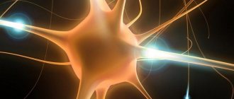
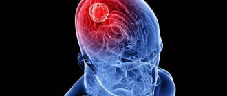
Acoustic neuroma (synonyms acoustic neuroma or schwannoma, as well as vestibular schwannoma) is a benign tumor of the VIII pair of cranial nerves.
The incidence is approximately 1 case per 100 thousand population per year. While remaining one of the most common brain tumors, occupying, according to various sources, from 10 to 15% of all brain tumors. It occurs more often in young (30-40 years old) people; cases of development in young children are extremely rare.
Pathophysiological characteristics
Neuroma is formed as a result of disruption of the mechanisms of cell division and growth in Schwann cells.
Since this is a benign tumor, some of the cell's functions are preserved; in structure and composition it also resembles a healthy nerve sheath cell. As the tumor grows uncontrollably, it puts pressure on the nerve, causing problems with the organ that communicates with the brain through that nerve.
When the 8th pair of cranial nerves is damaged, hearing and vestibular disorders occur (the so-called acoustic neuroma).
What are the causes of acoustic neuroma?
As is known, tumor neoplasms are divided into benign and malignant, that is, cancerous. Fortunately, acoustic neuroma is a benign tumor, meaning it does not metastasize and is usually not fatal. The consequences and manifestations of this disease are not pleasant, and among them are: frequent dizziness, headache, impaired coordination of movements and even complete loss of hearing.
Scientists have not yet established the exact causes of acoustic neuroma, but there are assumptions about the genetic nature of the disease. In addition, a relationship was found between the occurrence of this tumor and regular irritation of the plantar nerve, which occurs if you constantly wear uncomfortable or tight shoes. The disease can occur if the musculoskeletal system is constantly overloaded or due to a foot injury.
Risk factors include only complicated heredity when there is a genetic disease such as neurofibromatosis type II. With this disease, benign tumors called neurofibromas form in various parts of the body, including the auditory nerve. This usually leads to bilateral acoustic neuroma. It should be remembered that if pathological changes affected the chromosomes of both parents, and they both have neurofibromatosis, then their child will get sick with a 50% probability.
Provoking factors
The causes of schwannoma are not well understood. The main theory is the influence of gene mutations and hereditary predisposition. This is confirmed by a study that discovered the connection of neuroma with a mutation of part of the gene material of chromosome 22. These genes encode a protein necessary to limit the growth of Schwann cells.
The cause of the mutation may be:
- exposure of the body to large doses of radiation in early childhood;
- exposure to chemical toxins;
- the presence of benign formations in other parts of the body;
- predisposition to tumors;
- neurofibromatosis in the patient or his parents.
The last factor is evidence of the hereditary nature of neuroma. Neurofibromatosis is a disease that also develops due to a mutation of chromosome 22. If it is observed in one of the patient’s parents, the probability of a tumor occurring exceeds 50%.
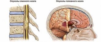
Locations of neuroma and who is susceptible to it
The most common location of neuroma is the vestibulocochlear nerve or auditory nerve, followed by the trigeminal nerve.
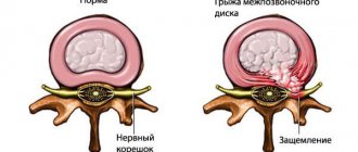
But, in principle, neuroma can appear on the membranes of any nerves. Neuroma (or schwannoma) is a benign neoplasm that arises in Schwann cells of the spinal, cranial, or peripheral nerves. Essentially, this tumor is a new growth in the cellular structures that line the nerve canals. These tumors have capsule-like lobular or round shapes. Tumor cells and fibers form “palisade” structures (Verokai bodies, nuclear palisades) with areas that consist of fibers.
Types and classes of tumors
According to their structure, neuromas are divided into:
- Epileptoid . The tumor is a dense body with a large number of fibers.
- Angiomatous . They are characterized by abnormal expansion of blood vessels, due to which a large number of cavernous cavities are formed.
- Xanthomatous . They are distinguished by a high content of pigment, which colors tumor cells yellow, yellow-gray, and green.
From the point of view of localization, the classification of tumors does not make sense, since any nerve in the brain can become a site for the growth of a tumor. However, the VIII pair of cranial nerves, the auditory nerves, are most often affected.
In turn, brain neuromas can be divided into:
- benign - develop slowly, retain their structure, do not penetrate into surrounding tissues due to the protective capsule around each cell;
- malignant - grows quickly, affects neighboring tissues, and gives metastases.
The photo shows what a neuroma looks like on a CT scan.
Recommendations
Consultation with a neurosurgeon and magnetic resonance imaging of the brain are recommended.
| • | Leading specialists and institutions for the treatment of this disease in Russia: |
| Alexander N.K., Director of the Research Institute of Neurosurgery named after. Burdenko N.N. | |
| • | Leading specialists and institutions for the treatment of this disease in the world: |
| Professor Duke Samson, USA. |
Symptoms and diagnosis of tumor formation
Symptoms vary depending on the location of the neuroma and the size of the tumor; in the case of schwannoma of the brain, cranial symptoms develop.
As the brain neuroma grows, other cranial nerves are affected:
- Trigeminal nerve - affected in 15% of cases. In this case, disturbances in facial sensitivity, muscle weakness, changes in taste and olfactory sensations, pain, and hallucinations may be observed. The last symptom manifests itself when the temporal lobe of the brain is damaged; smells and tastes are usually pleasant.
- Facial and abducens nerves . Possible visual disturbances - strabismus, double vision.
In advanced stages, neuroma affects the brainstem and cerebellum . When these structures are compressed, vital functions may be impaired. Breathing, swallowing, and speech suffer. Possible mental disorders.
Since the auditory nerve is most often affected, it is important to know the symptoms that indicate the development of an acoustic neuroma:
- ringing in the ears - manifests itself at an early stage of the disease, when the tumor has not yet reached large sizes;
- hearing impairment - begins with the loss of the ability to distinguish high tones, develops gradually as the tumor grows;
- disturbances in the functioning of the vestibular apparatus - loss of coordination of movements, dizziness, nausea, fainting - appear in the later stages, when the tumor grows and begins to compress the vestibular nerve along with the auditory nerve.
In 25% of cases, schwannoma develops in parallel with neurofibromatosis - in this case, symptoms of both diseases are observed.
First of all, a neurological examination is used for diagnosis, which helps to roughly determine the stage of the disease and includes an assessment of the following symptoms:
- nystagmus - involuntary vibrations of the eyeballs;
- dysfunction of the vestibular apparatus;
- hearing loss;
- impaired sensitivity of facial skin;
- double vision;
- damage to the facial nerve;
- weakening of reflex reactions.
To confirm the diagnosis use:
- examination using or MRI - the image reveals a round formation with clearly defined edges, as well as secondary manifestations - for example, enlargement of the ear canal;
- audiogram.
Using a biopsy, the malignant nature of the tumor is excluded and the composition and structure of the tumor is determined.
Treatment
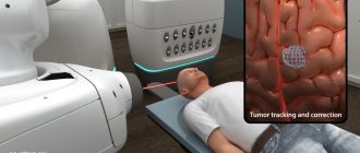
For the treatment of malignant and benign brain tumors located in hard-to-reach places, which also include vestibular schwannoma, the CyberKnife radiosurgical system is used.
How is radiosurgery performed using the CyberKnife system?
Radiosurgical system CyberKnife G4
CyberKnife irradiates the tumor remotely, without contact with the skin, without violating the integrity of body tissues; radiosurgical treatment does not require the use of anesthesia. That is why, throughout the world, radiosurgical treatment of acoustic neuroma is preferred in most cases. The most modern method of radiosurgery is CyberKnife.
Before starting treatment of vestibular schwannoma on CyberKnife, using instrumental diagnostic tools, a spatial model of the relative location of the tumor and healthy tissue is formed. During the treatment process, in the volume indicated in the three-dimensional model, with the help of many thin beams of ionizing radiation, a dose of radiation will be generated that is destructive for the tumor. The accuracy of delivery is ensured by a high-precision software and hardware complex, which operates strictly according to a given treatment plan. This allows for maximum protection of healthy tissue and uniform coverage of the entire tumor with a high dose of radiation.
Vestibular schwannoma: treatment in Kyiv on CyberKnife without surgery
Treatment of vestibular schwannoma with CyberKnife, unlike traditional surgery, does not require putting the patient under anesthesia. This treatment takes place without blood or cuts. In this case, the tumor is removed in just a few sessions.
In cases where it is impossible to use the CyberKnife because there are restrictions on the volume of the tumor, a neurosurgical operation is performed.
Health care
For the medical treatment of neuroma, the following drugs are used:
- Mannitol is a diuretic used to lower ICP. It is administered by drip in combination with glucocorticoids. It is used until the effect of the latter appears or before the start of radical therapy.
- Glucocorticoids - Prednisolone, Dexamethasone. The dosage of the drug is reduced after surgery or radiation therapy.
- Cavinton, Nicergoline - medications that improve blood circulation in the vessels of the brain .
Tumor surgery
The main treatment method for neuroma is surgery. This allows you to preserve the integrity of the nerves and avoid complete loss of facial sensitivity and hearing.
Indications for the operation:
- large tumor size;
- patient age under 60 years;
- increase in tumor size;
- serious condition of the patient.
The tumor can be removed in different ways:
- translabyrinthine;
- suboccipital;
- transverse temporal.
If it is impossible to completely remove the neuroma, its resection is performed.
Removal of neuroma:
Be careful, video of the operation! Click to open
Radiation therapy
Using a gamma knife is an innovative way to remove tumors
Small tumors and neuromas that cannot be removed for technical reasons can be treated with radiation therapy. Irradiation is carried out using:
- gamma knife;
- cyber knife;
- linear accelerator;
- proton accelerator.
The equipment is selected by the doctor based on the individual characteristics of the patient.
Malignant tumors require complex treatment - a combination of radiation and chemotherapy.
Forecast and consequences
Radiation therapy is highly effective - small neuromas that are treated in time disappear without a trace. More serious cases, even after treatment, may have noticeable consequences:
- permanent hearing loss due to atrophy of part of the auditory nerve;
- paresis of the facial nerve, leading to facial asymmetry, loss of sensitivity of taste buds, and impaired salivation;
- paralysis;
- disorders of cerebellar function;
- increased intracranial pressure;
- hydrocephalus.
If therapy is started in a timely manner, no consequences are observed.
Prevention of neuroma is impossible. However, you can avoid serious consequences of the disease if you consult a doctor in time when the first symptoms appear - hearing impairment, ringing in the ears, loss of balance and coordination in space.
Diagnostics
Diagnosis of neuroma of the eighth pair consists of conducting a thorough otoneurological examination, including neurological, otorhinolaryngological examinations, audiograms, impedance measurements and auditory evoked potentials. It is also necessary to conduct an MRI examination (if there are contraindications to the examination, conduct an MSCT scan). After identifying the presence of a neoplasm, a consultation with a neurosurgeon is necessary.
Also quite characteristic is the presence of an increased content of cerebrospinal fluid in the cerebrospinal fluid.