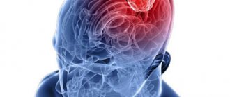
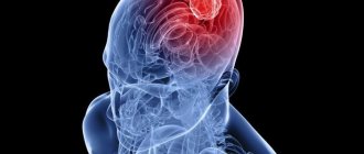
Characteristics of the pathology
In a newborn, the cerebellum weighs about 20 g - 5% of body weight.
By five months the weight triples. At the age of 15, the cerebellum reaches 150 g and no longer grows. In appearance, it resembles the hemispheres of the brain, for which it is also called the small brain. Located in the posterior cranial fossa. On top it is covered by the occipital lobes of the brain; under the cerebellum there is the medulla oblongata and the pons. Through its white matter fibers, the cerebellum is connected to all parts of the cerebrum. It has three departments:
- The most ancient in origin is the hook.
- The old one is a vermis that is located in the midline of the cerebellum.
- New - two hemispheres that resemble large hemispheres. Evolutionarily, this is the most developed part. Each hemisphere has three lobes, and each of them corresponds to a section of the vermis. The cerebellar hemispheres have gray and white matter. Gray – bark, white – fibers with cores: spherical, jagged, tires. These nuclei serve to conduct impulses and play a large role.
Cerebellar ataxia is a pathology that manifests itself primarily as a violation of motor coordination and walking. The ability to walk is built on the interaction of functions:
- Locomotion is a movement aimed at moving one’s own body in space.
- Maintaining balance.
- Adaptive reactions. Timely response to the presence of obstacles, regulation of movements taking into account changing external conditions.
There is a generalized form, when motor activity is disrupted in all parts of the body. In the local form, motor disorders affect individual parts of the body or functions - legs, arms, speech function, organs of vision. Pathology can manifest itself in one half of the body (with damage to the cerebellar tissue on one side) or in both.
Ataxia associated with damage to the cerebellum is a pathology that reflects malfunctions in the mechanisms responsible for anti-inertial regulation, which makes normal movement characteristics impossible (smoothness, regularity, accuracy, uniformity). Any normal movement is the result of the coordinated activity of several muscle groups (extensors, flexors).
Coordination of muscle activity is carried out by the cerebellum through its two-way interaction with other parts of the brain involved in the performance of motor functions and parts of the peripheral nervous system. The cerebellum is closely connected with the basal ganglia, cortical structures, and brainstem nuclei.
The transmission of information is carried out by motor neurons located in the spinal cord and proprioceptive (associated with the peripheral elements of sensory organs) neurons located in the muscles and ligaments. The cerebellum is the main coordinator of motor activity; it receives information about the planned movement, the slightest change in muscle tone and the position of parts of the body. The correct functioning of this part of the brain ensures the clarity and precision of small and complex movements.
Diseases affecting this part of the brain cause desynchronization of muscle contractions, which negatively affects the performance of any movements. Even small foci of infarction and hemorrhage in the cerebellum are life-threatening conditions that often provoke obstructive (associated with impaired drainage of cerebrospinal fluid) hydrocephalus.
The cerebellum is a special structure of the brain located in the posterior cranial fossa of the occipital and temporal lobes. The organ performs one of the main functions - maintaining balance and coordinating movements. It contains about 50% of the brain's neurons. In order to simply stand up or sit down, you need to use many muscles, the cerebellum is responsible for the coordinated work of them.
Cerebellar ataxia is a syndrome related to neurological disorders. The disease occurs quite often and is almost always associated with various injuries and diseases. Recently, specialists most often diagnose a hereditary form of pathology, and in more rare cases, an acquired form.
Classification of ataxias
Clinical medicine distinguishes several variations of ataxia: sensitive, frontal, cerebellar, vestibular, functional and hereditary, such as Friedreich, Pierre-Marie, and Louis-Bar ataxia.
The sensitive form is caused by damage to deep varieties of sensitivity. In the analyzed form, there are malfunctions in joint-muscular susceptibility, which receives information about the placement of the subject’s torso in space, malfunctions in vibration sensitivity, and disturbances in the sensation of pressure and weight.
O, expressed in excessive flexion of the limbs and excessively strong gait. A sick individual often loses the idea of the location of the elements of his own body in space and does not feel the direction of motor acts.
Subjects suffering from this type of pathology try to compensate for motor dysfunctions through vision: while walking, they always look at their feet, which helps reduce the feeling of ataxia.
However, patients are unable to move with their eyes closed.
The key differential diagnostic criterion for the described type of ataxia is the appearance of symptoms or a significant increase in symptoms in the absence of control through vision, for example, with eyes closed, in the dark.
This form can be found in the upper extremities, resulting in pseudoathetosis, which is a slow, worm-like movement of the hands with loss of visual control.
Pseudoathetosis and the sensitive form of ataxia are always accompanied in the limbs by disorders of deep sensitivity.
Doctors classify ataxia depending on which area of the brain is affected. There are four forms of the disease in total.
Other reasons for the development of the disorder
The disease is a symptom complex that consists of disorders of dynamic and static human motor skills. The acquired form of pathology develops in the presence of the following factors:
- head injury;
- multiple sclerosis;
- cerebral stroke (ischemic, hypertensive);
- viral or infectious diseases;
- heavy metal intoxication;
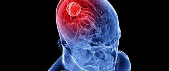
- brain tumors;
- obstructive hydrocephalus;
- Guillain–Barre syndrome;
- acute vitamin B12 deficiency.
The listed syndromes lead to the development of cerebellar ataxia with an acute onset. The disease, the symptoms of which begin to appear within a few weeks, has a subacute onset. The main causes of the development of pathology are endocrine disorders, metabolic disorders, malignant tumors, and toxic poisoning.
Progressive chronic ataxia occurs against the background of chronic alcoholism and atrophic processes affecting the cerebellum.
In some cases, cerebellar damage is not a consequence, but a symptom. This applies to oncological diseases of the following organs and systems:
- lungs;
- brain;
- ovaries;
- lymph nodes.
The “first bell” of brain cancer can be cerebellopontine angle syndrome. As a result of the growth of a malignant tumor, parts of the brain are compressed and nutrition and neural communication in their cells are disrupted.
With prolonged alcohol dependence, substance abuse and drug addiction, irreversible damage to the cerebellum occurs. Ataxia can also become a hereditary disease. In this case, therapy must be selected in a special way.
General description of the pathology
The cerebellum is a special structure of the brain located in the posterior cranial fossa of the occipital and temporal lobes.
The organ performs one of the main functions - maintaining balance and coordinating movements. It contains about 50% of the brain's neurons. In order to simply stand up or sit down, you need to use many muscles, the cerebellum is responsible for the coordinated work of them. Cerebellar ataxia is a syndrome related to neurological disorders. The disease occurs quite often and is almost always associated with various injuries and diseases. Recently, specialists most often diagnose a hereditary form of pathology, and in more rare cases, an acquired form.
Functions of the cerebellum
The main function of the cerebellum:
- motor coordination and maintenance of musculoskeletal tone;
- smoothness and proportionality of movements;
- body balance is constant;
- center of gravity;
- muscle tone is regulated and correctly redistributed.
Due to the cerebellum, the muscles work harmoniously and can perform any everyday movements. For the most part, the cerebellum is responsible for the tone of the extensor muscles.
In addition, the cerebellum is involved in unconditioned reflexes: through its fibers it is connected to receptors in different parts of the body. When exposed to any stimulus, a nerve impulse is sent from the receptor to the cerebellum, after which a response is given in the cerebral cortex with lightning speed.
With atrophy, nerve fibers are damaged. Coordination, gait and body balance are impaired. These characteristic symptoms are combined under the general term “cerebellar syndrome.”
This syndrome is characterized by disorders of a vegetative nature, motor sphere, and muscle tone, which immediately worsens the patient’s quality of life.
Features of the disorder
The peculiarity of this disease is that there is a disconnection of deep tissue impulses that send signals along the columns of the spinal cord.
And this can manifest itself in all the arms and legs, or in any of the limbs. A person has difficulty maintaining his balance while standing with his legs together and his arms extended forward.
Sometimes the cortex of the parietal lobe of the brain and peripheral nodes are affected.
If a patient has ataxia in the lower extremities, which arose due to loss of sensitivity in the joint-muscular sense, then he becomes extremely unstable and his gait takes on a stamping appearance, that is, there is excessive bending of the legs and stepping on the floor with force.
The patient constantly tries to control his movements visually and looks at his feet when walking. With severe damage to the posterior columns, all ability to move and be in an upright position is lost.
When patients close their eyes, the manifestation of this nervous disease is further aggravated.
Hereditary type of disease
Cerebellar ataxia of Pierre Marie is a hereditary disease that is dominant in nature. That is, it develops when receiving a defective gene from one of the parents. Skipping generations is extremely rare. It is one of the forms of a hereditary type of disease. Various factors can provoke the appearance of the first signs of pathology: severe infections (diphtheria, salmonellosis, typhoid fever), injuries, pregnancy, intoxication. Appears after age 20.
The symptoms of a hereditary disease constantly increase and progress. There are no periods of exacerbation and remission in such cases. Some diseases can change the course of the pathology, which significantly complicates timely diagnosis.
Hereditary cerebellar ataxia is divided into several forms:
- Friedreich's ataxia;
- non-progressive congenital ataxia;
- Holmes cerebellar atrophy;
- spinocerebellar ataxia;
- late cerebellar atrophy;
- Machado–Joseph disease.
Ataxia
Ataxia is a motor disorder, a disorder of movement coordination. The disease is characterized by a slight decrease in strength in the limbs, problems with balance when walking or standing. The movements of a patient with ataxic syndrome become awkward, imprecise, and their sequence and continuity are disrupted.
Features of the development of ataxia
Any movement is carried out as a result of a series of sequential contractions of different muscle groups (antagonists and synergists). For alternation to be normal, the work of three elements of movement coordination is necessary:
- structures and connections of the cerebellum (the central organ of movement coordination);
- receptors that perceive the state of stretching of tendons, muscles, joint capsule and their conductors at each specific moment in time,
- impulses coming from the organ that assesses the position of the body in space (vestibular apparatus).
Depending on which of these coordination elements is affected by disturbances, a different type of ataxia develops. According to the classification, there are:
- Sensitive ataxia – disturbances in the conductors of deep muscle sensitivity;
- vestibular ataxia – damage to the vestibular apparatus;
- Cerebellar ataxia (cerebellar) – damage to the cerebellum;
- Frontal ataxia (cortical) – damage to the cortex of the frontal or temporo-occipital region.
It is customary to classify hereditary ataxias as a separate group:
- Friedreich's familial ataxia,
- cerebellar ataxia of Pierre-Marie,
- ataxia telangiectasia, or Louis-Bar syndrome.
Causes of ataxia
Ataxia may be a consequence of:
- severe head injury (traumatic brain injury);
- accumulation of fluid in the cavities of the brain, spinal canal (hydrocephalus);
- congenital malformation of the brain/skull;
- infectious disease of the brain (encephalitis);
- cerebrovascular accidents (ischemic stroke);
- cerebral palsy;
- epilepsy in children;
- malignant neoplasms;
- abscesses.
Sensitive ataxia syndrome develops as a result of damage to:
- posterior brain stems,
- peripheral nodes,
- posterior nerves, optic thalamus,
- parietal lobe of the brain.
Cerebellar ataxia is a consequence of damage to the cerebellar vermis, its peduncles and hemispheres. Occurs in encephalitis, sclerosis.
- The vestibular form of the disease is caused by damage to any part of the vestibular apparatus - nuclei in the brain stem, cortical center in the temporal lobe of the brain, vestibular nerve, labyrinth.
- Cortical ataxia occurs when the frontal lobe of the brain is damaged, which is caused by disturbances in the functioning of the fronto-pontine-cerebellar system.
- Hereditary types of ataxia are transmitted in an autosomal dominant or autosomal recessive manner.
Symptomatic manifestations
Atrophy of the cerebellum of the brain is destructive for the patient, because with the death of nerve cells, pathological processes become irreversible.
Cerebellar disorders include several groups of disorders:
- First group. Impaired smooth movements of the limbs (mainly the arms). This is manifested by trembling of the hand at the end of any purposeful movement.
- Speech disorders.
- Voluntary movements and speech become slow. Next, the handwriting changes. Since the cerebellum is associated with motor acts, disturbances in its functioning are movement disorders.
Symptoms of cerebellar atrophy: asynergy of the muscles of the legs and torso, with difficulties arising when the patient attempts to rise from a lying position and sit down. These are very common signs of an affected cerebellum, and they indicate a disorder in the synergy of muscles (coordination of work) belonging to different muscle groups when they participate in the same motor act. The combination of simple and complex movements is completely disordered and disrupted.
Signs of cerebellar atrophy:
- The occurrence of incoordination of movements, the appearance of paralysis and various speech disorders. People cannot move smoothly, they sway in different directions, their gait becomes uncertain.
- Tremor and nystagmus (involuntary oscillatory movements of the eyeballs when they are abducted). Tremor is constantly present - during movement and at rest. Speech becomes scanned and dysarthric. What does this mean? A person with dysarthria has difficulty pronouncing words or distorts them due to unclear pronunciation.
- Scanned or telegraphic speech is possible. It is rhythmic, but the stresses are not placed according to the meaning, but correspond only to the rhythm.
- Muscle tone decreases due to atrophy of nerve fibers.
- Dysdiadochokinesis is a lack of coordination when the patient cannot perform rapid alternating movements.
- Dysmetria – the patient cannot control the amplitude of movement, i.e. accurately determine the distance between an object and himself.
- Hemiplegia arises from paralysis.
- Ophthalmoplegia is paralysis of the eyeballs, which can be temporary.
- Hearing impairment.
- Swallowing disorder.
- Ataxia - unsteadiness of gait; may be temporary or permanent. With such a drunken gait, the patient drifts towards the lesion.
- Severe cephalgia, with nausea and vomiting, dizziness due to increased intracranial pressure (ICP), and drowsiness are also possible.
- Hyporeflexia or areflexia is a decrease or complete loss of reflexes, urinary and fecal incontinence. Mental deviations are often possible.
The pathology has quite specific symptoms that are immediately noticeable. Patients make sweeping movements while walking. At the same time, their gait is shaky and uncertain. The legs are spread wide apart and practically do not bend at the knees when moving. Therefore, during the movement, the patient seems to sway from side to side (cerebellar gait). The symptoms are especially pronounced when the patient tries to stand up suddenly and start moving.
Involuntary stopping of movement even before reaching the goal is another characteristic symptom of the pathology. Patients with cerebellar ataxia are unable to quickly perform contralateral movements. When trying to turn, the patient “goes” to the side and may even fall. Speech with ataxia loses fluency and becomes scanned.
Dynamic cerebellar ataxia in humans is associated with damage to the cerebellar hemispheres. The main signs of the disease include tremors in the limbs at the end of any action, impaired handwriting, involuntary movement of the eyeballs (reminiscent of trembling), and movement disorders during multidirectional movements.
Primary symptoms of cerebellar (cerebellar) ataxia include an unsteady, unstable, unsteady gait with a noticeable expansion of the support base, lack of coordination, angularity, and clumsiness of movements. Other signs:
- Dysarthria. Scanned, slow, abrupt speech.
- Dysmetria (excessive or insufficient range of motion).
- Oculomotor disorders. Nystagmus (usually horizontal) – fast, frequent oscillations of the eyeballs, dysmetria of saccades (fast, coordinated eye movements).
Patients usually stand with their feet wide apart. An attempt to bring the feet closer to each other ends in loss of balance, swaying, and sometimes falling. Due to imbalance regulation, it is necessary to rely on surrounding objects or the support of another person. Mechanical movement in a straight line helps to identify the slightest gait disturbances.
Symptoms of cerebellar (cerebellar) ataxia are often supplemented by such signs as muscle hypotonia (weakness), slowness during movements, intention tremor (a disorder of fine motor skills of the limbs, which manifests itself as trembling and occurs after the completion of a voluntary movement), which is taken into account when determining treatment tactics. Symptoms are often observed:
- Asynergy. Deterioration in the ability to perform voluntary movements of a combined (consistent) nature.
- Strengthening postural reflexes (a set of reactions that occur to maintain a given position in space of a part of the body or the entire body).
- Deterioration of cognitive abilities, affective disorders provoked by damage to the ischemic etiology of the posterior parts of the cerebellum.
Impaired motor activity in cerebellar ataxia often does not correlate with muscle weakness, hyperkinesis (pathological, sudden movements provoked by an erroneous command from the brain), spasticity. However, such manifestations of pathology complicate the clinical picture. If the damage to the cerebellar vermis is partial, a pathological change in the function of standing and walking is observed.
Rostral lesions (the initial parts of the worm) lead to deviation of the body and a fall in the forward direction. Caudal lesions (caudal sections of the worm) initiate deviation of the body and fall in a backward direction. In the unilateral form, patients stagger and their ipsilateral shoulder (located on the side of the affected part of the cerebellum) is slightly drooping. When moving, patients deviate towards the affected area.
When neuronal pathways are damaged, rough tremor of the upper extremities is more often observed in a standing position with straight arms extended forward. Often, such destructive processes are associated with multiple sclerosis, which leads to damage to nerve endings and a deterioration in the transmission of nerve impulses.
Pronounced visible symptoms allow a person to “sound the alarm” and consult a specialist. Timely diagnosis will allow us to determine the exact cause of the disease and the extent of the damage.
Common symptoms include:
- intentional trembling of fingers and hands;
- "drunk" gait;
- asynergia (inability to perform combined movements);
- adiadochokinesis (inability to perform alternating rapid movements);
- nystagmus (high frequency oscillatory eye movements);
- hypotonicity of arm muscles;
- difficulty speaking with division into syllables;
- change in handwriting.
The manifestation of these symptoms may be mild. A rapid disturbance of gait and speech is typical only in cases of traumatic brain injury, when damage to the cerebellum occurs abruptly.
For diagnosis, a specialist may prescribe magnetic resonance imaging of the head and neck. This diagnostic method is currently the most common and accurate.
Cerebellar ataxia: types and classification, including Pierre Marie, causes, symptoms and treatment
Cerebellar ataxia (CA) is characterized by impaired ability to move and coordinate due to pathological processes in the cerebellum. The term “ataxia” is adopted to refer to any coordination disorders.
Description and history of the disease
The cerebellum is the part of the human brain located at its base. In the structure of the organ, two hemispheres, responsible for the accuracy of movements, are separated by the worm, which ensures stability and balance.
One type of ataxia is cerebellar (G 11.1-11.3 according to ICD-10), in which these problems are caused by pathology of the cerebellum. To describe a characteristic set of symptoms (gait disturbances, nystagmus, finger tremors, speech and handwriting disorders, etc.), the term “cerebellar ataxia syndrome” is used.
Depending on which part of the cerebellum is affected, ataxia is divided into static-locomotor (the vermis is affected, which is manifested by a disorder of gait and stability) and dynamic (the hemispheres are affected and the ability to make voluntary movements of the limbs is impaired).
Similar problems occur when standing for a static form and when walking for a dynamic form. According to various sources, the frequency of such pathology can reach 23 people per 100 thousand.
Causes of cerebellar ataxia
In most cases, cerebellar ataxia is a genetic disease, less often - acquired.
In the latter version, the pathology is a consequence of:
Hereditary forms of pathology
Hereditary forms are associated with gene mutations and are classified into:
- congenital non-progressive MA, caused by the absence or underdevelopment of cerebellar structures;
- autosomal recessive (Friedreich's ataxia), the symptoms of which appear in children from an early age (or up to 25 years), characterized by abnormal formation of the skeleton (this form develops when receiving a defective gene from both parents);
- recessive, X-chromosomal with progressive cerebellar insufficiency, low prevalence mainly in men;
- Betten's disease, congenital MA with slow development of the child, but with the patient's adaptation to his condition as he grows older;
- hereditary ataxia of Pierre Marie, refers to late autosomal dominant cerebellar ataxia, the signs of which (“chopped” speech, trembling, decreased mental abilities, changes in tendon reflexes, vision problems, strabismus, etc.) appear after 25 years (for the development of this form For ataxia, it is enough to receive the gene from only one of the parents).
Lecture on Friedreich's cerebellar ataxia:
According to the nature of the course, MA is divided into:
- acute (acute cerebellar ataxia develops, as a rule, due to stroke, encephalitis and other causes);
- subacute (caused by the action of tumor processes, multiple sclerosis, etc.);
- chronically progressive, lasting many months and even years;
- paroxysmally episodic (children, adults).
Symptoms and manifestations
Static MA disorders are characterized by a specific posture: the patient stands with his legs spread wide apart, balancing with his arms, trying not to turn or tilt his torso and head.
When trying to move the legs (Romberg's pose - feet together, arms extended forward, head raised), or a slight push (symptom of pushing), the patient falls without realizing it.
Dynamic disturbances manifest themselves in a “cerebellar gait”, reminiscent of the movements of a drunken person: the patient moves as if on stilts, with tense, straight legs spread wide apart.
His body is also straightened (Tom's symptom), slightly thrown back. It is difficult for the patient to make a turn, he skids to the sides, and may even fall.
The people were taken aback! Joints will recover in 3 days! Attach...
Few people know, but this is exactly what heals joints in 7 days!
As MA progresses:
- the ability to alternate movements is lost, to touch the tip of the nose alternately with the index fingers of different hands;
- handwriting and speech are impaired;
- facial expressions become poorer (mask-like face);
- pain develops in the lower back, limbs, and neck;
- muscle tone increases;
- convulsive twitching appears;
- ptosis, strabismus, nystagmus, and blurred vision develop;
- there are difficulties with eating and swallowing;
- hearing is impaired;
- Depression develops and the psyche changes.
All types of MA have a violation of the coordinated work of muscles when performing a motor act.
For congenital forms the following are typical:
- disproportionate effort to the action performed;
- impaired coordination of individual muscles;
- trembling;
- a rhythmic deviation from the correct trajectory of movement that increases as you approach the destination;
- vertical instability;
- abrupt speech, with syllable stress;
- nystagmus;
- late start of sitting, walking, unsteady gait in children;
- delayed mental and speech development (compensation is achieved by approximately 10 years).
Diagnosis of cerebellar ataxia
- Assessment of tendon reflexes.
- Functional tests.
- Spinal puncture and cerebrospinal fluid analysis.
- Blood and urine tests.
- MRI/CT.
- Dopplerography of the brain.
- Ultrasound.
- DNA research.
- Collecting information about diseases of close relatives.
Treatment
Treatment for cerebellar ataxia depends on its cause. If it is possible to eliminate the cause, then the disease is curable.
Thus, viral MA can go away on its own over time, while bacterial MA can go away with antibiotics.
For MA caused, for example, by multiple sclerosis, as well as hereditary ones, there is no specific treatment.
Therefore, for most forms of pathology, symptomatic therapy is used:
- drugs that improve cerebral blood flow (cinnarizine, piracetam, etc.);
- nootropics;
- muscle relaxants;
- betahistine preparations (Betaserc);
- anticonvulsants;
- physiotherapy;
- massage;
- occupational therapy;
- speech therapy exercises;
- adaptive devices (canes, modified utensils, etc.)
In the treatment of Pierre Marie's disease, melictin, baclofen, condelfin, and B vitamins are used, that is, means of reducing muscle tone.
Homeopathic treatment is prescribed by a homeopathic doctor based on the patient’s subjective description of his feelings, so there is no specific drug for all forms of MA.
Physical therapy for MA involves performing exercises with jerks, sharp blows, as well as repeated repetition of everyday skills (pouring liquid, turning pages, fastening a zipper).
Consequences and prognosis
Cerebellar ataxia is incurable, except for the form caused by an infectious process.
Rapid progression, dysfunction of many organs, and deterioration in quality of life are unfavorable prognostic signs.
The table below presents the main consequences of cerebellar ataxia:
Prevention measures
The only preventive measure is to vaccinate children against viral diseases that increase the risk of developing MA.
Families in which there are patients with hereditary MA are recommended to refrain from childbearing. Any closely related marriages should also be excluded.
Thus, cerebellar ataxia, disrupting coordination of movements and stability, creates a lot of intractable problems for the patient.
Therefore, if you have the slightest suspicion of such a pathology, you should immediately consult a doctor and take all possible measures to make life easier with this diagnosis.
In the video, neurologist M.M. Sperling will tell you a few more words about cerebellar ataxia:
Source: https://gidmed.com/bolezni-nevrologii/narushenija-dvigatelnih-fynkcij/mozzhechkovaya-ataksiya.html
Does the disease occur in children?
Improper coordination of movements in children can be caused by genetic predisposition, birth injuries, hepatitis, infectious pathologies, cerebral palsy, and brain hernias. In pediatric practice, both static and dynamic types of the disease are encountered. Diagnosing ataxia in the first year of a baby’s life is quite problematic, because during this period the development of gait occurs.
If you notice characteristic symptoms, you should definitely show your baby to a specialist and undergo an MRI of the brain. A puncture will also help diagnose the disease, during which the fluid surrounding the brain and spinal cord will be taken. Cerebellar ataxia in children due to another disease responds well to treatment.
Symptoms of the disease
With cerebellar ataxia, the patient experiences the appearance of sweeping, uncertain, asynergic movements. He has a characteristic unsteady gait. To ensure stability, a person spreads his legs wide while walking. If the patient tries to walk in one line, then his body sways in different directions. An increase in ataxic disorders is observed if a person abruptly changes the trajectory of movements or begins to walk quickly after sitting.
A person with cerebellar ataxia involuntarily stops the motor act even before reaching the goal. The disease is accompanied by dysdiadochokinesis, in which a person is unable to perform contralateral motor movements . Tardive cerebellar ataxia is accompanied by problems with coordination, which leads to handwriting problems. Patients experience the appearance of markography. Handwriting is characterized by sweeping and unevenness.
Cerebellar syndrome is accompanied by a delay in mental and speech development. In a pathological condition, coordination in individual muscles is impaired. Patients are diagnosed with the occurrence of vertical instability. If a child has this syndrome, he begins to sit and walk late. The disease is accompanied by abruptness of speech and vertical instability.
With cerebellar ataxia, patients experience nystagmus or dysartery. Patients experience problems with speech, which is characterized by scantiness and intermittency. Late cerebellar ataxia is characterized by unification of facial expressions, the appearance of pain in the lumbar region, and the occurrence of convulsive twitches.
With pathology, vision deterioration and the development of strabismus are observed. A person cannot fully swallow and eat food . The hereditary type of the disease is characterized by an altered psyche. Patients experience depression and hearing loss.
The symptoms of cerebellar ataxia are pronounced, so when they appear, the patient is advised to consult a doctor who will correctly diagnose and prescribe rational treatment.
Diagnostic measures
First, the neurologist conducts a reflex study to identify the location of the central nervous system lesion.
Also prescribed:
- MRI of cerebellar atrophy allows you to clarify in detail all the changes in the cortex and subcortex. The diagnosis can be determined in the early stages of the disease. This method is the most reliable.
- CT gives a complete picture of changes after strokes, identifies their cause, and indicates the location of cystic formations, i.e., all the causes of tissue trophic disorders. Prescribed for contraindications to MRI.
- Ultrasound examination is used to diagnose extensive brain damage due to strokes, TBI, trauma and age-related changes. Can identify the area of atrophy and determine the stage of the disease.
There is no specific prevention as such. Complete cure is excluded.
With good care and supportive treatment, the patient's life can be brought just a little closer to normal and extended as much as possible.
Creating comfortable conditions for the patient depends only on close people, if someone in the family is sick. And doctors can only help prevent the disease from progressing quickly.
Other diagnostic methods
There are several other diagnostic methods that can help detect vestibular ataxia. The patient's brain is examined using electroencephalography, MRI, and electromyography. In some cases, DNA diagnostics and additional tests are performed:
- laboratory blood test;
- examination by a psychiatrist;
- neurologist and ophthalmologist.
The totality of data obtained from all of the above studies provides a complete picture of the patient’s condition and allows us to judge the extent of damage to the vestibular apparatus. Based on them, the attending physician makes a diagnosis and prescribes treatment appropriate to the patient’s condition.
Diagnostics
To make an accurate diagnosis, a specialist usually only needs to perform a few simple tests. One of them is an attempt to stand in the Romberg pose. To do this, the patient is asked to stand with his toes and heels together and his arms extended forward with palms facing down and fingers spread.
Babinsky asynergy helps to identify cerebellar ataxia syndrome. In a standing position, the patient is asked to bend back and throw back his head. Normally, to perform this action, a person will need to straighten at the hip joint and slightly bend his knees. With cerebellar ataxia, such an attempt ends in a fall.
Ataxia occurring in an acute form requires immediate diagnostic examination, mainly in the format of MRI and CT. During imaging, the localization of lesions is revealed and the nature of the damage to the cerebellar tissue is determined. Other research methods:
- Electroencephalography. Shows the bioelectrical activity of brain structures.
- Electroneuromyography. Shows the functional state of muscles and peripheral nerves.
- Biochemical screening. Allows you to identify diseases associated with metabolic disorders and accompanied by symptoms characteristic of damage to the cerebellum and motor parts of the cortical structures.
- Dopplerography. Study of the state of cerebral vessels.
When a hereditary form of the disease is suspected, the diagnosis is made based on the results of DNA analysis. In patients with a chronic form of the sporadic type, during diagnosis, somatic diseases are identified (neoplastic diseases, disruptions in the endocrine system), which can cause cerebellar ataxia syndrome.
Symptoms of ataxia
Symptoms of sensory ataxia include:
- instability when walking, which is accompanied by excessive bending of the legs at the hip and knee joints;
- "stamping gait";
- reduction of instability when visual control is turned on.
Intention tremor is not typical for this type of ataxia.
At the same time, patients are able to hold their limbs in extreme positions of flexion or extension better than in average positions.
Cerebellar ataxia in humans. How to treat?
In cases where ataxia manifests itself as a secondary disease, treatment should begin with eliminating the underlying disease. Infectious and inflammatory processes require antibacterial and antiviral therapy. If the etiology of the pathology lies in vascular disorders, normalization of blood circulation will be required. For this purpose, drugs from the category of anticoagulants, thrombolytics, vasodilators, and angioprotectors are prescribed.
It will not be possible to completely get rid of the hereditary form of the disease. With this type of disease, the only help will be metabolic therapy - the use of B vitamins, preparations based on ginkgo biloba, nootropics.
A mandatory part of the treatment process is massage and physical therapy. A properly selected set of exercises will normalize the condition of muscle tissue, restore tone, and coordinate movements.
Medicines from the category of nootropics are able to influence the higher functions of the brain and increase its resistance to the influence of negative external factors. They stimulate the brain, improving mental activity and memory.
One of the effective representatives of the group of nootropics is the drug “Phenibut”. In its structure, the drug is a derivative of gamma-aminobutyric acid.
The drug has a corrective effect on impaired cortical functions of the brain, eliminates symptoms of physical and mental asthenia, has antidepressant properties, has a positive effect on memory, and reduces increased emotional excitability.
It is recommended to take the drug for dizziness caused by cerebellar ataxia, skull injuries, and infectious pathologies. Indications for use include pathologies such as memory disorders, anxiety disorders, insomnia, Meniere's syndrome, alcohol dependence, and increased anxiety.
The medicine should be taken in long courses to achieve positive dynamics. The daily dose of medication for adults is 0.75-1.5 g.
Diagnosis of pathology
The disease can be identified through a simple examination by a neurologist, but confirmation will require additional instrumental examinations. Instrumental research is necessary to determine the source and cause of ataxia.
To do this, an electroencephalogram is prescribed to study brain activity, as well as MRI and. Moreover, tomography is necessary not only of the brain, but also of the spine. If a hereditary form of the disease is suspected, DNA analysis is prescribed with the assistance of a geneticist.
Principles of treatment
Treatment of cerebellar atrophy is only symptomatic and is aimed at correcting existing disorders and preventing their progression. Patients are unable to care for themselves, they need outside care, and they are issued with disability and benefits.
Diagnosis and treatment of such patients after examination is best done at home. Familiar surroundings make the patient feel better, while novelty leads to stress.
Care must be thorough. It is strictly not recommended to self-medicate and use traditional medicine recipes. This will only make the condition worse. At home, the patient should not only lie down, he must be stressed mentally and physically. Of course, within the limits feasible for him.
It is advisable for the patient to move more in order to occupy himself with something and find work, and to lie down less during the day.
Inpatient care is needed only for acute forms of atrophy.
If there is no one to care for the patient, social care authorities are obliged to register him in a specialized boarding school. That is, in any case, the development of the disease cannot be left to chance.
A balanced diet and a clear daily routine are important. Naturally, quitting smoking and alcohol is necessary. Treatment is also required to restore movement and reduce tremors.
According to indications, surgery may be necessary - this will be determined by the doctor. It is imperative to prescribe medications that improve blood supply to the brain and improve metabolism in order to provide nutrition and oxygen to nerve cells.
There are a lot of such drugs - nootropics, angioprotectors, antihypertensives, and so on.
There are no methods for eliminating cerebellar atrophy, because the nerve tissue is not able to recover.
To eliminate psychotic disorders, psychotropic drugs can be prescribed: Teralen, Alimemazine, Levomepromazine, Thioridazine, Sonapax. They will help the patient reduce tension, relieve fear and anxiety, and improve mood, since such patients feel incompetent.
Regular examinations and examinations by a neurologist are necessary. This will allow you to monitor the effectiveness of the treatment. This is also necessary to check the patient’s condition, provide him with recommendations, and, if necessary, adjust treatment.
Treatment of ataxia of cerebellar origin is carried out taking into account the causes of the development of the disease. Direct treatment for cerebellar ataxia is usually not carried out, because this condition is always a consequence of other destructive processes and pathologies.
Cerebellar ataxia of degenerative etiology in humans is treated with drugs such as Amantadine (an antiparkinsonian drug), Buspirone (a tranquilizer), L-5-hydroxytryptophan (a precursor of the neurotransmitter serotonin), Pregabalin (an antiepileptic drug), Thyrotropin-releasing factor (a hypothalamic hormone).
However, the therapeutic effect after using these drugs has not been confirmed by randomized clinical trials. The tremor that occurs with this condition is eliminated with the help of anticonvulsants (Carbamazepine, Clonazepam) and the drug Isoniazid (an anti-tuberculosis drug).
In some cases, the doctor recommends a stereotactic radiosurgery procedure in the area of the thalamic nuclei. Physiotherapeutic procedures are the most important component of the treatment program. Physiotherapy is aimed at preventing complications (muscle atrophy, contractures - limitation of passive movements in the joint area), improving motor coordination and walking function.
Treatment of ataxia
In order to decide on a therapeutic course, you must first understand what ataxia is in humans. The essence of the pathology under consideration is a serious mismatch of motor acts and a coordination defect that is not caused by muscle weakness.
The described pathological symptoms can be observed on the limbs or affect the entire body. In addition, with ataxia, speech defects and a number of autonomic dysfunctions are common.
This disease is a syndrome of a local nature that grows over time throughout the human body, giving rise to other pathologies.
Therefore, when choosing a therapeutic strategy, it is important to direct it towards eradicating the essence of the problem that caused ataxia.
What ataxia is in humans should be understood in order to competently deal with it.
Ataxia is a complex of defects that lead to chaotic motor acts of the patient. Individuals suffering from the described destruction are unable to control their own movements.
Treatment of ataxia is a complex process, since corrective action should be aimed not so much at eradicating symptoms and weakening manifestations, but at eliminating the etiological factor.
In order to achieve this, it is often necessary to resort to surgical intervention, for example, to eliminate a cyst or tumor process.
For example, with Louis-Bar ataxia, a specific symptom is observed - telangiectasia, which has no effective correction methods.
Here only competent care, correction of immunodeficiency and support from relatives are possible.
If intoxication processes give rise to ataxia, then the first step is to eliminate the cause of poisoning and nourish the body with vitamin preparations.
For disorders of the vestibular analyzer, the use of antibacterial agents and hormones is practiced.
In Friedreich's ataxia, agents that can support the functioning of mitochondria (Riboflavin, succinic acid, vitamin E) play an important role in reducing symptoms.
Symptomatic therapy includes the following algorithms. First of all, general strengthening measures are carried out, including vitamin therapy and the prescription of anticholinesterase drugs, a specially developed system of exercises aimed at strengthening the muscles and reducing the phenomena of incoordination.
The prescription of B vitamin preparations, Cerebrolysin, immunoglobulins, ATP, Riboflavin is also indicated.
The prognosis of ataxias of hereditary origin is unfavorable, since there is a progression of nervous disorders and mental dysfunctions.
Working capacity decreases. Symptomatic therapy can prevent recurrent infectious diseases and intoxication processes, which will allow patients to live to old age.
Drug therapy
Treatment of cerebellar ataxia requires the mandatory use of the following categories of medications:
- muscle relaxants (help eliminate muscle spasms);
- nootropics (improves brain activity);
- drugs that help cope with vestibular disorders;
- antidepressants;
- B vitamins, vitamin C and PP;
- anticonvulsant medications.
For cerebellar ataxia, drugs such as Betaserc, Betagistin, Tagista, Betaver, Westinorm, Phenibut, Piracetam, Cerebrolysin, Mexidol, Sermion, Cavinton are used ", "Neurobeks", "Milgamma", "Mydocalm", "Pregabalin".
About treatment
Vestibular ataxia is not a completely curable pathology. The result of the therapy used depends on the individual characteristics of the body; it can occur spontaneously in the form of adaptation to the changes that have occurred. Parallel symptoms of vestibular ataxia (sleep disturbances, depression, tremor, psychological difficulties) can be corrected using special regimens.
Speech therapy and vocational adaptation are popular. The resulting curvatures of the spine require orthopedic intervention, and the need to restore mobility can be met with the help of physiotherapeutic procedures. Full recovery from vestibular ataxia requires vitamin therapy and a special diet.
Medications are used to relieve muscle spasms, boost the immune system, and prevent uncontrolled movements of the eyeballs. It may be necessary to cope with dizziness
What are the forecasts?
Today there is no way to prevent the disease. The prognosis for cerebellar atrophy is disappointing, since the nerve cells have died and will not recover. But today it is possible to prevent their further degradation.
If radial (clearly targeted) treatment is carried out, for example, correction of deficiency of vitamins B6 and B12, surgical removal of tumors located in the cerebellum, antibacterial therapy for infectious lesions, detoxification therapy for poisoning with ethanol, bismuth, mercury, there is a high probability of inhibiting the progression of the disease and restoration of functions.
If it is impossible to completely eliminate the causes of pathological processes, for example, with diagnoses of multiple sclerosis or mitochondrial encephalomyopathy of genetic etiology, the prognosis is unfavorable. Life expectancy depends on the nature of the disease and the rate of progression of pathological syndromes.
For prevention, it is recommended to limit the consumption of alcoholic beverages and promptly treat systemic diseases that can provoke cerebellar dysfunction.
Cerebellar ataxia is a destructive process that manifests itself as motor dysfunction, oculomotor disorders, and speech impairment. Modern diagnostic methods make it possible to identify the causes of pathology and provide correct therapeutic or symptomatic treatment.
Diagnosis of ataxia
Sensitive ataxia is diagnosed based on tests:
- finger-nose and heel-knee tests to identify dysmetria (insufficiently accurate and increased in volume sweeping movements, misses);
- for adiadochokinesis (inability to perform alternating, rapid movements), which include pronation and supination of the hand, flexion and extension of the fingers;
- Romberg (simple).
All tests are performed with both open and closed eyes.
For additional diagnostics that help identify the cause of the pathology, EEG, MRI, EMG and DNA examination are performed.
Medicine "Betaserc"
The French drug Betaserc is used for disorders of the vestibular apparatus and is a synthetic histamine substitute. The active ingredient in the medication is betagestin, a substance that can activate H-1 and H-3 histamine receptors in the central nervous system. The component affects blood flow, improves microcirculation, and stabilizes endolymph pressure.
With regular use of the drug, the frequency of dizziness is significantly reduced, ringing and tinnitus are reduced, and hearing is restored. The duration of the therapeutic effect of the drug ranges from several minutes to 24 hours.
An adult patient can take no more than 48 mg of betagestin per day. The dose of the drug is divided into 3 doses. A more precise dosage is determined by a specialist individually.
It should also be remembered that the drug can cause side effects: nausea, vomiting, dyspepsia, bloating, severe headache, allergic reactions.
Treatment and prognosis of patients with sensitive ataxia
Sensitive ataxia syndrome is not an independent disease as such: it is rather a symptomatic manifestation of other diseases or injuries of the central nervous system, therefore the specific treatment, severity of the pathology, as well as its prognosis depend on each individual case, the age of the patient, the disease - the root cause and the level of damage .
Sometimes sensitive ataxia syndrome goes away completely after its cause is eliminated, and sometimes it remains for life and leads to deep disability.
Treatment of the syndrome itself begins after eliminating its causes, while the doctor tries to annihilate its symptoms as much as possible, prescribing general strengthening and developmental therapy in the form of body-supporting procedures, vitaminization, physiotherapy, therapeutic exercises that promote the patient’s adaptation, stimulation of the immune system, including such a powerful agent as immunoglobulin, as well as nootropic drugs that stimulate the activity of neurons, and anticholinesterase drugs that enhance the transmission of nerve impulses.
If, after eliminating the root cause of sensitive ataxia syndrome, its manifestations do not disappear, then, most likely, drug therapy will not help get rid of them, since human nerve cells cannot divide and restore broken parts of the brain or nerves. But with proper therapy, partial recovery is possible not only by teaching the patient to regain control of his body, but also by building up new neural connections that will partially take over the functions of his dead brothers.
The drug "Tagista"
The Russian analogue of Betaserk is the drug Tagista, which differs from the original only in cost. The price of the substitute is 80-90 rubles per package. While an imported product will cost the buyer 640-690 rubles.
The medication is often prescribed for cerebellar ataxia. Treatment is carried out only under the supervision of a specialist. Tablets are available in dosages of 8, 16 and 24 mg of betahistine.
Indications for the appointment of "Tagist" are such pathological conditions as Meniere's syndrome, vestibular vertigo, vestibular neuritis, hydrocele of the inner ear. It is forbidden to use the product if you have pheochromocytoma or hypersensitivity to the components in the composition. The medication is not prescribed to children under 18 years of age.
Depending on the severity of the condition, Tagista tablets may be prescribed in 1-2 pieces. (at a dosage of 8 mg) three times a day. An improvement in the condition is observed after the first dose of the drug.
How to distinguish ataxia? Symptoms
The symptoms of cerebellar ataxia are very similar, and often the differences are minimal, which makes diagnosis very difficult. The main symptoms are:
- imbalance in the Romberg position (the patient stands with his eyes closed and his arms extended in front of him, his feet should be together);
- change in gait (the patient spreads his legs wide when walking, staggers, cannot move quickly);
- speech is impaired (dysarthria appears - the speech of such a patient is monotonous, abrupt);
- dysmetria (that is, impaired coordination of movement in the limbs. Dysmetria is detected using the finger-to-nose test - the patient with his eyes closed tries to hit the tip of the nose with his index finger and the knee-heel test - an attempt to move the foot along the other leg from the knee down);
- decreased muscle tone in the limbs;
- intention tremor (appears when approaching a target, absent at rest);
- nystagmus (involuntary pendulum-like movements of the eyeballs);
- trembling of the torso and head (titubation);
- oculomotor disorders;
- violation of synchrony when performing fast alternating movements (test for diadochokinesis - the patient is asked to make movements with his hands, as if he were screwing in a light bulb).
Symptoms characteristic of the disease that caused cerebellar ataxia also appear.
A separate group consists of hereditary cerebellar ataxias. Their main feature is inheritance either by an autosomal dominant type (one or both parents are necessarily sick) or by an autosomal recessive type (the parents are carriers of the pathological gene and will be healthy, in this case the children are sick).
The most common variant of autosomal recessive ataxia is Friedreich's ataxia (mainly the pathways from the spinal cord to the cerebellum are affected). It is characterized by:
- onset at the age of 8 - 15 years;
- impaired coordination of movements in the trunk and limbs is combined with the absence of tendon reflexes;
- spasticity develops in the legs;
- kyphoscoliosis;
- Friedreich's foot (high arch of the foot with hyperextension of the toes);
- change in handwriting;
- dysarthria;
- cardiomyopathy (myocardial disease).
The prognosis for this disease is disappointing; after about 10–15 years, the patient loses the ability to care for himself independently.
Louis-Bar syndrome, or ataxia-telangiectasia, in addition to progressive cerebellar ataxia, has the following symptoms:
- early onset (sometimes before 4 years);
- telangiectasia on the skin and in the eye area (a kind of clearly visible vascular mesh);
- impaired immunity (decreased levels of IgA and IgE);
- mental retardation;
- premature aging of skin and hair.
Pierre-Marie ataxia is one of the variants of autosomal dominant cerebellar ataxia. Its features are:
- onset of the disease at the age of 20 - 30 years;
- impaired motor skills and coordination, accompanied by increased tendon reflexes and the appearance of pathological foot signs;
- dysarthria.
Specific signs of sensitive ataxia are deterioration in the patient’s walking in the absence of vision control (in the dark, with eyes closed). Since this group of patients has impaired joint-muscular sensation, they do not feel their movements. Often such patients complain that they walk as if on a “cotton pillow.” Therefore, they are characterized by a slow gait with their heads down (they try to control each step with the help of vision), and they lower their legs to the floor with force, as if pressing down the foot (“stamping gait”).
The main symptoms of vestibular ataxia are dizziness, accompanied by nausea and sometimes vomiting; nystagmus, hearing loss on one side. Loss of coordination increases with eye and head movements. Patients avoid sharp turns and change body position very carefully. There is no dysmetry in such patients.
Cortical ataxia is manifested by weakness in the leg of the opposite side, that is, if the left frontal lobe is affected, the right leg will suffer. Control of vision while walking does not affect the severity of ataxia. Patients are usually thrown towards the pathological focus. Other symptoms characteristic of damage to the frontal lobe are also identified: personality changes, aggressiveness, impaired sense of smell, etc.
Prognosis and prevention
Treatment of cerebellar ataxia will be successful only if the true cause of the development of the disease is correctly established. The disease can be completely cured only in rare cases. Most often, patients remain with symptoms characteristic of the disease, which periodically worsen. In cases where it was possible to start therapy for the underlying disease in a timely manner, the prognosis will be as positive as possible. The patient’s age, general health, and the form of manifestation of cerebellar ataxia have a significant influence.
There is no specific prevention against the development of ataxia. Experts recommend timely treatment of infectious and viral pathologies, vaccination against serious diseases, and monitoring the functioning of the thyroid gland and cardiovascular system. Before planning a pregnancy, you should definitely consult a geneticist if there are cases of this disease in the family.
Complications of ataxia
Treatment of ataxia must be timely, as otherwise serious complications are possible. One of the most dangerous complications of ataxia is heart failure.
This is an acute or chronic condition, which is accompanied by shortness of breath, swelling, and frequent fatigue. The danger of the pathology is that it can cause pulmonary edema.
Today, heart failure is one of the common causes of death. No less dangerous complications of ataxia are repeated infectious diseases and respiratory failure.
Cerebellum, or cerebellum: general concepts
In a newborn, the cerebellum weighs about 20 g - 5% of body weight. By five months the weight triples. At the age of 15, the cerebellum reaches 150 g and no longer grows. In appearance, it resembles the hemispheres of the brain, for which it is also called the small brain. Located in the posterior cranial fossa. On top it is covered by the occipital lobes of the brain, under the cerebellum there is the medulla oblongata and the pons.
Through its white matter fibers, the cerebellum is connected to all parts of the cerebrum. It has three departments:
- The most ancient in origin is the hook.
- The old one is a vermis that is located in the midline of the cerebellum.
- New - two hemispheres that resemble large hemispheres. Evolutionarily, this is the most developed part. Each hemisphere has three lobes, and each of them corresponds to a section of the vermis. The cerebellar hemispheres have gray and white matter. Gray – bark, white – fibers with cores: spherical, jagged, tires. These nuclei serve to conduct impulses and play a large role.