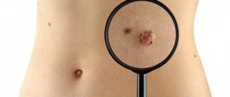
Moles present on human skin can be dangerous (they can become malignant over time) and harmless. Intradermal nevus is a benign formation that does not threaten human health. But in some cases, it can degenerate into a dangerous neoplasm, so you need to monitor external changes in birthmarks.
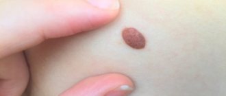
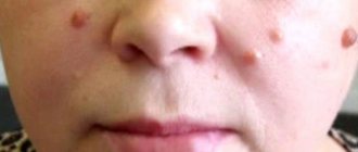
This type is distinguished by a dark shade (brown or dark brown), defined boundaries and softness. At the same time, there are no inflammatory processes in it, and in appearance it is similar to a wart and has a bumpy surface. Intradermal nevi can be single or multiple, the size of small neoplasms is about 3-5 millimeters, large ones reach 5 cm. Moles of a non-pigmented form are rare.
Types of pigmented nevus formations
Let us dwell in more detail on the features of the most common or most dangerous types.
Border
Borderline pigmented nevus, synonymous with junctional nevus, is the initial stage of the development of skin lesions. It is localized in the uppermost layer of the skin - the epidermis. The formation is congenital or acquired. It often develops in childhood and adolescence, localizing on the palms or soles. Microscopic analysis reveals well-defined clusters, called nests, of nevocytes in the lower epidermal layer at the border with the dermis, hence the name. This type is melanoma-dangerous because it can often degenerate into a malignant tumor.
It is a flat pigment formation. For a long time, pigmented nevi are small in size - up to 3 mm, which gives a person the impression that he has had these spots since birth. In some cases, borderline nevi begin to grow rapidly - 1 mm per year, growing in a few years to a size of 5-6 mm. With such a significant increase in the spot, it is necessary to show such a growing mole to an oncologist. It can transform into a dysplastic nevus, a precursor to skin cancer.
Intradermal
The most common form of pigment spots is intradermal pigmented nevus. It is otherwise called a “resting”, inactive, mole or birthmark. It is characterized by a variety of shapes and colors. A mole is the most common benign formation. It has the appearance of a flat plaque with a diameter of up to half a centimeter, any shades of brown, evenly colored, with a dense texture, rounded shape and clearly visible boundaries. The outer layer of such a formation can be convex, in the form of a wart or a hemisphere. In this case, a papillomatous melanocytic pigmented nevus of the skin is formed.
Nevocytes are located deep in the dermis, its middle and lower layers, forming clusters there. The cells have different shapes and are quite large in size. If they have small nuclei arranged in the form of rosettes, this confirms the benign nature of the formation. Intradermal pigmented nevus may be visible at birth or appear at an early age.
Reasons for education
At present, experts cannot name the exact cause of the formation of this type of nevus. There is only a version that explains how an intradermal mole can appear on the body.
During intrauterine development of the fetus, so-called nevus cells are formed - melanoblasts, which participate in the formation of nerve ganglia, meninges, and adrenal glands. These cells also produce melanin, which gives the skin its tint. Some melanoblasts do not have time to mature, remain in the dermis and begin to accumulate. As a result, birthmarks form on the body, one of the varieties of which is intradermal nevus. As the name suggests, the cells from which this type of mole is formed are located deep in the inner connective layer of the skin - the dermis.
An important role in the appearance of this type of nevus is played by the hereditary factor. Most often, birthmarks, which are located deep in the dermis, become noticeable after 10 and even after 30 years. This is explained by the fact that the transformation of a nevus into an intradermal one takes place in several stages:
- In childhood, birthmarks are less noticeable. They are superficial, located not deep under the epithelium;
- Over time, the cells begin to transform and sink into the deeper layers of the skin. First, they penetrate into the boundary zone between the epithelium and dermis. Borderline moles appear;
- After a certain time, melanoblasts penetrate even deeper into the dermal layer and enter the final development phase. The mole becomes more noticeable - intradermal. At this stage, a stalk may appear, and the mole takes the form of a wart - a papillomatous nevus is formed;
- Plunging into the dermis, pigment cells stop synthesizing melanin and some intradermal nevi lose color and become colorless.
Dysplastic nevi
Any pigmented formation can degenerate into a tumor with signs of malignancy (uncontrolled growth, the possibility of metastasis) - melanoma. The risk of such transformation is especially high if there is a dysplastic pigmented nevus.
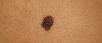
The link between hyperpigmentation and skin cancer has been discovered for almost 200 years, but it wasn't until 1978 that the true precursor to melanoma was found. Such formations (dysplastic nevi) are otherwise called VK moles (after the letters with which the surnames of the very first families studied then began). They are larger than normal birthmarks, their diameter can exceed 12 mm. Outwardly, they look like flat plaques; their surface may be uneven and their shape may be irregular. They rise slightly above the surface of the skin and are unevenly colored in brown shades. Sometimes such structures do not contain melanin.
On histological examination, the formation looks like a complex or mixed pigmented nevus, but there are signs of infiltrative growth, that is, penetration into the surrounding layers of the skin. The shape of the cells also changes: they become angular, with uneven contours, with brightly colored (hyperchrome) nuclei. The condition that allows melanoma to be excluded is the absence of penetrating (infiltrative) growth towards the upper layer of the skin - the epidermis.
VK moles can appear at any age and on any part of the body. Sometimes their number exceeds several dozen. If there are already such cases in the family, the probability of such a nevus appearing over the age of 59 years exceeds 50%. In this case, the disease is transmitted in an autosomal dominant manner. In this case, they talk about FAMMM syndrome (Family atypical multiple mole melanoma syndrome). This is the main factor in the development of skin cancer.
If a dysplastic nevus occurs in isolation, without a hereditary predisposition, then it has a benign course.
Juvenile melanoma; Dysplastic nevus
Distinctive features of intradermal nevus
Intradermal moles have characteristics that distinguish them from other formations:
- constant uniform color;
- clear boundaries;
- soft to the touch;
- There are no inflammatory processes.
It is probably rare that there are no moles on the human body; usually there are several of them and they can be of different types. For example, if the nevus is flat, then it is a non-cellular or pigmented type. A nevus that looks like a blackberry and protrudes above the skin is called papillomatous.
If we talk about size, moles can be very small from 2 millimeters and quite large at several centimeters. They also differ greatly in color and can be pale, that is, without color at all, they are called depigmented. They can be light brown, brown, pink and, finally, very dark, black.
Intradermal nevi
Pigmented nevus of the conjunctiva
This formation is practically not dangerous. It can be located on the conjunctiva (mucous membrane) and is then clearly visible. Also, a pigmented nevus of the eye can lie on the choroid of the eyeball and can only be detected when examining the fundus of the eye by an ophthalmologist. This spot is formed by accumulations of melanocytes and looks like an ordinary mole.
Eye nevi can be stationary (not changing) or progressive (growing). If a lesion on the choroid grows, over time it can cause a narrowing of the visual field, reduce its acuity and cause other unpleasant symptoms. Therefore, it is removed using microsurgery or laser therapy.
Spitz nevi against cancer: what you need to know
Spitz nevus is a type of rare, non-cancerous skin growth that usually affects people under 35 years of age.
Spitz nevus growths, or Spitz nevi, were once considered a type of skin cancer. They have since been proven to be benign, non-cancerous and relatively harmless.
Spitz nevi were also initially considered to develop only in children. Although 70 percent of cases develop in people under 20 years of age, they can affect anyone at any age.
Treatment of nevi
Treatment of pigmented nevus is carried out in cases where the formation is melanoma-dangerous, grows rapidly or is located in an area where it is constantly injured (for example, on the chin, temples, neck, lower back). If a person has age spots, he should regularly visit a dermatologist to have them examined and monitored.
Removal of a pigmented nevus is performed using surgery or minimally invasive intervention.
Removal of a pigmented nevus surgically is carried out if there is a risk of malignancy of the formation. Excision of the spot using a scalpel is carried out within healthy tissue, after which a scar may remain.
Minimally invasive methods are more often used on open areas of the skin, when the removal of age spots is primarily for aesthetic purposes. Cryotherapy, electrocoagulation, laser treatment, and removal using the so-called radioknife are used.
Diagnostics
To diagnose the formation and distinguish it from skin diseases, a differential approach is used. To do this, examine the mole according to the ABCDE system - asymmetry, uneven bumpy borders, different color areas, diameter (critical is considered to be over 6 mm). To check the malignancy of the formation, a smear is taken for microscopy, and sometimes a luminescent study is performed. Histological biopsy is prohibited as a diagnosis, since it provokes the degeneration of a benign mole into a malignant formation.
Prevention of malignancy
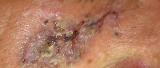
The most likely malignant degeneration of large, unevenly colored, numerous formations. You should consult a doctor if a mole begins to grow, its surface or shape changes, redness or skin itching appears around it, bleeding, peeling, or sudden hair growth on the surface.
To prevent melanoma (skin cancer), you should follow simple rules:
- Avoid skin exposure to direct sunlight from 11 a.m. to 4 p.m.;
- refuse to visit the solarium;
- prefer light clothes with long sleeves, wear wide-brimmed hats in summer.
Using sunscreen does not reduce the risk of developing melanoma.
general description
This pathology is often a benign neoplasm on the skin, which mainly rises above its surface. In some cases, it may take on a lumpy shape, and sometimes remains a small spot of different color.
Intradermal nevus has a painless surface and soft structure. The color of the neoplasm can be different: pink, red, brown and even black.
An intradermal nevus can be located on the neck, scalp, or face. Very rarely, the formation occurs on the body or limbs. It should be borne in mind that if the tumor is benign, then it practically does not cause pain. It degenerates into a malignant form, melanoma, in only 20% of cases.
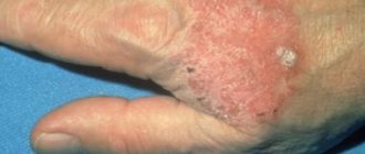
If there is some discomfort in the area of the skin lesion, or the intradermal nevus begins to grow rapidly, you should immediately consult a doctor.
The most dangerous species
Some age spots and nevi have an increased risk of degeneration into a malignant tumor. These include the following types:
- border;
- blue or light blue;
- VK mole;
- giant;
- nevus of Ota.
It is almost impossible to determine on your own what it is, a pigmented nevus or a dangerous tumor. In addition to external signs, it is necessary to take into account the nature of the location of the neoplasm, its composition and cell type. Only a dermatologist-oncologist can make a diagnosis of “pigmented nevus” and select therapy.
Kinds
Classification of melanocytic nevi according to J. Bhawan (1979) depending on the origin:
| View | Subspecies |
| Epidermal | Intraepidermal, borderline, complex. This group also includes special forms: spindle cell or epithelioid, nevus of balloon-forming cells, halonevus |
| Dermal | Mongolian spot, nevus of Oto, nevus of Ito and blue |
| Mixed dermal and epidermal | Combined |
| Melanoma precursors | Congenital, dysplastic |
Formations of epidermal origin are usually acquired and occur in adolescence.
Size classification:
- small (0.5–1.5 cm) – only observation is indicated;
- medium (1.5–10 cm), planned surgical removal of the tumor is optimal;
- large (over 10 cm) planned palliative therapy is indicated (surgical treatment due to the large area is not always indicated);
- giant – occupy one or more anatomical areas; Only palliative therapy and observation are indicated (surgical treatment is not used).
Papillomatous nevus in children
The formation of a mole can occur immediately after birth.
Expert opinion
Sakania Luiza Ruslanovna
Dermatovenerologist, cosmetologist, trichologist
Ask a Question
Such neoplasms do not pose a danger only if its properties do not change and the nevus itself is not subject to mechanical damage.
If a mole is regularly injured and bleeds, you should contact a specialist who may decide to remove the injured nevus.