There are diseases whose origin and “deep” causes have not been sufficiently studied. One of them is vitiligo or leukoderma. It comes from nowhere and disappears into nowhere. This is a serious, but absolutely not contagious skin pathology.
If white spots become noticeable on the body (one suddenly discovered non-pigmented area is enough), you need to find information and photos on the Internet where you can clearly see what vitiligo looks like, and immediately consult a dermatologist (you can even see a venereologist).
Tests confirming the diagnosis
A special examination - ultraviolet radiation with a Wood's lamp - can easily detect a bluish-white color of the skin in the armpits, groin, and under the hairline. This means that the natural pigment melanin in melanocytes - cells with large dark nuclei located in the basal layer of the skin - has begun to degrade.
Malfunction and death of melanocytes stops the accumulation of melanin and discolors the affected areas of the skin.
Biopsy is rarely used. To do this, scrape the epidermis along the edges and in the center of the spot. Test results often confirm doctors’ fears, although they exclude the patient’s infection with syphilis, leprosy, lupus or lichen.
Such a negative result is a great joy, since there is no unnecessary and contagious muck in the body.
Preventive recommendations
Preventing a disease is much easier than treating it. In the case of vitiligo, the risk of its development can be reduced by:
- proper, balanced nutrition;
- timely elimination of infectious and viral diseases;
- prevention of worms;
- active lifestyle;
- playing sports.
You should also protect yourself from nervous tension and stressful situations. It is important to monitor your immune system and be careful when taking any medications.
Injuries and burns lead to the death of melanocytes and loss of natural pigment, so you need to be as attentive to your health as possible.
Nutrition, as well as following a number of simple rules for vitiligo, can cure the disease using non-invasive methods without surgery.
Everyone in the world gets sick - both adults and children.
Vitiligo is indecently indiscriminate because it makes no distinction at all between the racial and national characteristics of people, their children, parents and friends.
Vitiligo is diagnosed in 1% of the world's population aged 10 to 30 years, that is, in both adults and children. This pathology is never found in infants and young children.
The first distinctive sign of the disease is a rounded or uneven-edged spot on the mucous membranes or skin (“circled” with a darker outline than a healthy area of skin), which constantly increases and merges with other light islands.
How to diagnose
In a hospital, a doctor can easily diagnose vitiligo, but it is better, of course, to donate blood and scrapings for analysis. The lesion does not contain melanin pigment; it is concentrated at its border, within a range above normal. Some diseases can also manifest themselves as similar spots:
- similar spots can cause sexually transmitted diseases, especially during syphilis. They often give additional symptoms, itching in the anus and irritation of the mucous membrane;
- lichen versicolor can also leave characteristic spots that disappear over time. At the site of formation of such spots, vitiligo may develop, then the skin will quickly lighten;
- leprosy forms light patches, not as light as vitiligo, but can spread widely. An important feature of leprosy is that the places where it is affected are devoid of sensitivity: with strong pressure, the skin does not react.
To clarify, you need to take the appropriate tests to the laboratory and consult with a dermatologist.
The path to recovery is long and difficult
A dermatologist, having determined the degree of vitiligo damage, selects a set of medications and an individual treatment regimen for each patient. The main effective methods of treating vitiligo:
- hormone therapy in the form of tablets. These are “Prednisolone”, “Betamethasone”, “Diprospan”;
- calcineurin inhibitor ointments - “Protopic”, “Elidel”;
- ointments that enhance the absorption of ultraviolet rays by the skin - “Melaginin”, “Psoralen”;
- gels and tablets of complex action - “Vitix”, “Viticolor”;
- immunosuppressants - Levamisole, Isoprinosine;
- light therapy of the affected areas with medium-length waves (type B);
- PUVA therapy together with photosensitizers (100-200 sessions);
- irradiation with infrared and red laser beams;
The course of treatment is at least 6 weeks, sometimes up to 3 months.
Classification
Based on the location of the spots, vitiligo is divided into three forms:
- Localized (segmental) - individual areas of the skin are discolored.
- Generalized - discolored areas are scattered throughout the body.
- Universal – about 80% of the entire skin is discolored.
By type of discoloration:
- Tricolor vitiligo - the appearance of spots appears in the form of a three-color fill (discolored skin, healthy epidermis and an area of medium pigmentation).
- Four-color vitiligo - a zone of hyperpigmentation is added to the three-color spot.
- Blue vitiligo - discolored spots with a bluish tint.
- Inflamed vitiligo - discolored spots surrounded by a rim of inflammation. Its structure slightly rises above healthy and discolored skin.
Classification of the skin defect helps to develop therapeutic regimens.
Additional wellness activities
Treatment of vitiligo with folk remedies at home is not contraindicated:
- St. John's wort oil (stimulating pigment production);
- black cumin oil (with a general strengthening effect);
- tincture of swamp duckweed (containing a lot of bromine and iodine);
- rubbing depigmented areas with birch tar;
- using alcohol tincture of walnut (to treat white spots);
- to strengthen hair – apply 2% Sulsena paste to hair (for half an hour).
The professionalism of the doctor and the competent behavior of the patient are 2 determining and effective factors in how to properly treat vitiligo.
A scientifically based approach to the problem guarantees a complete cure for leukoderma, although the disease can return if you do not follow a balanced diet, sunbathe for a long time in the solarium and on the beach, damage the skin with hard washcloths and the friction of tight clothing, continue to work in hazardous chemical industries, drink, smoke and lead an antisocial lifestyle.
Treatment of dermatitis - professional methods for effectively getting rid of the disease in different areas of the skin (105 photos)Treatment of varicose veins - which specialist to contact and how the treatment procedure occurs. Stages of recovery and description of treatment (140 photos + video)
Allergy pills are the most effective broad-spectrum medications. The best manufacturers and characteristics of optimal anti-allergenic products
Stages of vitiligo
The disease usually begins unnoticed. Rarely, the onset of the disease is preceded by slight itching, hyperesthesia and paresthesia in the form of tingling, crawling or primary redness of the skin (“pink vitiligo”).
Vitiligo spots are clearly defined, have smooth or scalloped edges, often round in outline. The color of the spots can range from white to milky white. The skin in the lesions (except for discoloration) is externally unchanged, without signs of atrophy and peeling.
Often, in spots located on open areas of the body (face, neck, chest, limbs) and exposed to frequent ultraviolet irradiation, not only redness, but also peeling and even thickening of the skin can be observed. The size, outline, number and location of spots are individual for each patient.
Once they appear, the symptoms of vitiligo can exist for years without changing, or the process on the skin progresses, accompanied by an increase in the size of the spot, the merging of old lesions and the appearance of new ones.
Spots often appear at the site of skin trauma (Koebner phenomenon).
There may also be cases of spontaneous disappearance of vitiligo lesions. Spontaneous repigmentation (usually partial and temporary) is observed in 20% of patients, mainly after sun exposure.
Important! It is not possible to predict how a patient’s vitiligo will behave throughout his life. It is not at all necessary that if a patient has a single focus of vitiligo, then in the future he will have multiple foci of depigmentation in all parts of the body!
The course of the pathological process with vitiligo is described as: stable vitiligo (the white spot that appears remains unchanged for many years); progressive vitiligo (the process of depigmentation constantly progresses slowly or quickly); unstable vitiligo (some of the white spots increase, while the other part may regress).
How the disease behaves at the time the patient contacts the doctor is important for prescribing one or another set of treatment, which I talked about in my previous articles on my blog!
As I already said, during the course of vitiligo (with treatment, and sometimes without it), some of the spots may, to a greater or lesser extent, restore their original color, i.e. repigment. Repigmentation is evidenced by the appearance of sharp scalloping of the edges (marginal type of repigmentation), smoothing of the outline of the boundaries, as well as the appearance of pigment inclusions in the focus of depigmentation in the form of small dots (perifollicular type of repigmentation), which is caused by the migration of melanocytes from the hair follicle.
Marginal repigmentation
Perifollicular repigmentation
Sometimes there is a combination of vitiligo with:
Setton's nevus (halonevus) is a pigmented nevus surrounded at the periphery by an area of depigmented skin;
achromotrichia - discoloration of strands of hair on the head, in the area of growth of eyelashes, eyebrows, etc.;
leukonychia - the formation of white spots on the nails.
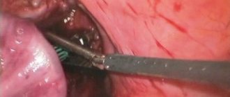
Surgery
Radical methods are used when other methods fail to achieve positive dynamics, when white areas of skin bring severe moral discomfort to the patient.
In this case, the problem can be solved in different ways:
- Segmental extraction of the depigmented area and replacement of it with a healthy flap of skin.
- Transplantation of micrografts (healthy areas of the epidermis several millimeters in size) onto bleached surfaces.
- Transplantation of cultured epidermis (artificially grown in laboratory conditions from cells of a sick patient).
- Transplantation of melanocytes (cultured and uncultured).
When choosing a radical method of treatment, you need to be prepared for the fact that even limited surgical intervention can provoke the formation of scars, lead to the formation of a cobblestone effect (uneven pigmentation), and the emergence of the phenomenon of Kobnerization (exacerbation of pathology). Any operation described above is dangerous because after its completion, implant rejection may begin.