Symptoms
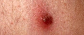
The most common type is nodular. It is characterized by a painless course of the pathology. First, several tubercles up to 3 mm appear, which are yellow in color. Then the size increases, and a depression develops inside, covered with a crust of skin. When removed, bleeding begins. The infiltrative type looks like a nodule that gradually grows. After 5 months, ulcers with raised edges appear. They hurt and bleed when touched. An unpleasant putrid odor appears. Plaque cancer grows quickly and infects other tissues. It looks like a red, painful lump.
Papillary is rare. It looks like a nodular one. Slowly developing. Its subtype, verrucous, is characterized by slow growth and rare metastasis. Glandular squamous cell carcinoma spreads to the mucous membrane, most often to the uterus. This is a formation that has a jelly liquid inside. Invasive is characterized by the rapid growth of metastases and their penetration into organ tissues and lymph nodes.
Stages
In order to better select a treatment plan for the disease, the disease is classified into the following stages:
- 1 – initial . The lesions are minimal, their size does not exceed 1-2 cm. Despite the fact that the base of the formation is not motionless, it is located within the affected area and does not injure neighboring tissues. There are no symptoms, metastasis is not detected;
- 2 – this stage is characterized by the rapid growth of the anomaly. The disease progresses, the area of distribution of mutating cells increases. However, this course cannot yet be considered active, since the tumor can be controlled.
In some cases, distant single metastasis and damage to lymph node connections located in the immediate vicinity may be observed;
- 3 – cancer spreads to most of the lymphatic system , affects surrounding formations, tissues and actively grows into neighboring organs and systems. Treatment at this stage is difficult, but there is still a chance for an optimistic prognosis. Symptoms are severe and often painful;
- 4 – final stage of the disease. Not only soft tissues, but also bone tissues and cartilage joints were subjected to irreversible processes. Even with a relatively small tumor, it is capable of producing multiple metastases.
Almost all joints become immobilized. The patient endures stage 4 extremely difficult. Treatment is no longer effective. The only thing doctors can do is to alleviate the symptoms and somewhat prolong the patient’s life.
Treatment of the disease
Oncology is treated depending on the location and stage of the disease. It is important to identify the problem in time, since the disease is prone to rapid metastasis. First, doctors surgically remove malignant tumors, and then carry out therapy to destroy the remains of cancer cells.
Medications
In the early stages, when the cancer pathology has not yet grown and secondary lesions have not appeared, chemotherapy is administered. This is a treatment with strong chemicals that are injected into the body. The method is also used before surgery to shrink cancerous growths. The following medications are used for therapy:
Radiation therapy
Used together with chemistry in the first stages or before surgery. This method also has a beneficial effect on lesions that are located deep under the skin. If the cancer is determined to be at the last stage and it is necessary to stop its development, radiation therapy is also used. It affects cells and stops the spread of possible metastases.
Surgical intervention
The tumor is removed along with the skin at a distance of 2 cm from it to eliminate possible malignant cells in the pathological area. If the formation is in an accessible place, cryodestruction, curettage or electrocoagulation is used. But such methods are ineffective if the squamous cell carcinoma is located in the hairline. If it is necessary to preserve an area of skin near the tumor, cryosurgery is used.
Folk remedies
These are additional therapy measures that are prescribed in conjunction with medications and surgery. Traditional medicine cannot be used as the only way to get rid of cancer. But to improve the condition, you can treat the ulcers with an ointment made from pomegranate seeds and honey. The lesions are wiped with lotion made from verbena and vinegar or from birch buds. A product made from oil and ground walnuts is applied spot-on.
Forecast
Squamous cell carcinoma with keratinization has a difficult prognosis even if detected in a timely manner. Only treatment carried out in the early stages can improve the situation.
Thus, the five-year survival threshold against the background of intensive therapy and depending on the stage of development of the anomaly is overcome:
- 1 – more than 92%;
- 2 – about 60%;
- 3 – about 35%;
- 4 – no more than 12%.
Photo: the process of restoring the skin area after tumor removal
Regardless of the not so optimistic statistics, it is necessary to understand that persistent ignoring of the problem makes these figures even more pessimistic - in an untreated state, the disease quickly progresses and claims a person’s life 2 to 3 years after the onset of the disease.
If you find an error, please select a piece of text and press Ctrl+Enter.
>
Survival prognosis
A patient’s standard of living after cancer depends on the type of cancer and the speed of its development. At the first stage, the forecast is 90%, at the second it drops to 60%, and at the third it drops to 30-35%, and at the fourth it does not rise above 10%. If cancer is detected at an early stage and the patient is provided with timely and comprehensive treatment, then the prognosis is 98% that the cancer is completely cured. Therefore, with any manifestation of neoplasms on the skin, you should consult a doctor for timely removal of dermatitis.
Squamous cell carcinoma, or squamous cell carcinoma, is a histological type of malignant tumor, such a diagnosis is made by the results of a biopsy after examining a sample of tumor tissue under a microscope. The neoplasm is formed from flat epidermal cells that look like scales. It can occur on the skin, in the oral cavity, in the larynx, trachea, bronchi, esophagus, genitals, and rectum.
At the European Oncology Clinic, the diagnosis and treatment of squamous cell carcinoma is carried out by expert doctors who have extensive experience working in leading oncology centers in Moscow. A team that includes oncologists, dermato-oncologists, surgeons, chemotherapists, radiotherapists and other specialists works with the patient. We use innovative treatment methods, the latest generation of drugs, and conduct antitumor therapy in accordance with leading international recommendations. The European Oncology Clinic is the first Russian private oncology center where you can receive effective palliative treatment in late stages, even in cases where the patient was abandoned in other clinics.
Treatment and prognosis of the tumor
Any location of squamous cell oncology involves the use of the following therapeutic techniques:
- Chemotherapy – involves the use of antitumor drugs; Radiation therapy - based on the use of gamma rays in treatment; Surgical intervention involves removal of the tumor itself and metastatic lymph node structures.
For small, superficial tumors, alternative therapies such as electrocautery, photodynamic therapy, or cryotherapy may be used. After treatment, the patient periodically visits the oncologist to avoid relapse.
Prognosis for squamous cell carcinoma is determined by the location of the oncological process and its stage:
- cervical cancer at the first stage has a 90% survival rate, at the second stage – 60%, at the third stage – 35%, at the fourth stage – 10%; Pulmonary squamous cell carcinoma is characterized by survival rate at the first stage - about 35-40%, at the second - 15-30%, at the third - 10%; Skin cancer at stages I-III – 60% survival rate, at stage 4 – 40%.
Tumors detected early are usually easier to treat and have a better prognosis than advanced cancers.
Causes of squamous cell carcinoma
The causes of squamous cell carcinoma are the same as for other types of malignant tumors. Certain mutations occur in cells that lead to malignant degeneration. “Incorrect” cells lose the external features and functions of normal ones, begin to multiply uncontrollably, and acquire the ability to spread throughout the body.
The main risk factors for squamous cell carcinoma:
- On the skin, such tumors often arise due to the action of ultraviolet rays. Exposed areas of the body are the most vulnerable.
- Squamous cell carcinoma of the genitals, head and neck is caused by certain types of human papillomavirus.
- The risk of developing squamous cell carcinoma is increased in smokers and people who drink a lot of alcohol.
- The likelihood of developing cancer increases with age as mutations accumulate in the cells of the body.
- Scars, burns, chronic inflammation.
- Exposure to certain carcinogenic substances, for example, if a person works in an industrial environment and comes into contact with chemicals.
- Decreased immunity.
None of these factors is guaranteed to lead to the disease - each of them only increases the likelihood to a certain extent.
Causes
The main provoking factors that can cause the development of a squamous cell tumor with keratinization are:
- incorrect exposure to the sun - direct radiation flows are extremely aggressive and can deeply affect tissues, destroying their structure at the cellular level. At the same time, the qualitative content of the cells undergoes a change, causing their degeneration;
- thermal or chemical burns - such phenomena completely break the structural molecular lattice, resulting in the appearance of abnormal, chaotically multiplying cells of a malignant nature, which, in fact, is cancer;
- direct contact with dangerous chemical components - during the production process a person may be forced to receive a certain toxic dose, which, accumulating in the body, gives rise to pathological formations;
- skin diagnoses – Paget virus, Bowen virus, xeroderma pigmentosum – the chronic course of these diseases can cause microtraumas of the surface and qualitatively change the content of tissues.
The usual processes of cell division in the lesion are disrupted and the process becomes uncontrolled. After a little time, this zone becomes malignant and a precancerous anomaly forms;
- smoking - carcinogens, penetrating into the blood, create favorable conditions for the development of cancer processes in the most vulnerable places of the human body.
What does tonsil lymphoma look like: photo and description.
In this article we will tell you how to detect Bartholin gland cancer at the beginning of its development.
What does cervical cancer look like? Photos are posted here https://stoprak.info/vidy/zhenskix-polovyx-organov/shejka-matki/kak-vyglyadit-izobrazheniya-s-opisaniem.html.
What are the types of squamous cell carcinoma?
Malignant neoplasms of this histological type are found on different parts of the body. Depending on the location, their properties, approaches to diagnosis and treatment, and prognosis for the patient may differ slightly.
Skin cancer
Malignant skin tumors are represented by squamous cell carcinoma in approximately 20% of cases. Much more often, patients suffer from basal cell carcinoma, which originates from cells located in the lower layer of the epidermis.
Squamous cell carcinoma is more aggressive than basal cell carcinoma. It is more likely to grow into the deeper layers of the skin and spread throughout the body with the formation of distant metastases. However, this happens quite rarely. Most often, the tumor can be detected and removed at an early stage.
As a rule, squamous cell carcinoma occurs on the skin of the face, ears, neck, back of the hands, and less commonly in the genital area. Often, a neoplasm develops where scars and chronic damage are located.
Squamous cell carcinoma of the red border of the lips
Malignant lip tumors account for no more than 1–3% of all cancers. In most cases (95%) they are represented by squamous cell carcinoma, which comes in two types:
- Squamous cell keratinizing carcinoma does not behave as aggressively, grows slowly, and rarely forms distant metastases.
- Nonkeratinizing squamous cell carcinoma grows rapidly, ulcerates earlier, and metastasizes more often.
Research shows that this type of cancer is 3 to 13 times more common in men than in women. This is probably due to the fact that males are more often exposed to sunlight at work, and smoking and drinking alcohol are more common among them.
Oral cancer
Oral cancer is a malignant tumor that occurs on the mucous membrane of the lips, cheeks, gums, the anterior two-thirds of the tongue, the palate, and the floor of the mouth (located under the tongue). In 90% of cases they are represented by squamous cell carcinoma, of which 5% are keratinizing squamous cell carcinoma, which is less aggressive, less likely to grow into surrounding tissues, spread to lymph nodes and metastasize.
Esophageal carcinoma
The mucous membrane of the esophagus is lined with stratified squamous epithelium, and squamous cell carcinoma can develop from it. Most often, such tumors are located in the cervical esophagus and the upper two-thirds of the thoracic region. In the lower third of the organ, adenocarcinomas, malignant tumors of glandular cells, are more common.
Laryngeal cancer
In laryngeal cancer, the tumor almost always develops from squamous epithelium and is a squamous cell carcinoma. Typically, the appearance of a tumor is preceded by precancerous changes - dysplasia. The cells that are located in the lesion do not look like normal ones, but they also differ from cancer cells. In some cases, dysplasia does not lead to the development of cancer and even goes away on its own, especially if its cause is eliminated, for example, a person quits smoking. But in some people, precancerous changes lead to “cancer in situ” and then an invasive tumor.
Trachea and bronchus cancer
Squamous cell carcinoma is the most common type of malignant tumor in the trachea. It usually occurs in the lower part of the trachea, grows quite quickly, invades its wall, leading to ulceration and bleeding. This is a rare type of cancer and its main cause is smoking.
The most common lung cancer is non-small cell cancer - it occurs in 80% of cases and in 30% of cases it is squamous cell carcinoma. Often these tumors are located in the bronchi.
Cervical cancer
The cervix consists of two parts. The exocervix is located outside, in the vagina, this is what the gynecologist sees during the examination. The endocervix is the canal of the cervix, it connects the uterus to the vagina. Normally, the exocervix is lined with squamous epithelium, and the endocervix is lined with glandular epithelium. The place where they meet is called the transformation zone.
Squamous cell carcinoma represents 90% of malignant tumors of the cervix. Most often, the neoplasm occurs in the area of the transformation zone. Cancers that develop from the glandular cells of the endocervix are called adenocarcinomas.
In rare cases, glandular squamous cell carcinoma occurs in the cervix.
Vulvar cancer
The vulva is the name given to the external female genitalia: the vestibule of the vagina, the labia majora and minora, and the clitoris. The majority of cancer types that develop in this area are squamous cell carcinoma (70–90%). They are divided into two groups:
- A large group are tumors whose origin is unknown. Most often they are diagnosed in older women.
- A smaller group are malignant tumors caused by the human papillomavirus.
Rectal cancer
In most cases, malignant tumors of the rectum are represented by adenocarcinomas - glandular cancer. Squamous cell carcinoma in this organ is very rare and accounts for 10 to 25 cases for every 100 thousand cases of colorectal cancer.
Squamous cell carcinoma accounts for 90% of all malignant neoplasms of the anal canal, the narrow passage that connects the rectum to the anus.
Tonsil cancer
A person has four types of tonsils: palatine (when they become inflamed, tonsillitis develops), tubal (located in the pharynx near the openings of the auditory tubes), lingual (behind the tongue) and pharyngeal (in children this causes adenoids). Most often, malignant tumors develop in the tonsils. In most cases it is squamous cell carcinoma. It is difficult to diagnose, so it is often detected in late stages.
Skin tumor
Of the total number of malignant tumors, skin carcinoma is approximately ten percent. Dermatologists are currently reporting an upward trend in incidence, with an average annual increase of 4.4%. This cancer most often develops in older people, regardless of their gender. Light-skinned people are especially predisposed to the disease, as well as people who live in conditions of strong insolation (high mountains and hot countries) and are outdoors for a long time.
Among the total number of phenomena of such oncology, from 11 to 25% are squamous cell forms and from 60 to 75% are basal cell cancers. Since the development of basal cell and squamous cell skin cancer occurs from epidermal cells, such diseases are also classified as malignant epitheliomas.
Types of disease diagnosis
The oncologist prescribes certain types of diagnostics to the patient, depending on which organ the malignant tumor is located in:
Location of cancer
In all cases where a pathological formation is detected, a biopsy is performed - a study during which a fragment of suspicious tissue is obtained and sent to the laboratory for histological and cytological examination. Biopsy is the most accurate method for diagnosing cancer. It helps not only to reliably establish a diagnosis, but also to determine the histological type of the tumor. In order to check the extent of cancer spread in the body and clarify the stage, the doctor may prescribe additional tests:
- computed tomography, MRI;
- X-ray of the chest, bones;
- PET scan;
- Ultrasound and endoscopic examination of organs into which cancer could have grown.
Diagnostics
Diagnostic tests for squamous cell oncology include the following procedures:
- PAT; Endoscopic examination; Cytogram; X-ray diagnostics; Laser confocal scanning microscopy; CT, MRI; Biopsy with histology; Laboratory diagnostics with identification of tumor markers, etc.
The tumor marker for squamous cell type cancer is the SCC antigen. If its concentration in the blood exceeds 1.5 ng/ml, then the patient has a high probability of such a cancerous lesion.
In such cases, the patient is prescribed a thorough endoscopic and tomographic examination to identify the location of the cancer focus.
Survival prognosis for squamous cell carcinoma
The prognosis depends on where the cancer began to grow, at what stage the diagnosis was made and treatment started. For example, often the survival rate for cancer of the skin and red border of the lips tends to 100%, because such tumors, as a rule, can be detected early enough, and they are not very aggressive. If distant metastases appear, the chances of remission become extremely low. But such patients can still be helped: to slow down the progression of squamous cell carcinoma, prolong life, improve their general condition, and relieve painful symptoms.
Skin carcinoma symptoms
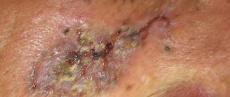
Squamous cell skin cancer is characterized by rapid spread and growth both in depth and along the surface of the epidermis. The growth of a tumor into the tissues located under the skin (cartilage, bone, muscle), or the addition of an inflammatory process is accompanied by the occurrence of pain syndrome. Squamous cell skin cancer appears as a nodule, plaque, or ulcer.
The ulcerative form of squamous cell skin cancer is externally a crater-shaped ulcer, which is surrounded, like a roller, by tight, raised and abruptly ending edges. The ulcer has an uneven bottom, it is covered with crusts of dry bloody-serous exudate. It smells quite unpleasant. The plaque of squamous cell skin cancer has a bright red hue, a lumpy surface and a dense consistency. It often bleeds and increases rapidly.
In squamous cell carcinoma of the facial skin, the coarsely lumpy surface of the nodule makes its shape resemble a mushroom or cauliflower. Characterized by a brown or bright red color and high density of the tumor node. Its surface may become ulcerated or eroded.
Prevention
Basic measures to prevent squamous cell carcinoma:
- Quitting smoking and drinking alcohol.
- Protecting your skin from ultraviolet rays is the most important measure for preventing skin cancer. You should not visit solariums or go to the beach from 10.00 to 16.00, when solar activity is highest. Clothes with long sleeves and trousers, a wide-brimmed hat, and sunglasses help protect yourself.
- Preventing infection with HPV, which leads to the development of cancer: you need to avoid promiscuity and use condoms. There is currently a vaccine against human papillomavirus infection. It is recommended that all adolescents be vaccinated before becoming sexually active.
Squamous cell carcinoma develops from keratinocytes (one of the types of epidermal cells - stratified squamous keratinizing epithelium). Affects the skin and mucous membranes.
Reasons for the development of pathology
The main reason for the development of squamous cell skin cancer is considered to be genetic predisposition. It can be hereditary or acquired and is expressed in:
- Damage to cellular DNA under the influence of certain factors, resulting in a mutation of the “TP53” gene, which encodes the “p53” protein. The latter, as a regulator of the cell cycle, prevents tumor transformation of cells. "TP53" is one of the main genes involved in blocking the development of malignant tumors.
- Disorder of the functions of the immune system directed against tumor formations (antitumor immunity). Many cellular mutations constantly occur in the human body, which are recognized and destroyed by cells of the immune system - macrophages, T- and B-lymphocytes, natural killer cells. Certain genes are also responsible for the formation and functioning of these cells, mutations in which reduce the effectiveness of antitumor immunity and can be inherited.
- Disorders of carcinogenic metabolism. Its essence lies in the mutation of genes that regulate the intensity of the function of certain systems, which are aimed at neutralizing, destroying and quickly removing carcinogenic substances from the body.
Favorable background for the development of squamous cell skin cancer are:
- Age. The disease is extremely rare among children and young people. The percentage of cases increases sharply among people over 40 years of age, and after 65 years of age this pathology occurs quite often.
- Skin type. People with blue eyes, red and blond hair, and fair skin that is difficult to tan are more susceptible to the disease.
- Male gender. Among men, squamous cell carcinoma develops almost 2 times more often than in women.
- Skin defects. Cancer can develop on clinically healthy skin, but much more often - against the background of freckles, telangiectasias and genital warts. precancerous diseases (Bowen's disease, Paget's disease, xeroderma pigmentosum), in the area of scars formed as a result of burns and radiation therapy, after which cancer can occur even after 30 years or more, post-traumatic scars, trophic changes in the skin (with varicose veins), fistulous openings passages in osteomyelitis of the bone (metastasis rate is 20%), psoriasis. lichen planus. lesions in tuberculosis and systemic lupus erythematosus, etc.
- Long-term decrease in general immunity.
Among the provoking factors, the main ones are:
- Ultraviolet radiation with intense, frequent and prolonged exposure - sunbathing, PUVA therapy with psoralen, carried out for the treatment of psoriasis and also desensitization for allergies to sunlight. UV rays cause a mutation in the TP53 gene and weaken the body's antitumor immunity.
- Ionizing and electromagnetic types of radiation.
- Prolonged exposure to high temperatures, burns, prolonged mechanical irritation and damage to the skin, precancerous dermatological diseases.
- Local exposure over a long period of time (due to the specifics of professional activity) of carcinogenic substances - aromatic hydrocarbons, soot, coal tar, paraffin, insecticides, mineral oils.
- General therapy with glucocorticoid drugs and immunosuppressants, local therapy with arsenic, mercury, chlormethyl.
- HIV and human papillomavirus infection types 16, 18, 31, 33, 35, 45.
- Irrational and unbalanced nutrition, chronic nicotine and alcohol intoxication of the body.
The prognosis without treatment is unfavorable - the incidence of metastases is on average 16%. In 85% of them, metastasis occurs in regional lymph nodes and in 15% - in the skeletal system and internal organs, most often in the lungs, which always ends in death. The greatest danger is from tumors of the head and facial skin (70% affected), especially squamous cell carcinoma of the skin of the nose (dorsum of the nose) and neoplasms localized in the forehead, nasolabial folds, periorbital areas, in the area of the external auditory canal, red border of the lips, especially the upper one, on the auricle and behind it. Tumors that arise in closed areas of the body, especially in the area of the external genitalia, of both women and men, are also highly aggressive in terms of metastasis.
Localization and symptoms of the tumor
Squamous cell skin cancer
Most often, according to statistics, people with light skin color and red hair get sick. This is explained by the fact that this type of skin has the least protection against ultraviolet radiation. Squamous cell carcinoma occurs in areas of the skin that are exposed to sunlight frequently and for long periods of time.
Squamous cell skin cancer can develop on a healthy area of the skin, but more often develops during the progression of precancerous conditions: focal and cicatricial atrophy of the skin after burns and injuries, at the site of genital warts, against the background of a number of diseases - lupus erythematosus, psoriasis, scrofuloderma, tuberculous lupus, pigmentary xeroderma, idiopathic skin atrophy.
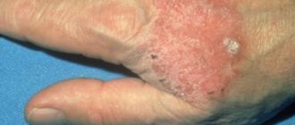
Most often, affected areas are found on the upper half of the torso and arms. The lesion looks like a red spot or plaque with peeling in the center. The scales easily peel off from the surface with the appearance of weeping. The plaque grows and rises above the skin level, and when zones of hyperkeratosis and growths appear, it acquires a heterogeneous mottled appearance. Bowen disease often transforms into squamous cell carcinoma of the skin.
The risk of developing squamous cell carcinoma is higher in patients with immunosuppression, in HPV carriers, or in those who have had any form of the disease.
exophytic or endophytic in its growth pattern . There are usually no difficulties in diagnosing the exophytic form - the tumor looks like a solitary node (in some cases there may be several nodes) rising above the surface of the skin.
The nodule grows over time, becomes lumpy, taking on the appearance of a “cauliflower” . Blood vessels grow in the node, due to which it can acquire a purplish-red or brown color. Sometimes the tumor looks like a yellowish-red or reddish-brown lesion with areas of hyperkeratosis and warty growths on the surface. This form is called verrucous .
The endophytic form of growth is characterized by frequent relapses and metastasis. A node formed on the skin grows into the subcutaneous fat and ulcerates. The bottom of the ulcer is rough, covered with a whitish film. Smaller nodes may form around the main node, also ulcerating, they increase the area of the affected skin. The tumor quickly grows into the underlying tissue and migrates hematogenously to other organs (usually the lungs).
Surgical treatment is used for small tumors. It is based on excision of the tumor within healthy tissue. Surgical treatment methods include electrocoagulation and curettage.
Surgical treatment of skin cancer requires the formation of a cosmetically satisfactory scar.
Chemotherapy, photodynamic therapy, and laser-induced light-oxygen therapy are also used to treat squamous cell skin cancer .
Photodynamic therapy is also common; it requires additional treatment of the tumor with a substance.
The prognosis for squamous cell skin cancer and its timely detection is relatively favorable. Tumors rarely metastasize and recur. The most dangerous location of the primary lesion is in the area of the ear canal, orbit, postauricular area and in the area of the nasolabial folds.
Squamous cell breast cancer
Macroscopically, this cancer is a whitish-yellow node with cavities inside, which determine the size of the formation.
Tumor cells have brightly colored nuclei of different shapes and sizes, located scattered or in random groups. Surrounded by either inflammatory cells or necrotic cells. Some of them may become keratinizing.
Oral squamous cell carcinoma
More than half of the cases of oral cavity diseases are tongue cancer. Since the main cause is alcohol consumption and smoking, the majority of people with this form of cancer are men. Risk factors for the development of squamous cell carcinoma are poor oral hygiene and inflammatory diseases of the oral cavity.
Healthy mucous membranes rarely transform into cancer.
In the early stages, the disease is asymptomatic. As the formation increases in size, swallowing and chewing may be impaired, and speech may change.
Detection of a tumor of the tongue, oral mucosa, and lips is possible both by the patient himself and during a routine examination by a dentist, during which the doctor can scrape off the area of the affected mucosa, examine and detect altered cells. Clarification of the nature of the tumor is possible only after a morphological study.
Lower lip cancer develops most favorably and rarely metastasizes. Upper lip cancer has a more serious prognosis and often metastasizes to regional lymph nodes.
Squamous cell carcinoma of the larynx
Other risk factors for development are: male gender (in recent years, due to the increase in the number of smokers, men and women develop laryngeal cancer with approximately the same frequency), age 30 - 60 years, history of HPV disease, immunosuppression (including HIV), professional hazards (contact with chemicals or radiation), poor nutrition (lack of vitamins and microelements).
Precancerous conditions are:
- with a high incidence of malignancy: leukoplakia, papillomatosis, pachydermia;
- with a low incidence of malignancy: fibroma, scar after burns or previous infections.
Squamous cell keratinizing carcinoma has a relatively benign course, develops slowly and rarely metastasizes. Squamous cell nonkeratinizing laryngeal cancer, on the contrary, grows quickly and metastasizes early.
Classification
Squamous cell oncology is classified according to several principles.
According to the prevalence, cancer is invasive and microinvasive.
According to the degree of cell differentiation, keratinizing, non-keratinizing and poorly differentiated squamous cell carcinoma are distinguished.
Squamous cell carcinoma is also classified by stage, tumor shape, etc.
Squamous cell keratinizing carcinoma
This oncoform is characterized by slow development and growth. Its main difference is the presence of differentiated cancer cells, of which this cancerous tumor consists. It is formed from “pearls” - limited structures with a grayish-white shiny surface.
From a prognostic point of view, this form of squamous cell carcinoma is conventionally considered the most favorable.
Keratinizing cancer can have a highly or moderately differentiated form. Moreover, with an increase in the degree of differentiation, the favorableness of the prognosis also increases, because such formations progress much more slowly.
Another characteristic manifestation of differentiated squamous cell carcinoma is the presence of scaly, keratinized particles located on the outer side of the formation and forming a yellowish border.
The keratinizing form of squamous cell oncology almost always forms on the surface of the skin, although in exceptional cases it can also be found in other structures of the body.
Non-keratinizing
The non-keratinizing form of squamous cell carcinoma consists of a cluster of undifferentiated cellular structures, which is why it has the highest rate of malignancy, an aggressive course and rapid progression.
This cancerous form can form on any organ, but it is still more often found on mucous tissues. On the skin, such an oncoform is detected in only 1 case out of 100, i.e. in 10%.
Poorly differentiated
Low-grade squamous cell oncology has much in common with sarcomatous formations, since it consists of spindle-shaped cell structures.
This type of cancer is characterized by increased malignancy and rapid progression.
Microphoto of poorly differentiated squamous cell carcinoma
Poorly differentiated squamous cell cancer is characterized by localization predominantly on the mucous tissues of various organic structures.
Glandular
Glandular squamous cell oncology is usually formed on organic structures that, in addition to mucous membranes, have a branched glandular network, for example, in the uterus or lung tissues.
The tumor, in addition to the squamous epithelial component, also contains glandular structures, which negatively affects the course of the oncological process.
Most often, such a squamous cell oncoform is localized in the tissues of the uterus, is characterized by aggressive and rapid progression, and has an unfavorable prognosis.
Invasive
A high rate of invasiveness indicates the ability of the oncological process to grow into structures adjacent to the tumor and local lymph nodes.
Invasive cancer has a less favorable prognosis than non-invasive cancer, but if detected early, it responds well to specific complex antitumor therapy.
It is difficult to accurately determine the causes of squamous cell cancer. Of considerable importance in this process is pathologically low resistance to cancer cells and the presence of specific damaging factors such as:
Radioactive exposure (in persons employed in nuclear production, through abuse of diagnostic procedures using X-rays, gama rays, etc.); Aggressive ecological environment (polluted atmosphere near industrial enterprises, as well as in large cities); Abuse of ultraviolet radiation (frequent and many hours of exposure to the sun or solarium causes genetic mutations that provoke the appearance of abnormal malignant cells); Nicotine addiction and alcoholism; Genetic predisposition; Taking immunosuppressive drugs that depress the immune system (Mercaptopurine or Azathioprine); Unhealthy eating habits; The presence of industrial hazards (among miners, chimney sweeps, metallurgists or woodworking industries); Infectious lesions (HIV or HPV); Age characteristics (after 65).
In addition, various types of precancerous pathological skin conditions such as Bowen's syndrome, xeroderma pigmentosum, Paget's pathology, cutaneous horn or senile keratosis, contact dermatitis, keratoacanthoma, etc., increase the likelihood of squamous cell carcinoma.
The clinical manifestations of squamous cell oncology are determined by the specific localization of the tumor process, however, all types of such cancer have some common characteristics.
Squamous cell carcinoma can develop in several clinical forms: infiltrative-ulcerative, papillary or mixed.
- The infiltrative-ulcerative or endophytic clinical form of squamous cell carcinoma is distinguished by the presence of ulcerations of the primary nodular lesion, on which a large ulcer forms. It is characterized by irregular outlines, the edges are denser and located above the center, the bottom of the ulcer is whitish, rough and exudes a stench. The tumor process grows and spreads invasively, that is, it grows deep into the tissue, so the ulcer practically does not grow in appearance. But muscle and bone tissues, nearby structures, etc. are quickly affected. The papillary or exophytic clinical squamous cell oncoform is distinguished by the presence of a nodular lesion clearly demarcated from neighboring structures, which gradually grows, acquiring ever larger sizes. As a result, a tumor of a red-brown hue, similar to cauliflower, is formed. It has lumpy, uneven surfaces with a clearly visible notch in the center. Such tumors are pedunculated or broad-based; they can gradually grow and develop into an infiltrative-ulcerative clinical form.
The remaining clinical signs are determined by the location of the tumor process. For example, squamous cell skin cancer is characterized by a painful, itchy tumor that bleeds, with swelling and redness around the lesion.
Lung cancer of this type is accompanied by a hoarse voice and incurable cough, causeless weight loss and painful sensations in the chest, discharge of mucous and bloody sputum and constant hyperthermia.
And squamous cell cancer of the uterus is characterized by the presence of uterine bleeding and leucorrhoea, pain in the abdomen and pelvis, extending to the lower back and perineum, chronic fatigue and general weakness of the body.
The development and course of squamous cell carcinoma occurs in several stages:
zero – characterized by the absence of a primary tumor focus, metastases in the lymph nodes and other organs; The first stage is when the tumor is up to 5 cm in size, and there are still no metastases in the lymph nodes and other organic structures; The second stage is a tumor larger than 5 cm or it is of any size and has grown into nearby structures, although there is no metastasis; Third stage - determined for any tumor size in the presence of lymph node metastasis, but there are no metastases in other organs; The fourth stage is detected if the tumor is of any size, can grow into neighboring tissues, with or without lymph node metastasis, but with obligatory metastases to other distant organic structures.
Treatment of squamous cell carcinoma
As mentioned above, treatment of squamous cell carcinoma of any location does not come down to the choice of a specific group of methods, but is carried out in combination: surgical and therapeutic treatment .
Surgical treatment consists of removing the organ, part of the organ and regional lymph nodes. But it is worth remembering that the extent of surgical intervention depends on the stage of the disease and the possibility of conservative therapy (preoperative and postoperative).
Symptomatic treatment plays a major role in the treatment of squamous cell carcinoma in late stages (III-IV). It consists of psychological support for the patient and relatives, elimination of pain, and elimination of hemorrhagic syndrome (bleeding). This also includes a complete diet, balanced in the content of proteins, fats, carbohydrates, rich in vitamins and ions necessary for the body. In addition, symptomatic therapy includes fighting infection and maintaining the function of all body systems.
Treatment
Surgical methods are used to treat the disease.
Surgical and therapeutic methods of treatment are used to treat verrucous carcinoma.
The most radical method is complete excision of the carcinoma. Before the operation, the patient is sent for X-ray examinations or magnetic resonance imaging. These studies will make it possible to clarify the scope of education and its boundaries.
The tumor is removed using microscopic tissue control, this is necessary to completely remove tissue with altered cells.
If verrucous carcinoma is located on the skin of the face, then conventional surgery, if possible, is replaced by laser excision of the tumor. For small tumors, cryotherapy is sometimes used, that is, the affected tissue is removed by freezing with liquid nitrogen. Both of these methods are good because after them there are no noticeable scars left on the skin.
If there are contraindications to surgery to remove carcinoma, the following treatment methods are used:
- Iontophoresis in the area of the tumor focus;
- Chemotherapy;
- Treatment with retinoids.
Radiation therapy is usually not used in the treatment of verrucous carcinoma, since radiation can lead to the spread and acceleration of the malignant process.
conclusions
Squamous cell carcinoma is a malignant neoplasm of the skin and mucous membranes originating from keratinocytes. Tumor cells are capable of forming horny substance. The most malignant is non-keratinizing low-grade cancer, the least is keratinizing well-differentiated.
Risk factors for squamous cell carcinoma in most cases are smoking or drinking alcohol, exposure to carcinogens (as an occupational hazard), exposure to radiation and sunlight. Precancerous conditions (Bowen's disease), autoimmune diseases (lupus erythematosus, for example) and viral infections (oncogenic human papillomavirus) play a major role.
The main treatments for squamous cell carcinoma differ slightly for different locations, but are always used in combination with each other. The prognosis and course of the disease depends on the location, stage and morphological characteristics of the tumor.
We have put a lot of effort into making sure you can read this article and would welcome your feedback in the form of a rating. The author will be pleased to see that you were interested in this material. Thank you!
Basal cell skin cancer.
Basal cell skin cancer (basal cell carcinoma) is the most common. Usually, it poses no danger. Since it is easy to treat, it grows for a long time, and practically does not metastasize. However, there are varieties of basal cell skin cancer (see photo) with blurred boundaries, unclear contours, and simply similar to completely different diseases. Due to their invisibility, they can gradually grow to very large sizes and penetrate the bones, ear, eye socket, cartilage, and nerves. It is often impossible to remove such basalioma. As you can see in the photo, skin cancer is of the basal cell variety and takes various forms. These can be light pearlescent or, as it were, made of wax cones. Often with a visible pattern of blood vessels. The ears, neck, and face are favorite places to appear. The tumors may appear as flat, scaly, flesh-colored or brown patches on the back or chest. Less commonly, in the form of a pale waxy scar.
The photo shows basal cell skin cancer of the superficial variety, initial stage. It has characteristic external features: slightly raised roller-shaped edges with a pearly sheen.
Basal cell skin cancer of the nodular variety. The photograph shows characteristic symptoms: dilated blood vessels, pearlescent shine, small bloody crusts.
Photo of basal cell skin cancer of the pigmented variety. It has dark spots, which makes it look like melanoma.
In the photo, skin cancer (second stage basal cell carcinoma) of the sclerosing variety looks like a scar. It is very dangerous due to its mild manifestations and inconspicuous course. And at the same time, it gives a large number of relapses and grows deeply.
Symptoms
Typically, keratinizing squamous cell cancer of the cervix is accompanied by clinical manifestations at the second or third stage. A significant portion of the time, the dangerous disease progresses asymptomatically. In the absence of symptoms, it is possible to detect an oncological process with regular preventive examinations.
The appearance of signs is associated with the growth of the tumor and poisoning of the body with its waste products. Gynecologists call the following symptoms of squamous cell keratinizing cervical cancer.
- Pain syndrome. The appearance of this symptom is caused by compression of surrounding tissues and organs by the neoplasm and involvement of nerve endings. The pain can be localized in the lower abdomen, rectum and back. Pain accompanies urination, defecation and sexual intercourse.
- Discharge. In reality, discharge can be of a different nature. During the reproductive cycle, women experience intermenstrual discharge and bleeding. Sometimes there is an increase in discharge during menstruation. When the tumor decomposes, the discharge has the consistency and appearance of meat slop and is accompanied by a putrid odor. If infection occurs, the discharge may be purulent in nature. If the lymphatic capillaries are affected, the woman notices the occurrence of profuse leucorrhoea.
- Impaired functioning of organs in the pelvis. This phenomenon occurs due to compression by a growing tumor. Frequent urination and constipation may occur, as well as blood in the urine and stool.
- Swelling. Typically, swelling is associated with damage to the lymph nodes, where metastasis begins early.
- General signs of illness. A woman may experience weakness, low-grade fever, loss of weight and appetite. Anemia and associated consequences such as pallor, brittle hair and nails often develop.
Signs of keratinizing squamous cell carcinoma of the cervix are almost impossible to distinguish from manifestations of other diseases. To confirm or refute the diagnosis, you should consult a doctor who will prescribe the necessary examination.