General information
Aganglionosis of the large intestine (congenital megacolon) is one of the most common developmental anomalies of the digestive organs.
According to observational data, the prevalence of Hirschsprung's disease in the population of newborns ranges from 1:30,000 to 1:2,000. In 90% of patients, the disease debuts before the age of 10. In boys, the anomaly is detected 4.32 times more often than in girls. In 29.0-32.7% of cases, the pathology is associated with other developmental defects, while in 9% of patients, colonic aganglionosis develops as part of Down syndrome. The disorder was first described in the writings of Danish pediatrician Harald Hirschsprung in 1888. The relevance of timely detection of the disease is due to the severity of its complications.
Hirschsprung's disease
Common Causes
Many people believe that every child up to a certain age looks cute with a chubby belly. But still, its size, like that of a frog, should cause fear, not tenderness.
The work of a child’s body is significantly different from that of an adult. If a child has a big belly, the reason for this may be minor. For example, a baby is prone to frequent gas formation in the intestines or bloating.
As the child grows, the internal organs also develop, so abdominal pain is normal. Children often experience colic.
Impressive belly size in a three-year-old child
At 3–4 years old, the child’s body changes: muscles and bones become stronger, the stomach tightens, and therefore becomes smaller.
If the child himself is fat, then perhaps we are talking about obesity, and in this case it is necessary to establish the causes of this scourge. Endocrine problems may be the reason why your baby has a big belly. 3 years is a turning point for a child’s body.
There are many reasons why fatness appears in the abdomen, and each of them is very serious.
Pathogenesis
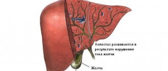
The absence of ganglion cells in the area proximal (upper) relative to the anus leads to denervation of this area. As a result, intestinal motility slows down, and the passage of feces further through the gastrointestinal tract becomes more difficult. All this leads to constipation and disruption of the frequency of bowel movements.
To push feces further through the intestine, the area proximal to the affected area experiences severe stress. Hypertrophy of its muscle layer occurs. Therefore, the upper healthy area of the intestine is always enlarged. For comparison, this can be clearly seen in the photo of a healthy person and one diagnosed with Hirschsprung’s disease. The patient will always have an overly protruded abdomen.
There are no ganglion cells not only in the muscle layer, but also in the mucous membrane. Therefore, not only peristalsis, but also normal absorption of nutrients is slowed down in the affected area of the large intestine. Which leads to disruption of metabolic processes in the body.
The development of Hirschsprung's disease is caused by a violation of embryogenesis, presumably at 7-12 weeks of gestation, when the nerve plexuses of Meissner (in the submucosal layer of the colon) and Auerbach (in the muscular layer of the intestine) are formed. Due to the premature cessation of neuroblast migration or their insufficient differentiation, instead of the typical submucosal and myenteric plexuses with ganglia, the intestinal neurostructures are represented by individual nerve fibers and glial elements.
The sooner the migration of neuroblasts is completed, the more extensive the aganglionic portion of the colon wall is. Acetylcholinesterase accumulates in the mucous layer, which causes intestinal spasm, which is a pathognomonic sign of Hirschsprung's anomaly. Due to tonic spasms and lack of peristalsis, the denervated segment becomes a functional obstacle to the movement of feces. Chronic retention of intestinal contents leads to constant constipation and significant expansion of the overlying intestine.
Big belly in a newborn
Abdominal bloating in newborns is considered a natural phenomenon. And all because of weak muscles and abdominal walls. The size of the liver also has an effect, because in a newborn baby it is larger in size than other internal organs. Intestinal colic occurs due to weak functioning of the gastrointestinal tract, as a result of which the stomach becomes rounded. Therefore, it is not a terrible situation when a child has a big belly at one year old.
But excessive belly size can also indicate health problems. The most common cause of this phenomenon is considered to be congenital pathologies, such as polycystic renal disease, liver cirrhosis or ascites. These anomalies can be detected in the maternity hospital during the first examination of the baby.
Many babies under the age of 2 look very touching with a protruding tummy. Those around you are touched and admired. This seems natural to parents, because the body requires a supply of energy to grow, but the muscular frame is not sufficiently developed, and it is difficult for it to hold the abdominal walls. In addition, a large belly is noticeable in the child immediately after eating food, as well as liquid, but it decreases as it is digested. And this is understandable. Most often, children under the age of one year are susceptible to this phenomenon.
But if the child is thin, but has an impressive abdomen, then parents should consult a doctor who will help exclude or diagnose the following diseases:
- Umbilical hernia. Experts say that it affects babies up to 3 months of age and, less often, up to a year. In the case of a hernia, the abdomen takes on a pointed shape and increases in size.
- Rickets and other diseases resulting from impaired metabolism. In this case, the child has a large belly, like a toad; it protrudes not forward, but to the sides. The cause of rickets is considered to be a lack of vitamin D in children's bodies.
- Improper activity of pancreatic enzymes. The body reacts sharply negatively to fructose and sweets.
- Reduced renal and adrenal function.
- Hepatitis. Children over three years of age are susceptible to inflammatory liver diseases.
Symptoms, signs, treatment and prevention of rickets in children
Causes
The cause of rickets in infants is a lack of vitamin D or calcium in the child's body. Vitamin D is produced naturally in a baby's body when bare skin is exposed to sunlight. Newborns can get vitamin D from formula, supplements, and small amounts of foods.
Calcium comes to the baby from food and drinks. Milk and dairy products are a good source, as are some green vegetables, sardines and canned salmon. For some foods, such as soy and others that do not contain dairy, be sure to add calcium. Both vitamins are vital for strong and healthy bones.
If a baby is a vegetarian or on a vegetarian diet, he or she may also be more likely to develop rickets because they are not getting enough calcium.
The connection between vitamin D and rickets has been known for many years and is well understood by doctors and scientists. If a baby does not get enough minerals or vitamin D to build healthy bones, they will eventually fail to form hard outer shells. This is why bones become soft and weak. As your baby grows and becomes heavier, weight gain can lead to crooked, irregularly shaped bones and deformities.
Symptoms of rickets in infants
Irregular shape or deformation of the skeleton, pain, brittle bones, poor growth and development. Rickets causes softening, weakness and poor bone formation in the baby, which can lead to their deformation:
- Irregular skeletal shape. Thickening of the ankles, wrists and knees (“inverted knees”). Legs and pelvis that curve outward (“X- and O-shaped”) or a chest bone that sticks out like a “chicken breast.”
- Pain. Bones affected by rickets often hurt, so the child does not want to walk and may get tired quickly. Brittle bones. Bones become weaker and more likely to break.
- Poor growth and development. The baby's skeleton does not grow and develop properly. The child may be short for his age.
- Dental problems, including weak tooth enamel and delayed teething.
- Soft bone tissue of the skull and delay in covering the crown of the baby (non-ossified area of the vault of the head).
Diagnosis of rickets
The number of children who develop the disease is unknown. However, research in the UK has found that approximately 1 in every 100 children who are not Caucasian suffer from rickets. English disease is more common in babies who have darker skin and those who are breastfed rather than formula-fed. African-American, Hispanic, and Asian children need more sun exposure than fair-skinned children. However, fair-skinned children can also develop rickets if they do not spend enough time in the sun or wear sunscreen.
There are other types of rickets that are not caused by a lack of vitamin D or calcium, although these types of rickets are rare. English disease can develop in children who have other diseases such as liver disease, kidney disease or kidney failure. There is also a type that is inherited (genetic). These types cannot be treated with vitamins alone and require hospitalization.
Prevention of rickets in children
As the baby grows, the bones also constantly grow and develop. How much bones develop and how strong they are depends only on the amount of minerals such as calcium and phosphorus. Such minerals form the hard outer shell of bones. This process is called mineralization. The child receives calcium and phosphorus from food and drinks.
Infants with rickets need vitamin D, because this vitamin helps the small body absorb calcium properly. And gets into bones or teeth. The type of illness caused by a lack of vitamins and other nutrients can be completely prevented by making sure that the child gets enough nutrients.
Newborns get vitamin D from breast milk or from formula with supplements. However, if the mother does not get enough of the vitamin, breast milk will not provide the baby at all. This means that parents should give their baby vitamin D supplements. When giving the baby vitamins, doctors may prescribe calcium supplements for the treatment of rickets in children. Seniors can receive vitamin supplements or by exposing the skin to sunlight.
You need to think about preventing rickets in a child even before birth. A pregnant woman should eat right. Research shows that getting enough vitamins during pregnancy is important for the baby's health. A pregnant woman should definitely take vitamin D supplements with food, especially when spending little time in the sun. By the way, after giving birth you also need to eat well. If the mother does not receive the required amount of nutrients and vitamins, this means that the baby will not receive them from breast milk.
How to treat rickets in infants?
When sick, infants may be prone to developing infections such as pneumonia and gastroenteritis (inflammation of the stomach and intestines). Symptoms of these diseases can be serious in infants and young children. Infants and children with rickets can easily break their hands.
The problem of the disease is still relevant today, but today there are many treatment methods. In preventing this disease, much is in the parents’ hands; all that remains is to try to provide the baby with everything necessary to avoid this dangerous disease. Sunbathing is an important component of successful treatment of rickets. It is only important to start prevention on time, then the children will be healthy!
We invite you to watch a video about rickets from the school of Dr. Komarovsky
Coxsackievirus in children.
Causes of jaundice in newborns.
Vitamin D during breastfeeding.
In the section - childhood diseases.
Classification
Systematization of Hirschsprung's disease is carried out taking into account anatomical and clinical criteria. Depending on the location of the aganglionic area, the most common form of the disease is distinguished: rectosigmodial, detected in 70% of patients, rectal (up to 25% of cases of anomaly), subtotal (3%), segmental (1.5%), total (0.5%). According to the location of the dilated intestine, megarectum, megasigma, left-sided, subtotal and total megacolon, megaileum are distinguished. When diagnosing intestinal Hirschsprung aganglionosis, the following clinical features are taken into account:
- Children's version of the disease. It is detected in almost 90% of patients. It is characterized by rapid development, an almost complete absence of independent defecation, and increasing signs of intestinal obstruction. Surgery is usually performed in two stages.
- Prolonged variant of aganglionosis. Debuts in children. Due to the small length of the denervated segment, it develops slowly. To correct constipation, enemas are used for a long time. It is possible to perform both two-stage and one-stage operations.
- Latent variant of the anomaly. Constipation appears in adolescence, and chronic colonic obstruction quickly develops. To relieve constipation, daily enemas are necessary. The passage of feces is usually restored in two stages.
With a compensated course, independent stool persists for many years; as the disease develops, 3-7 days of constipation occur, the resolution of which requires laxatives or an enema. In patients with a subcompensated condition, constipation lasts more than 7 days without the use of laxatives and enemas. Decompensation of the disease is indicated by the absence of independent defecation and the urge to defecate, compaction of intestinal contents and the formation of fecal stones, and the ineffectiveness of conservative methods of bowel movement.
Complications and consequences
Possible complications and consequences of the undergone manipulation are in the plane of the likelihood of infections entering the body.
Hirschsprung's disease in children, in the absence of proper treatment for a long time, results in a concomitant disorder in the form of dysbiosis. Protein metabolism is not happening correctly.
After surgery, the risk of infection in the intestines increases. Enterocolitis occurs.
In the period after surgery for Hirschsprung's disease, the body recovers completely within six months. Patients may experience spontaneous passage of stool. Sometimes constipation occurs. This condition indicates a slow recovery of the body. Individual characteristics emerge.
Children are under the foster care of a doctor for at least 6 months after surgical procedures. Parents maintain strict control over the baby's condition. With strict adherence to the regimen, the child begins to develop a bowel movement reflex. It is necessary to strengthen the immune system.
The body's adaptation to changes occurs gradually. The patient should not violate the prescribed regimen. If your health worsens, you should immediately consult a doctor. You cannot try to stop the condition on your own. Causes a sharp increase in complications and pathological conditions. Delay is especially dangerous for children.
In the case of Hirschsprung's aganglionosis, patients have impaired digestion and absorption of nutrients, which, in combination with a decrease in appetite, leads to severe malnutrition up to cachexia. Iron deficiency anemia is often detected. Children experience delays in growth and physical development. Long-term stagnation of feces in the intestine provokes dysbiosis and inflammatory changes in the mucous membrane, which can manifest as paradoxical diarrhea.
The most dangerous complications of Hirschsprung's disease are intestinal obstruction, bedsore of the wall with fecal stones and intestinal perforation. Toxic megacolon may occur, which is characterized by dilation of the proximal intestine and excessive growth of pathogenic bacterial flora. This condition often leads to peritonitis and sepsis due to the penetration of intestinal bacteria through the pathologically altered intestinal wall. In this case, sharp diffuse abdominal pain, repeated vomiting, and febrile fever occur.
Clinical manifestations of the disease in children
As mentioned above, the disease in children manifests itself depending on the affected area and the length of the area without innervation. The larger the affected area, the more serious the clinical symptoms of the disease become.
Hirschsprung's disease occurs in newborns quite unnoticed while they are breastfed, and the first clinical symptoms begin to appear when transferred to artificial feeding or with the introduction of complementary foods. In addition, in newborns on the first day of life, in the presence of the disease, there is a delay in meconium, which should be expelled within 24 hours after birth.
Depending on the location where the innervation of the intestinal region is disrupted, Hirschsprung's disease in children is divided into several anatomical forms.
- Rectal, in which a small part of the rectum is affected. Symptoms of this form of the disease are characterized by minor constipation due to poor diet and are rarely found in young children. True, this form of Hirschsprung's disease occurs in only 25% of cases and responds well to therapeutic treatment.
- Rectosigmoidal. It occurs much more often and accounts for approximately 70% of cases. Constipation when the rectosigmoid area is affected is more persistent, since in addition to the rectum, part of the sigmoid colon also remains without innervation. Feces do not pass through this area well and can accumulate there in large quantities, causing discomfort to the child and causing chronic pain. This form lends itself well to cleansing enemas only in the initial stages, then they become insufficient. Constipation appears already during the first weeks of life, especially on artificial feeding, and with the introduction of complementary foods, the clinical symptoms only worsen and the child stops going to the toilet independently. In addition to chronic constipation, the appearance of bloating is characteristic of the abdomen, which increases significantly in size and in the photo looks like a frog's belly, which is actually what it is called.
- Segmental, is quite rare and accounts for about 1.5% of all cases. The innervation of the area disappears in small intervals, and healthy parts of the intestine remain between them. The clinical picture will depend on the extent of areas not controlled by the nervous system. Symptoms can vary, from intermittent constipation to the complete inability to go to the toilet on your own, along with bloating and a “frog belly.”
- A type of disease that affects either the left or right side of the large intestine is called subtotal. It is also quite rare and accounts for 3%. This is one of the most severe forms of Hirschsprung's disease, in which treatment with enemas and laxatives is possible only at first, and surgical treatment can no longer be avoided, no matter what ideal diet the child follows. All symptoms characteristic of Hirschsprung's disease, and these are severe constipation, bloating, “frog belly”, lack of body weight, delayed physical and mental development, vitamin deficiency, anemia and fecal stones, will be especially pronounced in this form of the disease.
- Total, with damage to the entire large and occasionally small intestine. Fortunately, this common pathology is extremely rare and accounts for only 0.5% of all cases of Hirschsprung's disease. Severe constipation and bloating are characteristic from the first day of life, and the photo of the baby's abdominal cavity shows signs of intestinal obstruction.
In addition to the 5 forms, several more stages of Hirschsprung's disease in children are distinguished.
- The stage at which the child is able to go to the toilet at least occasionally, or cleansing enemas have a good effect is called compensated. With it, the child’s general well-being also does not suffer, he does not lag behind in development, there is no anemia or signs of hypovitaminosis.
- When clinical symptoms increase, cleansing enemas become less effective, and the general condition of the child begins to deteriorate against the background of prolonged constipation, they speak of the disease transitioning into subcompensation.
- The stage of decompensation develops at the slightest violation of the diet and is manifested by severe constipation, which is difficult to respond to drug therapy, and cleansing enemas do not completely empty the intestines. Sick children develop intestinal obstruction. Of course, the stage of decompensation is more typical for adults, and in childhood its development is possible only with total and subtotal anatomical forms of the disease.
- There is also an acute form of Hirschsprung's disease, which develops only when the diet is violated, and the rest of the time it practically does not manifest itself in any way and does not require constant treatment.
At 3–4 years old, the child’s body changes: muscles and bones become stronger, the stomach tightens, and therefore becomes smaller.
If the child himself is fat, then perhaps we are talking about obesity, and in this case it is necessary to establish the causes of this scourge. Endocrine problems may be the reason why your baby has a big belly. 3 years is a turning point for a child’s body.
There are many reasons why fatness appears in the abdomen, and each of them is very serious.
Symptoms and treatment of rickets in children under one year old
Protrusion of the parietal and frontal tubercles; deformation of the chest like a chicken breast; Harrison's groove; thickening of the lower ends of the forearm.
Rice. 4. Curvature of the spine (kyphosis) with rickets.
Rice. 5. Rachitic O-shaped legs (genu varum): 1 - in front; 2 - behind.
When examining a child or palpating the chest, thickenings on the ribs in the area where the cartilage joins the bone can be detected relatively early - the so-called rachitic rosary (Fig. 2). The ribs are soft and pliable, the chest is easily compressed from the sides. A transverse groove appears on the lateral surfaces, running from the xiphoid process to the axillary region, corresponding to the line of attachment of the diaphragm - Harrison's groove. In severe cases of the disease, the anterior part of the chest with the rib cage and adjacent costal cartilages protrudes forward, resembling a chicken breast (Fig. 3) or a ship's keel. Quite often, kyphosis develops (Fig. 4), an arched curvature of the spine posteriorly (rachitic hump) or curvature to the side - scoliosis. Later, deformities of the long bones of the extremities appear: thickening of the epiphyses of the bones of the forearm (rachitic “bracelets”), shins, phalanges of the fingers (“string of pearls”). The bones of the shoulder, forearm, and collarbone are bent. Even before the child begins to walk, curvature of the lower limbs occurs in the form of brackets or the letter O (genu varum; Fig. 5). Less commonly, older children experience an X-shaped curvature of the legs (genu valgum; Fig. 6). Deformation of the pelvic bones is now rarely observed.
Rice. 6. X-shaped curvature of the legs with rickets (genu valgum).
Rickets is characterized by muscle hypotonia, lethargy and flabbiness of the muscles, weakness of the ligamentous apparatus, resulting in loose joints. Muscle hypotonia explains a peculiar symptom: when lying down, the stomach expands to the sides (“frog belly”). There is a delay in static and motor functions: children with rickets begin to sit, stand and walk later than healthy ones. Their appearance is often delayed and the order of teeth eruption is disrupted. The functions of a number of internal organs are impaired. Sluggishness of the muscles of the abdominal wall and intestinal walls contributes to bloating; blood stagnation develops in the abdominal organs; the spleen and liver enlarge. The secretory, absorption and motor functions of the gastrointestinal tract change, creating conditions for the occurrence of frequent bowel movements, alternating with prolonged constipation.
Respiratory dysfunction is of great importance: shortness of breath is always noted, the soft chest collapses with each inhalation, and the diaphragm contracts less vigorously. As a result, the lungs do not expand enough, and atelectatic areas often form near the spine, predisposing to the development of pneumonia. Morphological changes in the lungs, decreased gas exchange with the development of hypoxemia also contribute to the development of pneumonia.
With rickets, the function of the circulatory system is also difficult. Due to insufficient expansion of the chest during inhalation, the suction effect on the blood flowing to the heart is very weak. In this case, the weakness of contractions of the diaphragm contributes to stagnation of blood in the liver, the liver increases in size, and stagnation develops in the area of the portal vein.
There is more or less pronounced hypochromic anemia in the blood, in some cases there is a classic picture of anemia of the Yaksha-Gayema type (see Anemia). Significant biochemical changes in the blood during rickets. In the initial period, and especially during the height of the disease, hypophosphatemia develops: the level of inorganic phosphorus may decrease (up to 3-1.5 mg%). The calcium content in the blood is normal or slightly reduced (up to 9-9.5 mg%). Alkaline phosphatase activity increases compared to normal. Reserve blood alkalinity is reduced. The amount of magnesium and citric acid decreases to 1-2 mg% (normal 3-4 mg%) in the blood serum. There are shifts in the vitamin balance: low levels of vitamins C, A and group B in the blood.
Congenital rickets is called rickets, which arose during the period of intrauterine development of the fetus due to poor nutrition and some diseases of women during pregnancy due to vitamin D deficiency and mineral metabolism disorders. About 20 cases are described in the literature, which, by the nature of the disease, can be classified as congenital rickets. Some authors dispute the possibility of developing congenital rickets.
Late rickets includes protracted rickets that began in early childhood with a recurrent course. Moreover, at the age of 4-5 years and later, under unfavorable conditions, an exacerbation of all symptoms of rickets can be observed.
Diagnosis . When making a differential diagnosis, it is necessary to remember that there are a number of diseases of young children whose symptoms are similar to rickets: scurvy, hypothyroidism, chondrodystrophy, congenital fragility of bones, hydrocephalus, tuberculous spondylitis, Down's disease and congenital dislocation of the hip joint.
The prognosis for rickets is usually favorable, but becomes serious when pneumonia is added. An unfavorable circumstance is the addition of tetany to rickets (see Spasmophilia).
Rickets is a widespread disease in infants that affects the growth and development of children's bones up to one year and up to 3 years of life. Rickets leads to softening and weakness of the bones, which subsequently become bent, i.e., deformed.
Prognosis and prevention
The outcome of Hirschsprung's disease depends on the time of its detection and the degree of damage to the intestinal nerve ganglia. The prognosis is relatively favorable in case of early diagnosis and surgical correction. Without treatment, infant mortality in the first months of life reaches 80%. Due to the congenital nature of the pathology, specific prevention measures have not been developed. When the first signs of the disease appear, you should immediately consult a doctor to avoid the development of severe complications.
Aganglionosis of the large intestine is a congenital disease, and therefore there are simply no methods of specific prevention. The most effective preventive measures can be considered early detection of the disease and prescribing the correct treatment. After all, only in this case will the prognosis be favorable and many serious complications will be avoided, and the quality of life of patients after surgery will significantly improve.
Methods of nonspecific prevention include:
- regular exercise (swimming and jogging are especially useful in this case);
- a balanced diet (more fresh vegetables and fruits, herbs, whole grain bread and excluding fried and canned foods, hot herbs and spices from the diet);
- you should eat in small portions;
- You need to drink at least 2-3 liters of clean water per day.
Following simple preventive measures will make life much easier for patients. The main thing is to identify the disease in a timely manner and carry out the correct treatment! Beginning of the form End of the form
Treatment of ascites in boys and men
One thing can be said with complete confidence: a disease such as ascites does not occur on its own. This is especially true for males. As a rule, this is the result of some complex disease. Usually these are diseases of those organs that are responsible for fluid exchange in the body (kidneys, stomach, liver, heart, etc.).
In folk medicine there are recipes from various herbs that can affect the entire body. Let's give one of them.
To prepare it you will need chamomile, medicinal sage, gray jaundice and black currant leaves. All these herbs must be taken in equal proportions and ground in a coffee grinder. Then take 1 spoon (teaspoon) of this mixture and pour a glass of boiling water. Leave to infuse, and then strain through a fine mesh or gauze. This infusion should be taken one glass per day in four divided doses. The course of treatment is one month, after which there is a week break.
The following folk recipes, which will use herbs, are intended only for males. They are intended to treat a very unpleasant disease - testicular hydrocele.
Enlarged internal organs
Due to the increased size of the internal organs, a large belly is noticeable in the child. You don't have to worry about this problem for 1.5 years. But if the child is more than 3-4 years old, then there is a reason for concern. There is a protrusion not of the entire abdominal region, but only of a certain part.
The cause may be inflammation or infection. In such a condition, you need to show the child to a doctor to identify the problem. Gradual enlargement of the abdomen can lead to rupture of the spleen.
With hepatomegaly (enlarged liver), a large belly is considered normal for a child. The following dimensions are considered acceptable: protrusion to the sides of the ribs by 2–3 cm.
This condition is observed in children aged 6 to 7 years. But if the child is much older, then the problem may lie in a certain disease.
An enlarged abdomen is possible with coprostasis (constipation). This is a disruption of the intestinal tract. Coprostasis is diagnosed when the intestines rarely empty their bowels, less than once every two days. This disorder occurs due to a disorder of motor activity and secretion of the colon.
Treatment of ascites
Ascites of the abdominal cavity requires complex treatment. Therapy begins with treatment of the pathology that provoked ascites. When such a disease cannot be treated, methods are adopted to alleviate the patient’s condition. In very severe cases, a liver transplant is prescribed.
Drug therapy
Drug treatment usually includes taking diuretics to normalize liver function and stop inflammatory processes. To improve lymphatic drainage, the patient must remain in bed or semi-bed rest.
In cases where the development of ascites is caused by portal vein hypertension, Albumin and hepatoprotectors are prescribed, as well as plasma administration.
If drug therapy does not bring the desired result and the amount of fluid has not decreased, the doctor prescribes laparocentesis.
Laparocentesis
This procedure involves piercing the abdomen with a thin needle. In this case, the accumulated excess fluid is removed through a special catheter. This operation is performed under local anesthesia. In one procedure, pump out no more than 5 liters of liquid. There should be 3-4 day breaks between operations.
Contraindications to laparocentesis:
- purulent infections;
- adhesive disease;
- perforation of the intestinal walls;
- pronounced flatulence.
Possible complications after laparocentesis:
- intestinal damage;
- peritonitis;
- collapse.
If fluid accumulates quickly, drainage catheters are installed to continuously drain it.
Is it possible to cure abdominal ascites using traditional methods?
The use of traditional methods is aimed at eliminating the causes that caused the formation of ascites. There are several folk recipes for treating the disease:
- Parsley decoction. You will need 800 g of fresh parsley and 1.5 liters of milk. Wash the parsley, chop finely and pour into a container. Pour milk over parsley and cook over low heat until the volume of milk is reduced by half. Strain the resulting broth and drink 2 tbsp. spoons once an hour. Store the product in the refrigerator.
- Infusion of birch buds and leaves. Pour boiling water over the ingredients in a ratio of 1:10. With every 200 ml of boiling water add 0.2 g of soda. Leave the product to infuse for 6 hours. Drink 100 mg per day, divided into 2 doses.
An infusion of birch buds and leaves is effectively used for taking baths for ascites.
Abdominal hydrocele and kyphosis
At the age of 6, a child’s body is susceptible to problems such as curvature of posture. Incorrect position of the back at the table, weak bone frame are the causes of childhood kyphosis. The disease also occurs due to weakness of the spinal muscles, which prevent the vertebrae from bending forward. As a result, the following problems appear:
- 2-4 folds on the stomach;
- protrusion of the abdomen (in an upright position).
Also, the volume of the abdominal cavity depends on the type of kyphosis. It can be genotypic, that is, hereditary. This is quite difficult to deal with.
Ascites (abdominal dropsy) is characterized by the accumulation of fluid. It cannot be called an independent disease, but rather a symptom, which is a signal indicating disturbances in the functioning of the body. The main causes of ascites include:
- kidney diseases, for example nephrotic syndrome;
- nutritional dystrophy;
- tumors in the abdominal cavity.
Ascites is divided into two types: acquired and congenital. If the belly gradually becomes larger, and a protruding navel is clearly noticeable in the horizontal position of the child, then a doctor’s consultation is definitely needed.
Rickets
A disease associated with insufficient bone mineralization and a disorder of the bone formation mechanism. The main factor influencing the development of rickets is a lack of vitamin D. It can occur for the following reasons:
- insufficient sun exposure;
- disruption of vitamin absorption processes;
- hereditary predisposition;
- prematurity of the baby;
- malnutrition of the child or mother.
Signs of the disease develop from 1-1.5 months, but they become noticeable by 3-4 months. Typical symptoms are loss of appetite, anxiety, itchy skin. Due to the fact that the child rubs his head on the pillow, the back of his head becomes bald. Treatment is specific: vitamin D supplements, diet adjustments, sunbathing.