The 16th week from the moment of conception refers to the second trimester of pregnancy and corresponds to the 18th obstetric week. Ultrasound diagnostics allows you to track the formation of the fetus, comparing the data with the norm.
The main goal of the procedure is to detect pathologies caused by chromosomal abnormalities and severe defects. At this stage, the main organs and systems of the fetus reach sufficient levels to detect disorders. The size of the baby allows for a thorough examination and assessment of development.
Why and to whom is ultrasound diagnostics recommended?
At 14–16 weeks, pregnant women are prescribed a routine hormonal status study (PAPP-A, hCG, estriol, ACE). The procedure is aimed at detecting fetal abnormalities. If the examination reveals a hormonal imbalance, the doctor prescribes an ultrasound scan for the patient at 16 weeks of pregnancy.
The procedure is prescribed by a doctor in a number of cases:
- Poor results of the first screening test.
- Negative hormone levels.
- Suspicion of a frozen pregnancy.
- Multiple births.
- A woman complains of pain.
- Miscarriage, fetal defects in a previous pregnancy.
- The presence of children born with genomic pathology.
- Pathologies of placental development, history of polyhydramnios or oligohydramnios.
Symptoms such as uterine tone and spotting are a reason for urgent hospitalization.
Important! Delayed diagnosis of a frozen pregnancy can lead to serious consequences.
If research data indicate a high probability of fetal defects, the patient receives a referral for amniotic fluid testing - amniocentesis.
Why is ultrasound prescribed?
Many mothers at 16 weeks feel fetal movements in the stomach. Those who already have a child will not confuse such sensations with anything.
If the baby is the first or the fetus is small, then movements may not be audible. During the ultrasound procedure, you can look at the child and see his movements.
Not only body movements are visible on the equipment monitor: it is possible to distinguish the baby’s facial expressions, the gender of the child, and monitor the cervix.
Video:
An ultrasound examination at week 16 is one of the best ways to examine the fetus, the condition of the cervix, and detect or exclude pathology (see video).
The basis of such research is ultrasonic waves. They are reflected from tissues that have different densities. The device analyzes the results obtained and displays them on the screen in the form of a specific photo.
An ultrasound at the sixteenth week can be performed in one of the following ways:
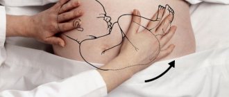
- Transabdominal - carried out with a special sensor, which is applied to the surface of the abdomen and slowly moved. Before the procedure begins, the surface of the abdomen is treated with a special gel, which helps ultrasound waves pass through the tissue better;
- Transvaginal - carried out with a sensor, which the doctor inserts into the vagina. With its help, you can qualitatively diagnose the condition of the cervix.
Ultrasound at 16 weeks of pregnancy is usually prescribed to women who, for some reason, missed the study in the 1st trimester.
In addition, a study may be prescribed to exclude hereditary diseases that can be transmitted from father to child, and cervical pathologies.
Transvaginal ultrasound may be prescribed for those women in whom the doctor suspects isthmic-cervical insufficiency.
With ICI, thinning and excessive softness of the cervix and its isthmus are observed, which leads to termination of pregnancy in the second and third trimester. At week 16, the doctor will be able to find out the sex of the fetus.
Video:
If at 16 weeks of pregnancy the doctor prescribed a transvaginal ultrasound, no special preparation is required, but for a transabdominal examination it is worth fulfilling some requirements.
This ultrasound is performed when the bladder is full. Otherwise, empty spaces can be a hindrance to quality research.
Video:
READ What will a photo taken on an ultrasound at 12 weeks of pregnancy tell you?
Before an ultrasound, a woman should drink 1 – 1.5 liters of water. Only if you follow all the doctor’s recommendations when preparing for an ultrasound, the procedure will be carried out correctly.
Then the condition of the cervix, the size of the baby, and possibly its gender will be studied with high accuracy.
Mother's condition and fetal development
Week 16 is marked by the baby’s first movements in the womb. Primipara, obese women usually feel fetal movements at a later date.
Body tissues accumulate fluid and sodium salts, which leads to swelling of the limbs. The expectant mother's kidneys work under increased load. The rapid growth of the uterus forces all systems and major organs to work in an enhanced mode. Pain occurs in the lumbar region.
A baby at 16 weeks of pregnancy from conception demonstrates excellent abilities:
- movement in amniotic fluid;
- swallowing amniotic fluid using breathing movements.
The child has periods of sleep and wakefulness. The baby's limbs have formed, his hands touch his tiny face and clench into fists. The mother's emotions are transmitted to the child, who may already experience anxiety.
What are they watching?
A woman can receive a protocol and photo of ultrasound results at 16 and 17 weeks of pregnancy. Based on the data, the doctor creates a biophysical profile of the fetus.
Important! The presence of genetic abnormalities requires the woman to make a decision to terminate the pregnancy for medical reasons.
Sufficient size makes it possible to diagnose malformations of the child with a high probability, such as:
- syndromes associated with genome disorders (Down, Edwards, other chromosomal abnormalities);
- malformations of the heart and brain;
- assessment of the size of tubular bones (humerus, femur), nasal bone;
- fruit size;
- amount of amniotic fluid;
- localization of the placenta.
Proper intrauterine nutrition of the fetus depends on the condition of the placenta.
Incorrect position of the organ (complete presentation) requires prompt resolution of labor, restriction of motor activity and sexual activity.
An ultrasound specialist assesses the condition of the cervix, which should normally be closed.
Ultrasound indications
By 16 weeks, the uterus is quite large. By this time, its weight is approximately 250 g. During the procedure, the doctor must examine the height of this organ.
If the entire distance below the navel is divided in half, then the place where the uterus is located will be determined - this location is considered the norm.
A woman will be able to determine the location of the uterus herself. To do this, you should put your hand on the lower abdomen, about 7 cm from the navel down. Incorrect location of the uterus indicates disturbances in the development of the fetus.
Video:
The baby in the womb is constantly developing and growing, and as a result, the uterus is growing. As it grows, it puts pressure on other organs - the intestines also become slightly compressed.
A woman may experience various unpleasant symptoms, such as constipation, bloating, heartburn, etc.
When performing an ultrasound, the doctor evaluates not only the condition of the cervix. At the 16th week of pregnancy, the internal organs of the fetus are examined and measurements are taken.
At this age, the baby weighs 90 - 110 g, abdominal circumference 88 - 116 mm, head - 112 - 132 mm, length of the fetus itself - 120 - 150 mm, lower leg - 15 - 21 mm, thigh - 17 - 23 mm. The sex of the child does not play a role in determining the size of the organs.
A fetus at 16 weeks has developed legs, already formed marigolds. Legs and arms are symmetrical.
The fetus is already able to hold its neck straight and also turn its head left and right. The eyes and ears of the fetus are located in their place.
Video:
The child's heart must work at full strength. At this stage of its development, it is capable of pumping about 25 liters of blood per day.
The liver, which previously performed the functions of hematopoiesis, is engaged in digestion, since the intestines, stomach, and gall bladder are just beginning to work, and therefore do not function at full capacity.
When performing an ultrasound, you can see its contents in the intestines. Black-green, dark green original feces consist of bile. The bladder is already working. It is emptied every 40–45 minutes.
Video:
The functions of hematopoiesis are performed by the bone marrow. At the sixteenth week of fetal development, the blood has a certain composition. It contains components inherent in the blood of a man or a woman.
READ Dates and standards for ultrasound screening of the 1st trimester
When performing an ultrasound, the doctor will be able to determine the group and Rh factor. The fetus lacks fetal hemoglobin. It will form a little later.
The replacement of fetal hemoglobin with regular hemoglobin will occur only 5-6 months after the baby is born.
The placenta should be formed by 16 weeks. The length of the umbilical cord should be about 50 cm, its diameter should be approximately 2 cm.
Photo of the fetus: is it possible to determine the sex of the child?
There is a real opportunity to determine the sex of the baby using an ultrasound. At week 16, the information is tentative.
The fetus moves freely in the amniotic fluid. Fingers have formed on the tiny arms and legs, which mom can see in the ultrasound photo. The child demonstrates excellent facial expressions, reflexively grimacing and smiling. A yawning baby is a wonderful moment that photography can capture.
The genitals are not yet sufficiently developed; the boy’s testicles are located in the scrotum. The girl's fallopian tubes and uterus have already formed. If the position is poor, the child may block the examination area with his leg or arm, which makes it difficult to determine the sex.
A specialized center equipped with high-precision equipment will most likely inform the mother who is preparing for birth.
Photo of a boy
Photo of a girl
Research standards: fetal size
Normal fetal sizes for 16 weeks of pregnancy are considered to be from 14 to 20 cm with a weight of 140 to 230 g.
Diagnostics are performed in accordance with the established protocol and take into account all the necessary parameters:
- Number of fruits.
- Cervix – at least 30 mm.
- BPR (distance between the parietal tubercles) – 37 – 47 mm.
- LZR (OFD, fronto-occipital size) – from 49 to 60 mm.
- Exhaust gas (HC, head circumference) – 131 – 161 mm.
- DP (HUM length of the humerus) – from 15 to 20 mm.
- Coolant (AC, abdominal circumference) – about 122 mm.
- DH (chest diameter) – about 41 mm.
- Femur length (FML) – from 23 to 31 mm.
- The size of the humerus is from 15 to 21 mm.
- Forearm bone – from 17 to 23 mm.
- Tibia length – 23 – 31 mm.
- Systole - diastolic ratio in the umbilical cord artery (USR) is from 4.55 to 4.67.
- Heart rate (HR) – from 140 to 160 beats per minute.
The protocol records the condition, degree of maturity of the placenta, status of the cervix and pharynx, and uterine tone. The placenta at 16 weeks has zero maturity. Areas of infarction and calcifications should be absent. The specialist determines whether the amniotic fluid corresponds to the norm.
Decoding the results
An ultrasound scan looks at the size of the fetus, the presence of formed organs, gender, the condition and size of the placenta, and the condition of the cervix.
Fetometry norm at 16th week:
- Weight from 75 to 120 g.
- Length from 10 to 15 cm.
- Transverse head size (TDS) – 31–37 mm.
- Head size in circumference is 112–136 mm.
- Abdominal circumference – 88–116 mm.
- Femur – 17–23 mm.
- Shin bone – 15–21 mm.
- Humerus – 15–21 mm.
- Forearm bones – 12–18 mm.
All major organs must be formed and correspond to the duration of pregnancy. On 3D ultrasound you can see facial expressions and purposeful movements. Heart rate is about 140–160 beats per minute; you can listen to the heartbeat during an ultrasound.
You can try to determine the sex of the child - the external genitalia are formed, but in boys the testicles are still in the abdominal cavity.
By the 16th week of pregnancy, the placenta is finally formed. The thickness of the placenta should be in the range of 13.8–24.3 mm, the echostructure should be homogeneous. The placenta should attach above the os, but even with low placentation, there is a chance that as the uterus grows, the placenta will rise.
The cervix is examined transvaginally - the cervical canal must be tightly closed, the length of the cervix is at least 30 mm.
To see what a fetal ultrasound looks like on the monitor:
3D ultrasound at 16 weeks: is screening carried out?
Modern ultrasound scanners with advanced capabilities provide a picture of the fetus in 3-D format, which allows the specialist and parents to observe a three-dimensional image of the baby. The procedure does not require special preparation and lasts up to 60 minutes. The method is characterized by data accuracy.
3D photo of the fetus
If there are no deviations from the norm, a routine ultrasound examination is carried out between 18 and 22 weeks of embryonic development. In case of poor results of the first study, with unfavorable hormonal status, the doctor recommends screening at 16 weeks.
The appointment is caused by the need to make a decision on termination of pregnancy for medical reasons, or preservation of the fetus. New data often refute concerns raised by the results of the first screening.
What happens at 16 weeks of pregnancy?
Your condition improves significantly compared to the first weeks of pregnancy, your appetite increases, and you can eat whatever your heart desires. You should eat in moderation and not be led by your appetite - after all, losing extra pounds during pregnancy is difficult, and they will greatly hinder you during childbirth.
You should know that at the 16th week of pregnancy a mother’s weight may increase by 2.5-3 kg. If you have gained significantly more, feel well and have no swelling, reconsider your diet: most likely, you eat too much sweets, flour and other foods that do not benefit the body of mother and baby.
Many mothers begin to behave too demanding and capricious, terrorizing those around them with constant complaints and requests. If you notice such behavior in yourself, try to change your attitude towards pregnancy and others - after all, in fact, your situation is a miracle and a holiday for the family, and not a reason for whims. Your relatives are just as worried about your health and your baby’s; you shouldn’t quarrel with them because of momentary mood changes.
How do they do it, do they need preparation?
At the 16th week of embryonic development, ultrasound is performed transabdominally. A sufficient amount of amniotic fluid eliminates the need to fill the bladder.
The abdomen is covered with gel, the smooth converter glides easily over the surface of the skin. The procedure does not require prior preparation.
Uncomfortable fetal positioning may require repositioning. The woman will be asked to stand up and move around a little.
If the fetus stubbornly refuses to change position, the study can be postponed to another day. Diagnosis of the placenta is carried out vaginally to accurately determine the distance between the placenta and the cervix.
Proper nutrition
The belly at the 16th week of pregnancy is still practically invisible, although with a second pregnancy its size may be larger due to sprained ligaments and tendons. The uterus still weighs only 250g, so it does not exert significant pressure on the internal organs.
Now is a very important time when active growth and formation of all the baby’s organs and systems is taking place, so the mother must eat well to provide the baby’s body with all the nutrients.
A pregnant woman should pay increased attention to the quality and quantity of food consumed. The composition of the menu matters - it should contain a sufficient amount of protein, healthy fats and carbohydrates, minerals and vitamins. You need to eat 4-5 times a day in small portions, the dishes should be varied and satisfying. Breakfast should be the most nutritious, lunch should include first courses, and dinner should be light so that the expectant mother can sleep peacefully.
Where is the examination done and how much does it cost?
The free procedure is carried out in a antenatal clinic as prescribed by a local doctor. To obtain accurate results, you can use the services of a specialized center staffed with qualified personnel and high-resolution equipment.
Gynecology and obstetrics centers offer examinations costing from 1000 to 1500 rubles. The service includes Doppler ultrasound, 3-D or 4-D examination with video recording on disk media. A regular two-dimensional ultrasound procedure costs from 600 to 800 rubles. A screening test to determine hormonal status and justify the risk of defects will cost from 2500 to 3600 rubles.