Trophic ulcers of the lower extremities require long-term complex treatment in combination with therapy of the underlying disease
Treatment of trophic ulcers is a long and difficult process that requires a balanced and detailed analysis of the cause of the disease. The doctor’s art lies in the correct combination of therapy for the underlying disease that has caused weakening of the body with the treatment of a skin defect.
A single cure for trophic ulcers (such as a miracle pill or super ointment) does not exist, and it is unlikely to appear in the foreseeable future, so the doctor’s skill is to correctly combine existing powerful medications.
Trophic ulcer - what is it?
Trophic ulcers on the legs photo
Despite the fact that this name is extremely widespread among doctors of all specialties, and they, without saying a word, understand what they mean, it is quite difficult to define a trophic ulcer.
Firstly, for the reason that such a diagnosis is not in the classification of diseases, and secondly, because this concept is collective in nature. Hemodynamic disorders, innervation disorders, lymphostasis and injuries lead to the appearance of tissue defects.
Therefore, in simple terms, trophic ulcers are deep defects that arise as a result of disturbances in innervation, hemodynamics, and lymphatic drainage, which reach a level below the basement membrane of the skin, are complications of many chronic diseases, and heal with the appearance of scar tissue.
This definition contains the most important facts, for example, the fact that if an ulcer has already appeared, then even if it heals, the appearance of a scar cannot be avoided. And for what reasons can such severe tissue nutritional disorders occur?
Treatment at home
Rules
Drug treatment of the disease can also be carried out at home with medications, subject to certain rules:
- A diet that involves limiting the number of carbohydrates consumed, avoiding spicy and spicy foods, and increasing the amount of fresh fruits and vegetables.
- Frequent rest.
- Performing exercises that prevent blood stagnation improves metabolic processes.
- Wearing special orthopedic shoes with high-quality soles to avoid tissue injury.
Medicines
Treatment of purulent diseases on the leg at home involves taking medications that improve the blood circulation process and also eliminate the cause of development:
- Aspirin (acetylsalicylic acid tablet): the drug has antiplatelet properties (blocks the processes of platelet aggregation and adhesion).
- Venotonic drugs: used for the development of varicose ulcers.
- Broad-spectrum antibiotics.
- Non-steroidal anti-inflammatory drugs.
- Antihistamines.
Local treatment for ulcers is aimed at cleansing the wound of dead skin and eliminating pathogenic microbes:
- Washing the wound with antiseptic solutions (chlorhexidine, potassium permanganate, furatsilin).
- Applying dressings using a medicinal gel (or using ointments): dioxykol, levomikol, streptolaven.
When ulceratively treating a diseased area of skin with antiseptic solutions, non-viable tissue areas must be removed as much as possible. Then apply a bandage. The dressing must be done once every three days. More frequent treatment can cause tissue injury.
Causes of trophic ulcers
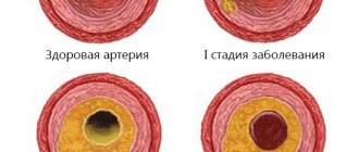
varicose veins, atherosclerosis and other causes
The main reason for their appearance is not trauma or injury, but long-term illnesses leading to chronic insufficiency of arterial and venous blood flow, and a disorder of autonomic innervation. The following are the most important reasons:
- "Varicose ulcer." It occurs in 2/3 of all cases. Chronic venous stagnation develops, which leads to trophic disorders;
- Arterial ulcers.
They occur much less frequently, as a result of severe ischemia of arterial thrombosis (occlusion). Most often they appear in the foot area, where the blood flow is weaker, and less often on the lower leg.
Almost all arterial lesions arise due to atherosclerosis, which occurs with obliteration of blood vessels, as well as due to thromboangiitis (Buerger's disease). Here the mechanism triggers a sharp decrease in pressure, leading to chronic hypoxia. At the same time, part of the blood flow is still preserved, since with complete thrombosis it is not an ulcer that occurs, but gangrene.
How arterial ulcers appear: they are preceded by chilliness and fatigue, cold limbs, the presence of intermittent claudication, then night pain appears. It all starts with a cut, bruise, or rubbing of the leg.
- Damages in diabetes mellitus.
Diabetic ulcers occur in 5% of all cases, but represent a very difficult problem to treat given the reduced innervation of tissue in the periphery as a consequence of diabetic polyneuropathy. With a diabetic ulcer, the arterial pulse is usually preserved and there is no intermittent claudication.
See also Diabetic Foot Syndrome
trophic ulcer on the leg with diabetes photo
The presence of polyneuropathy leads to the absence of pain even with deep and wide defects. This fact “puts vigilance to sleep” and often leads to very late visits to the doctor when urgent amputation is needed.
- Trophic ulcers that occur after thrombophlebitis of the legs. Their share in the total number is 6-7%, and very often they are adjacent to varicose veins, if phlebitis occurs in any long-term section of the vessel;
- Post-traumatic lesions. They arise as a result of injuries and wounds, and are often combined with the original trophic disorder. For example, if an injury occurs against the background of diabetes mellitus, which leads to a violation of trophism, then it can be very difficult to understand what caused the defect;
- Neurotrophic ulcers. They are of purely neurogenic origin and are recorded in no more than 1% of all cases. They arise as long-term consequences of injury to the spine and sciatic nerve. This is “to blame” for the damage or simply interruption of the vegetative fibers that regulate the vasomotor regulation of tissue nutrition;
- Finally, ulcerative-trophic lesions of mixed origin arise, which can be immediately classified into several categories.
There are also special forms, for example, hypertensive ulcer, or Martorell's lesion. As a rule, its occurrence is characterized by a combination of the following factors:
- Affects females, in adulthood – from 40 to 60 years;
- Patients have persistent and severe arterial hypertension, with a crisis course, which is also called “malignant”;
- Symmetrical lesions often occur;
- The peculiarity of this type of trophic disorder is severe pain in the legs, spread and progression, and a tendency to infection, that is, unfavorable factors.
Looking ahead, we’ll say right away that in this case the entire success of treatment and limb preservation depends on the success of treatment of the underlying disease, in this case arterial hypertension.
hypertensive ulcer in a woman
Doctors - surgeons and infectious disease specialists, radiology specialists also know even more rare causes of the development of lesions, for example, those that occur after frostbite and burns. Congenital and acquired arteriovenous shunts and fistulas, ulcers of syphilitic etiology, or those resulting from the action of ionizing radiation are extremely rare. How does an ulcer form?
Manifestations of tissue trophic disorders
In most clinical cases, tissue nutritional disorders are caused by venous diseases. Depending on the severity, there are several types of trophic disorders in venous insufficiency:
- Hyperpigmentation – darkening of the skin color of the lower leg.
- Lipodermatosclerosis is a local thickening of the skin and subcutaneous fat.
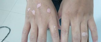
- White atrophy is a limited area of skin that is light in color. The lesion is usually located in the area of hyperpigmentation. Considered as a pre-ulcerative condition.
- A trophic ulcer is a defect of the skin and underlying soft tissues that occurs after the rejection of necrotic tissue.
Trophic ulcers can form with any type of trophic disorder. They are characterized by a sluggish course, a low tendency to heal and are prone to recurrence.
Stages of trophic ulcers, symptoms and photos
Of course, it never happens that a person falls asleep healthy in the evening, and the next morning finds himself with a festering ulcer into which you can put your fist. An ulcer, like many other pathological formations, has a clear stage of development. But before it occurs, there is a “pre-ulcer” stage with its own symptoms.
Signs of “pre-ulcer” - initial stage
initial stage of trophic ulcer photo development
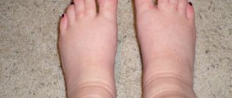
The symptoms of the initial stage of a trophic ulcer are as follows. At first, when there is no ulcer yet, you can detect induration, or thickening of the skin in the calves and legs, burning and itching, and swelling. In some cases, dilatation of small subcutaneous (subcutaneous) veins occurs.
The patient may experience leg cramps. With an arterial nature, chilliness appears and the color of the limb below the level of chronic occlusion changes to white and pale. The initial stage of a trophic ulcer on the leg may be accompanied by symptoms of the appearance of bluish or purple spots, which can merge into a single whole.
initial stage photo 2
Finally, the ulcer itself appears. There is no depression in the center yet, although everything is ready for its appearance, the tissues in the depths have already died or are dying, and it is a red (or brownish) wet scab of a certain area, with a “restless”, often painful periphery.
Stages of “life” - development of trophic ulcers
Now the defect begins to expand and deepen. In its development, it usually goes through three stages of life, which in uncomplicated cases take about 2-3 months. What happens during this time? These stages are called exudation, granulation and epithelialization:
- Exudation stage. It lasts up to 2 weeks.
Inflammation and perifocal edema occur along the perimeter. Tissue necrosis occurs in the center, that is, an ulcerative defect itself is formed. It is at this stage that a copious discharge appears from the wound, which has an unpleasant odor.
When conducting a bacteriological examination of the discharge, a significant appearance of colonies is revealed, which indicates strong microbial contamination of both the edges and the bottom of the defect.
In some cases, the process is not limited to one local zone. If lymphatic vessels - collectors and veins - passed through the ulcer, then lymphangitis and thrombosis of the veins (thrombophlebitis) occur. The appearance of secondary erysipelas, streptoderma, and erysipelas is common.
If the “rampant” local infection is not stopped, then gradual obliteration of the lymphatic vessels may occur with the development of persistent edema, especially in the feet, which is called lymphedema. Ultimately, this can lead to elephantiasis and permanent disability.
- Stage of repair, or granulation.
Continues for the next 2-3 weeks. All this time, the bottom and walls of the formation are covered with fresh granulations, and the wound is cleared of necrosis.
It is at this stage that wound cleansing with fly larvae can be used, which carefully eat only dead tissue without causing any harm to the living (no matter how shocking this may be to many).
Suppuration gradually decreases, the severity of swelling of surrounding tissues also decreases. Scanty, serous discharge continues to appear.
- Stage of epithelization of the ulcer.
This is a late stage of ulcer development, which is observed a month or later from the beginning of the development of the process of tissue destruction, which is accompanied by the formation of epithelium, where this is possible, and where not, a scar appears.
How to treat trophic ulcers? They need to be treated strictly following the principles to avoid complications. What does this mean?
Vacuum cleaning of wounds
Vacuum therapy or negative pressure therapy is a method of removing serous fluid and dead tissue from a wound or surgical site. Currently, vacuum ulcer debridement can be used on all types of wounds: acute, subacute or chronic. It reduces swelling, promotes rapid healing and the formation of young connective tissue.
The essence of the method is that a piece of porous sponge with silver ions is inserted into the wound, then the whole thing is covered with a transparent membrane. A hole is made in it and a drainage tube is inserted, which is connected to a vacuum source. The fluid is drawn from the wound through the sponge into a reservoir for subsequent disposal.
The membrane prevents air from entering and allows a vacuum to form inside the wound, reducing its volume and making it easier to remove fluid. Before starting the procedure, the ulcer should be washed.
The duration of treatment depends on the size and depth of the wound. The dressing is changed every 24-48 hours.
Principles of treatment of trophic ulcers on the legs, drugs
The most important thing is to determine the cause and begin intensive treatment of the underlying disease. Treatment of a trophic ulcer on the leg, while venous thrombosis continues in other areas, or blood sugar is “exorbitantly” elevated, is useless, since even if the defect is eliminated, several new ones will immediately appear in other places.
One should not hesitate to make a decision in favor of hospitalization, even to a surgical hospital, if treatment of trophic ulcers of the lower extremities at home was delayed and was ineffective. Currently, there are many options for operations that effectively eliminate the cause of the ulcer and preserve the limb, so there is no need to be afraid of surgeons.
The main stages are:
- Consultation with a phlebologist and a vascular surgeon to decide on a priority operation. (In the case of arterial ulcers, blood flow is always restored first with surgery);
- In the case of a conservative approach, staying in an upright position is sharply limited (and in the diabetic form, putting weight on the leg is completely prohibited);
- In case of varicose ulcers, compression garments are prescribed. Treatment of trophic ulcers of the lower extremities should be accompanied by improved outflow. According to indications, compression hosiery of high compression classes is prescribed;
- In the first stage of ulcer formation, infection control is required, taking into account the sensitivity of the pathogen to antibiotics;
- At subsequent stages, the fight against infection gives way to reparative and restorative therapy;
- Throughout treatment, the most careful care of the ulcer is needed, and urgent hospitalization if the condition worsens.
Treatment of trophic ulcers of the lower extremities at home
At home, you can successfully use medications for trophic ulcers, but only after consultation with a vascular surgeon and a specialized specialist, for example, a diabetologist. The purpose of this is not to “miss” the operation, which in some cases should precede treatment.
In some cases, treatment in a hospital is preferable, since hyperbaric oxygen therapy sessions, rational antibacterial therapy, and correction of heart failure and blood glucose levels can be prescribed there. If all this is not required, then only then can trophic ulcers of the lower extremities be treated at home, but under the supervision of a doctor. The main groups of drugs include:
- Reparants (applied upon completion of the first stage). These include “Solcoseryl”, “Actovegin”, “Methyluraciol ointment”, “Bepanten”. Promote regeneration;
- Phlebotonics for varicose veins - “Detralex”, “Eskuzan”, “Troxerutin”, “Phlebodia”, “Venoruton”. Diosmin can be used in micronized form;
- Drugs to improve microcirculation (“Hepatrombin”);
- Local use of antibacterial drugs - antibiotics, Levomekol, Vishnevsky ointment;
- Rinsing the ulcer and removing the discharge (hydrogen peroxide, chlorhexidine solution, furatsillin) with 2-3 times changing the dressing. You can use gelatin-zinc ointment Unna;
- General antiplatelet agents are used that improve blood flow and reduce thrombus formation (Trental, Pentoxifylline, acetylsalicylic acid;
- For symptomatic purposes, NSAIDs (ketoprofen, ketorolac) are used to relieve pain and reduce inflammation;
- For severe swelling and itching, antihistamines are used, both topically (Fenistil gel) and orally;
- At the stage of wound healing, vitamin preparations are used, as well as special wound coverings, for example, Algipor. This is an absorbable dressing made from seaweed with antiseptic properties.
In any case, treatment of ulcers at home, and especially the use of antibiotics, should be under the supervision of a doctor.
Treatment prognosis
With any long-term trophic ulcer, there is a threat of its malignancy, or transformation into a malignant tumor. This occurs in 3-4% of all cases, and is often an example of the abuse of ultraviolet radiation and irritating ointments.
The next danger is purulent-septic complications, which can result in dry or wet gangrene, and sepsis, which often leads to death, especially in conditions of multiple organ failure.
In conclusion, it must be said that the most effective way to treat ulcers is to prevent them. Our body gives us many signals about an approaching disaster. And, even in the case of diabetes, when the patient simply has no pain, blood sugar levels are a threatening symptom.
The emphasis in the fight against ulcers should shift from the effect to the cause - only then the patient can not be afraid of relapses and threatening complications.
Folk remedies
Treatment of trophic ulcers using traditional medicine is a slow process that requires patience. Here are the most effective means:
Golden mustache
Golden mustache is used to treat trophic ulcers with varicose veins on the legs of women. The plant leaf is washed under running water, torn into small pieces and kneaded in a mortar. The resulting pulp is spread on the affected areas, covered with a sterile gauze cloth and bandaged. Keep the compress for 3 hours and do it daily (until the condition improves).
Note! When applying a compress, a person may experience severe pain.
Cabbage leaf
A fresh cabbage leaf is soaked in sea buckthorn oil, applied to the sore spot and secured with a bandage. The procedure is done at night, the compress is removed in the morning. The product is used daily for 2 weeks.
Potato
Wash one raw potato, peel it and grate it on a fine grater. Apply the resulting mass to the affected area of skin (in a thin layer) and leave for 30 minutes. Then the foot must be rinsed with warm water. It is recommended to perform the procedure 2 times a day for 10 days.
Aloe for trophic ulcers
A freshly cut aloe leaf is washed under running water and passed through a meat grinder. The resulting mass is spread on a sterile gauze pad or cotton swab and fixed at the site of the ulcer with a bandage (so that the aloe pulp touches the wound). The compress must be kept for 1-2 hours and changed once a day. Treatment is not stopped until the wound heals.
Helpful advice! The remaining juice of the plant can be used to treat the ulcer in between compresses.
Treatment with honey
Mix 1 tbsp. l. liquid honey with egg white, beat the mixture and apply to the affected area. It is recommended to wrap the top of the leg with film and bandage it. The bandage must be kept on for 8-10 hours, so it is better to do it at night. Course of treatment: 7-10 days.
Birch tar
Birch tar can be purchased at a pharmacy. Method of application: a small amount of tar is applied to the trophic ulcer (so that it completely covers the wound), and a sterile gauze bandage is applied on top and the compress is kept for several hours. The procedure is performed once a day. The duration of therapy is 1-6 months.
Boric acid
A sterile cotton swab is moistened in an alcohol solution of boric acid and the sore spot is wiped daily (2 times a day). Treatment is carried out until complete recovery. It is important to prevent the wound from drying out too much.
Sea buckthorn oil
Sea buckthorn oil not only cleanses the wound from infection, but also promotes the regeneration of epithelial tissue. It is recommended to purchase the drug at a pharmacy. Application: dip a sterile cotton swab in oil and lubricate the wound. The procedures must be performed 2 times a day - morning and evening (for a month).
Sea salt
Prepare a saline solution (dissolve 100 g of sea salt in a liter of lukewarm water) and then perform the following steps:
- Rinse the wound with clean running water.
- Cover the ulcer with a dry sterile cloth (or gauze).
- Soak gauze folded in several layers in a saline solution and place it on top of the napkin.
- Secure with adhesive tape.
The bandage must be kept on for 1-2 hours. Perform the procedures daily for a week.