Features of the structure of the cervical canal
The cervical canal is the area of the cervix located between the uterus and vagina. In gynecological practice it is called the pharynx because it is presented in the form of an opening. The external pharynx has a diameter of about 2-3 mm in width and reaches 4 cm in length. In nulliparous women, it has the shape of a point. After childbirth, it acquires a slit-like shape, and its noticeable elongation can also be seen. The walls of the pharynx are represented by the endocervix, which performs the following functions:
- Provides the formation of a protective barrier. The mucus in the cervix acts as an obstacle and protects the uterus from the penetration of pathogenic microorganisms. It is the substances contained in the mucous secretion that prevent the proliferation of bacteria and viruses.
- Ensuring menstrual bleeding. The endometrium rejected by the uterus leaves the body through the cervical canal, so its complete or partial blockage is dangerous.
- Creating certain conditions for the penetration of sperm into the uterine cavity during ovulation. Against the background of changes in hormonal levels, mucus changes the acid level, this helps sperm get to the egg.
- Takes part in ensuring the process of labor. Against the background of contractions, a systematic expansion of the cervical canal is ensured. Together with other organs of the reproductive system, it undergoes changes and forms the birth canal for the exit of the newborn and the placenta.
The cervical canal also ensures normal pregnancy. After fertilization and attachment of the fertilized egg, it closes tightly and in most cases remains in this position until the end of the third trimester. Premature dilation is dangerous; most often it occurs against the background of a lack of progesterone, the pregnancy hormone. This condition can cause miscarriage or premature birth, so the doctor must monitor the condition of the woman’s cervix at every examination.
Why is cervicometry needed?
Ultrasound examinations of the areas of the uterine pharynx from the inside and outside, the size of the cervical canal in its closed space (the most important) are necessary to identify all existing risk factors for the threat of premature termination of pregnancy.
During cervicometry, the size of the cervix is measured and the structure of its tissues and mucous membranes is examined. The cervix is responsible for holding the unborn child in the uterine cavity; if muscle tone becomes too weak, this leads to the opening of the cervical canal, its shortening and the threat of spontaneous birth ahead of schedule.
All expectant mothers undergo an ultrasound scan during pregnancy, but for women at risk, the procedure is especially necessary. And it is very important that it be carried out methodically correctly, that is, by an experienced and qualified specialist using good equipment of the “expert” category. Ideally, this should be a color scanning device with a high screen resolution.
Changes in the cervical canal after fertilization
After successful fertilization and throughout pregnancy, the shade and structure of the cervical canal changes. The mucous membrane acquires a bluish tint. Many women consider such a change as a sign of pregnancy and can actually see it, because the blue discoloration can be seen not only on the cervix, but also on the labia.
Significant changes also occur inside the cervical canal. From the moment of pregnancy, a mucous plug begins to form in it, which will protect the fetus from external factors. The production of bactericidal mucus is ensured by the endocervix. That is why the absence of discharge during the first two weeks after expected fertilization may be considered a less accurate sign of pregnancy.
The width of the lumen of the cervical canal is approximately 3-8 mm. In women with a history of childbirth, such a hole has a slit-like shape, and in nulliparous women it has a pinpoint shape. If you suspect the development of pregnancy, you should consult a gynecologist. The doctor will be able to see signs of successful fertilization even with a minimal delay.
Attention! Such changes are natural and occur almost painlessly.
At the 4th obstetric week of pregnancy, you can undergo an ultrasound examination, which will confirm that the fetus is successfully attached to the wall of the uterus. At this time, you can see and evaluate the condition of the cervical canal, as well as detect signs of miscarriage or missed pregnancy.
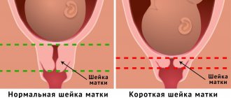
During the normal course of the gestation process in the early stages, the ends of the pharynx should close tightly. The length of the cervical canal during pregnancy should be about 40 mm. The doctor can predict the imminent onset of labor or identify the risk of early miscarriage based on the condition of the pharynx.
Norm indicators of cervical length during Cervicometry.
In our clinic in Pyatigorsk “Ultrasound 4D”, cervicometry is carried out by highly qualified specialists who master the research methodology at a high level. Specially trained employees will quickly and efficiently perform the cervicometry procedure, which will immediately identify abnormalities, if any, and make a final diagnosis.
The normal length of the cervix depends on what stage of pregnancy the patient is at:
- At 20 weeks of pregnancy the norm is 40 mm
- At 34 weeks – 34 mm
These indicators are considered the norm, but deviations from it are possible; a length of over 25 mm is considered normal. Such indicators are not considered a violation and do not indicate the presence of pathology in the body. For more accurate determinations, you need to resort to ultrasound and measurement of the cervix using cervicometry.
If the readings are lower, there is a risk of miscarriage. In such situations, measures are taken to prevent premature birth, and after stabilization of the condition, ultrasound cervicometry is performed every 2 weeks.
Examination during pregnancy
Already at the first appointment, the gynecologist will take a smear for bacterial culture. Such a study will be repeated several times during pregnancy. If detected early, many infections are not dangerous and can be cured before delivery. If treatment is not possible, vaginal birth is replaced by caesarean section to prevent infection of the baby.
Expectant mothers are concerned about the following questions:
- Does it hurt to take a smear for examination? Gynecologists claim that the procedure is completely painless. Biological material is collected during an examination on a chair using a special brush. If a woman doesn't concentrate on the process, she probably won't notice anything.
- Can the procedure cause an increase in uterine tone? No, the process of collecting material is not at all associated with any mechanical effect on the cervical canal and does not provoke an increase in the tone of the uterus.
- Is taking a smear dangerous for the fetus? Such an examination is absolutely safe for the fetus in the womb. The greatest risk occurs when infectious and inflammatory processes are detected.
Do not worry, the analysis will not harm the pregnant woman and will not affect the development of the fetus in the womb. In some cases, a short-term increase in tone is possible, but more often this is due to the attitude of the woman herself towards the examination procedure.
Norms during pregnancy
With the onset of pregnancy, the cervical canal and cervix close into a tight ring. This condition allows the female body to unimpededly bear the fetus until completion of gestation. During the period of preparation for childbirth, starting from the 36th week of pregnancy, a gradual softening and smoothing of the cervix occurs. This condition is necessary for the normal passage of the baby through the birth canal. If the canal remains narrow, the doctor can open it mechanically using special instruments.
Simultaneously with this process, the cervical canal expands. During the first contractions, its diameter is about 1 cm, and within a few hours the dilation reaches 10 cm. Depending on the internal diameter of the cervical canal, the obstetrician assesses readiness for childbirth. Full readiness is determined when the dilation is 10-12 cm. At this width, the birth canal is ensured, starting from the uterus, passing into the cervix and vagina.
Length and diameter of the cervical canal during pregnancy
Doctors consider the length or extent of the cervical canal as an important marker that allows them to confirm the natural course of gestation. If any deviations from the norm are detected, therapeutic measures are used; they allow the pregnancy to be maintained, especially in the early stages.
| Gestation period (weeks) | Length of the cervical canal in (millimeters) |
| 10-14 | 34-36 |
| 15-19 | 38-39 |
| 20-25 | 40 |
| 25-29 | 40-41 |
| 30-32 | 35-40 |
| 32-36 | 33-36 |
| 36-40 | 29-30 |
Such standards are reference indicators, so you should not try to interpret the doctor’s notes and information obtained during ultrasound diagnostics on your own. Much depends on the anatomical structure; it is important to understand that the cervical canal has different lengths in all women. If a girl is diagnosed with a short cervix, this is not a pathology, but is considered as an anatomical feature. The doctor will manage the pregnancy using a different protocol.
The diameter of the cervical canal during pregnancy should remain unchanged. The cervix closes tightly after the fertilized egg is attached to the womb and remains in this position until 34-36 weeks of pregnancy. In the later stages, the body starts the process of natural preparation for childbirth, so the canal may expand slightly. The beginning of this process is indicated by training contractions (in comparison with real ones, they are less painful).
Full opening of the cervical canal is ensured only by real contractions. As a rule, with a frequency of their occurrence once every 10 minutes, the clearance is about 10-20 mm. At this moment, you urgently need to call an ambulance and go to the maternity ward. It should be borne in mind that childbirth at 32-34 weeks is not a rare occurrence, especially with a repeat or multiple pregnancy.
A clearance of 1 mm in the cervical canal does not indicate the development of pathology, and such a disorder often occurs during repeated births. If the deviation is diagnosed early in pregnancy, doctors take a wait-and-see approach. By 12-18 weeks of pregnancy it can close on its own. If the diameter increases, intervention is indicated; the woman will be offered to install an obstetric pessary, allowing her to carry the pregnancy to term.
Prevention
If you want your pregnancy to pass without any worries, be sure to carefully monitor your health and regularly visit your doctor for a routine gynecological examination. Proper preparation for conceiving a child involves preliminary treatment of any gynecological diseases and endocrine pathologies. If you find any abnormality, do not ignore it. Consult your doctor to find out the best ways to solve the problem.
In addition, in the prevention of diseases of the cervix and cervical canal, it is important to pay due attention to daily intimate hygiene. You need to wash yourself twice a day and change your underwear once a day. If you are pregnant, avoid douching. Such external influence on the pharynx can negatively affect its functioning, and there is a danger of infection of the fetus or causing microtrauma to the mucous membrane of the cervical canal. The same consequences can occur after rough sex, so moderate your passion for a while.
Before pregnancy, many women do not even think about how complex their body is, for example, what the cervical canal is, where it is located and what functions it performs. But upcoming motherhood forces you to pay closer attention to your own body and its features. The cervical canal plays an important role during pregnancy, so all expectant mothers should learn more about it.
The outcome of pregnancy largely depends on the condition of the cervix and the canal located inside it. When labor begins, how it will proceed, whether there is a threat of the baby being born prematurely - all this can be suggested by the doctor during a gynecological examination.
The cervical canal is open during pregnancy
A slight opening up to 1 cm in diameter is not dangerous for the pregnant woman and the fetus. Provided that the length of the cervical canal is at least 30 mm. In this case, the change is not considered a pathology, but the woman needs constant monitoring; perhaps the doctor will recommend going to the gynecological department for preservation. Therapy consists of using progesterone drugs and antispasmodics to eliminate tone.
Those at risk for enlargement include:
- multiparous women;
- girls with multiple pregnancies;
- patients with elevated testosterone in the blood.
A more significant expansion may indicate the development of serious pathologies that interfere with the normal process of gestation.
Isthmic-cervical insufficiency
A similar diagnosis is given to women if the cervix dilates and softens prematurely. However, she cannot hold the fetus in the uterus. The disorder is often diagnosed in the second trimester of pregnancy, when the fetus rapidly gains weight in the womb. Pathology is diagnosed during a gynecological examination and ultrasound examination.
The problem is significant due to the growing risks of spontaneous abortion and premature birth. It is difficult to accurately determine the causes of the development of isthmic-cervical insufficiency; doctors often rely on hormonal imbalance and the predominance of testosterone in the hormonal background of a pregnant woman. Male hormones do affect the cervix and can cause it to soften prematurely.
Attention! ICI can occur due to abnormal development of the female genital organs. Often the cause is surgical abortion, resulting in cervical injury.
If pathology is detected in a timely manner, the risks present can be minimized. Operational tactics are often used. During the intervention, sutures are placed on the tissue around the cervical canal to prevent premature opening. They are removed several weeks before the expected date of birth, when the risks to the fetus are minimized.
In some cases, they resort to installing special obstetric devices (rings or pessaries). They are used after 24 weeks with diagnosed minor dilation. It is believed that suturing is a more reliable technique.
Polyps
Neoplasms on the cervix appear in women due to hormonal imbalance. This pathology does not prevent delivery, but requires constant monitoring. It is mandatory to undergo an examination to eliminate the risk of developing cervical cancer. Sometimes a biopsy is recommended.
You shouldn't worry too much about this. A polyp is a benign neoplasm, for which a woman will have to provide treatment after childbirth. In some cases, it resolves on its own, without the use of surgical or medicinal tactics.
Endocervicitis
Endocervicitis is an inflammatory process occurring in the cervical canal. Pathology can also be considered as one of the causes of noticeable enlargement, but it is not the main problem. The risk is that the causative agent of the pathology in the vast majority of cases is an infection, the treatment of which must be provided during pregnancy. The patient must undergo a smear of the cervical canal, based on the results of which the gynecologist will select the necessary treatment.
Attention! Against the background of endocervicitis, a woman experiences pain in the genital area.
The cervical canal is closed
Narrowing of the lumen of the cervical canal during pregnancy is normal. Such a conclusion can be obtained after performing an intravaginal ultrasound. Complete fusion (narrowing) during pregnancy is excluded, because such a pathology contradicts the possibility of fertilization. A woman will not be able to get pregnant because, due to stenosis, sperm will not be able to reach the egg. Consequently, a closed cervical canal is the norm and such a conclusion should please, and not frighten, the pregnant woman, because it confirms the natural course of the process.