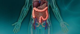
What are metastases in cancer?
Malignant cells from foci of primary localization spread through blood and lymph flow to other organic structures, forming metastatic foci, which are actually secondary localizations of cancer.
So, metastases are cancer cells that spread throughout the body from the main focus of the tumor process.
When cancer spreads into neighboring tissues, it is called regional metastasis. If malignant cellular structures penetrate into peripheral tissues through the bloodstream or lymphatic fluid, then distant metastasis occurs.
Metastases. Diagnostics
At the Yusupov Hospital, doctors diagnose metastatic cancer using modern research methods:
- tomographic (MRI, CT, PET-CT);
- scintigraphy of skeletal bones;
- Ultrasonography;
- various diagnostic punctures under visual control of a tomograph;
- full laboratory examination, including determination of the level of tumor markers in biological fluids, immunological and molecular genetic;
- endoscopic (gastroscopy, bronchoscopy, colonoscopy) diagnostics with biopsy;
Reasons for the spread
In general, metastasis is caused by certain oncological growth factors that stimulate the formation of capillary and vascular networks around the tumor formation.
As a result, a favorable environment is created for malignant structures, which provides them with the necessary nutrition. In this scenario, metastasis occurs throughout the body.
In general, the spread of malignant cells can occur in different ways:
- With the bloodstream - malignant cells spread hematogenously through veins, capillary structures and vessels throughout the body;
- With lymph flow. Lymph nodes act as a protective barrier for malignant structures and their partial destruction occurs in them. But when there are too many changed cells, macrophages cannot cope with them;
- Implantation or along the membrane of serous tissue.
Metastases of lymphogenous origin are most characteristic of cancer of the uterine cervix and stomach, larynx and colon, sarcomas and melanomas.
Hematogenous routes of metastasis are usually observed in late stages of chorionepitheliomas and sarcomas, pelvic and abdominal tumors, hypernephroma, etc.
Causes
The causes of spread are complications of cancer in the later stages of its development. They appear when a large number of pathological cells form in the tumor.
Development can be stopped by the good immunity of a sick person and high tissue resistance. The location depends on the location of the cancer, and the frequency of spread depends on the structure and growth of the tumor, and the age of the patient.
Note! Metastases are more dangerous than the primary malignancy.
Answering the question at what stage metastases appear, it should be noted that most often they appear at the third and fourth stages of cancer development. Sometimes they can appear in the early stages, but in this case they will be microscopic in size and located in the lymph nodes.
Note! Any examination by an oncologist begins with examining the condition of the lymph nodes, since this is where the process of metastasis most often begins.
A large number of abnormal cells that are formed during metastasis are eliminated by the human immune system. Some of them linger in the capillaries and become overgrown with tissue, so the immune system does not cope with them properly.
At what stage do they appear and how quickly do they spread?
If a cancer patient does not receive the necessary treatment, then metastases will occur over time in any cancer process, but the timing of appearance is not always clear.
In some oncological pathologies, metastasis occurs within a few months after the formation of the primary tumor focus, while in others it is detected only after several years. Therefore, it is impossible to even tentatively establish the timing of metastasis.
Considering metastasis in the lymph system, we can say that metastases are a sign of the transition of cancer to the second stage of development.
If hematogenous spread of malignant cells appears, then we are talking about the transition of oncopathology to stage 4. On average, metastases form at stages 3-4 of cancer. That is, in fact, the appearance of metastatic processes determines the stage of the cancer tumor.
Video about how cancer metastasizes:
Introduction to Cancer
Cancer, or a malignant tumor, is the appearance of new pathogenic cells in tissue due to its death or a defect caused by a genetic failure or environmental influences. Tumors grow on their own. Under the influence of cumulative factors, they mutate, turning into tumor, then malignant centers. They seize nearby organs, destroying them. They produce independent daughter cells that quickly spread through the blood or lymph to distant tissues throughout the body. The waste products of pathogenic organisms infect and poison vital organs. This situation leads to the premature death of a person. In the first stages, all tumors are curable.
Scientists have proven that certain types of cancer are a consequence of viral diseases (HPV, HIV, sexually transmitted infections). People who lead an unhealthy lifestyle or have a genetic predisposition are most susceptible to cancer.
People at greatest risk:
- Smokes tobacco products, inhales combustion products of solid fuels (smoke from a fire).
- Carelessly treats excess weight, suffers from obesity.
- Does not eat raw fruits and vegetables, which leads to a deficiency of vital vitamins and minerals.
- Leads a passive lifestyle (leads to deformation of internal organs, the appearance of defects and the formation of a favorable environment in the body, where malignant pathogenesis then develops).
- Frequent consumption of alcohol-containing drinks (detoxification occurs, and then pathogenic changes in organs).
- Infection with viral infections (HIV, HPV, hepatitis, sexually transmitted infections).
- Works at nuclear power plants (influence of radioactive and nuclear radiation).
Due to low public awareness, the initial symptoms are ignored by people. The tumor grows, metastasizes to other organs of the body, secondary and obvious symptoms appear (the affected organs simply hurt), which cannot be cured. It only manages to make it easier.
Types of cancer
Cells differ in their method of division and growth depending on the tissue that is the origin of the pathogenic process. The tumor is divided into types:
- carcinomas - originate from the integumentary tissue;
- melanomas - originate from the inner layers of the skin containing melanin;
- sarcomas - formed from connective germ tissue (in muscles);
- leukemia – begins with blood-forming cells;
- lymphomas – originate in damage to the lymphatic system;
- teratomas - grow from embryonic cells at the intrauterine stage;
- gliomas – come from glial cells;
- Choriocarcinomas - degenerate from placental tissue.
Stages
Depending on the stages of detection of a new pathogenic formation, treatment is determined and prescribed, and further predictions are made. The stage is influenced by multiple factors: location of the formation: size, location of metastases in cancer, presence and extent of damage to the immune system. Oncological tumor has five stages:
Development of a cancerous tumor
- Zero – the tumor has barely appeared, it cannot be determined, or is located in one place, the size is small. In this case, the greatest guarantees of treatment (100%) are that after therapy and surgery the person gets rid of the disease forever.
- The first one has fairly favorable prognoses. The process is localized in one place, does not have time to occupy neighboring organs, and does not affect the immune system, in particular the lymph nodes. It is possible to cure. The recovery rate reaches 90%.
- The second is the growth of pathogenic tissue and metastases to neighboring organs, but the lymphatic system is not yet affected. Treatment is possible, but a lot depends on the type of cancer, individual characteristics of the body, and early diagnosis. The prognosis is positive if distant organs survive.
- The third stage has little chance of recovery and survival, which is quite possible, however, with proper diagnosis and treatment. After all, the main processes are still occurring in the main focus and have spread only to nearby tissues. The pathological process is complicated by initial damage to the lymphatic system, all other systems are not affected. It is difficult to predict anything; it is not considered possible to completely get rid of malignant cells.
- Fourth and last: the chances of recovery tend to zero. Daughter independent cells spread throughout the body (distant organs are not affected, which is extremely rare), throughout the immune system. There are more pathogenic foci; this stage cannot be treated or corrected. Taking medications is limited to pain relief and slowing down the growth of malignant cells in order to prolong the patient’s life.
What affects healing?
Treatment depends largely on the stage, correctness of diagnosis, availability of specialized equipment, necessary funds, qualifications of the oncologist and the wishes of the patient himself. The mentioned process is protracted and requires enormous self-organization and precise implementation of medical prescriptions. Out of desperation, people begin to self-medicate, preferring folk methods and various magical rituals. They come to their senses and go to a medical facility when the time for treatment is lost and there is almost no chance of defeating cancer.
The higher the stage, the less chance of treatment. More money, effort and medicine will be spent to obtain positive results. Minimum chance of stage 4 cancer with metastases. Therefore, as a preventive measure, it is recommended to regularly undergo medical examinations (in Russia they are carried out once every four years), lead a healthy lifestyle, play sports, and get appropriate vaccinations against viral infections.
How do different types of cancer metastasize?
Typically, metastases are detected in the pulmonary structures, liver and lymph nodes. Much less frequently, metastatic foci are found in the heart and skeletal muscles, spleen and pancreas.
Experts have identified a certain pattern of cancer metastasis in different locations:
- Melanoma usually metastasizes to the lungs, liver, muscle, or skin;
- Pulmonary cancer - in a healthy lung, liver and adrenal tissue;
- A malignant tumor in the ovaries and uterus, stomach and intestines, pancreas usually metastasizes to the lungs, liver and abdominal cavity;
- Mammary, renal and prostatic oncology spreads mainly to bone, liver and lung tissues.
How are metastases diagnosed?
Plain radiography, ultrasound, radioisotope research, computed tomography, magnetic resonance imaging, positron emission tomography - all these techniques are essential in recognizing metastases. These techniques make it possible to clarify the size, prevalence and growth pattern of metastases, their disintegration, suppuration, and germination into neighboring organs and tissues. In addition, these same diagnostic techniques make it possible to monitor the effectiveness of treatment based on the degree of regression of metastases.
Conventionally, two stages of diagnosing metastases can be distinguished:
- Primary examination, when the main tumor is only diagnosed;
- Follow-up with an oncologist after treatment. If no metastases were initially detected, and the treatment was successful, you will still have to undergo periodic examinations in the future - there is a risk of relapse.
Varieties
Metastasis has many options and varieties that differ significantly from each other.
Virchowsky
Virchow's metastasis is localized in the supraclavicular region of the neck and occurs against the background of gastric cancer. This location of the secondary cancer focus is determined by the direction of lymph flow from the abdominal cavity.
Malignant cellular structures rise along the lymphatic pathways precisely to the cervical lymph node; they cannot pass further, so they begin to form into a secondary tumor. Virchow metastasis can occur due to cancer of the liver, pancreas and other abdominal structures.
Krukenbergsky
Such metastases are also characterized by lymphogenous origin and are localized in the ovaries. The share of such secondary tumors accounts for about 35-40% of the total number of ovarian metastases.
Krukenberg metastases are observed in malignant gastric, mammary glandular, intestinal or bile duct lesions, bladder or cervical cancer.
Schnitzler
Schnitzler metastases are the spread of a malignant process to the perirectal tissue and perirectal lymph nodes.
Such metastatic formations can be palpated during rectal digital examination and present as painless lumps.
Most often occurs against the background of stomach cancer.
Osteoblastic
Metastatic tumors that form in bone tissue and promote the activity of osteoblasts are called osteoblastic. Against the background of increased osteoblastic activity, increased calcium deposition occurs in bone tissue, which contributes to their rapid growth.
Such metastatic foci occur against the background of mammary glandular, thyroid or prostatic cancer, sarcomas and lymphomas. The prognosis is mostly unfavorable.
Solitary
Metastases of the solitary type are large-nodular single formations localized in the lung, brain and other tissues.
Osteolytic
Osteolytic secondary formations are also localized in bone structures, however, their effect on bones is of a slightly different nature. They destroy bone tissue and activate osteoclasts, which leads to destructive changes in bones.
Diagnostics
Metastases are a sign of advanced process. Detection of metastases is possible during professional medical examinations and medical examinations. With appropriate symptoms (for example, pain or pathological fractures due to metastases in the musculoskeletal system), the patient can actively consult a doctor, which will entail searching for the original focus and building a treatment strategy.
In some cases, the primary lesion may not be detected (for example, with a history of chemotherapy or radiation therapy).
Sometimes metastases themselves are diagnostically important in determining the location of the tumor. Their cellular structure can tell this.
As for regional metastases, their presence can be suspected when there are changes in the lymph nodes. Superficial lymph nodes are accessible for palpation; their enlargement allows one to suspect the presence of a tumor process.
Cervical groups are affected by breast cancer, oral cavity and tongue cancer, lymphomas and other hemoblastoses. Supraclavicular lymph nodes are susceptible to damage from neoplasms of the lungs, esophagus, colon and stomach. Axillary lymph nodes enlarge with diseases of the mammary glands. Inguinal - for neoplasms of the genitourinary system in men and women, and the rectum.
Metastases of various locations can be diagnosed using instrumental methods such as ultrasound, radiography with or without contrast, CT or MRI . Using these methods, it is possible to determine the size of the tumor and stage the disease.
An indispensable method of investigation is a biopsy of a suspicious area of tissue. In some cases, the process can be diagnosed by the presence of characteristic tumor markers found in the blood serum.
Symptoms and signs
The clinical picture of metastasis depends on its location and type of primary tumor. Typically, metastases lead to severe dysfunctional changes in the structures of the body.
- With hepatic metastasis, patients experience itchy skin, jaundice and liver failure;
- Cerebral metastatic processes lead to rapid encephalopathy;
- Pulmonary metastasis causes bronchopulmonary inflammation, respiratory disorders, etc.;
- Bone metastases are characterized by severe pain throughout the body.
On the skin
Skin metastases occur predominantly against the background of malignant lesions of the ovaries, lungs and kidneys. Metastatic processes on the skin are of lymphatic or hematogenous origin. In men, such metastases are localized on the abdomen and neck, chest and head, and in women on the chest and abdomen.
Signs of skin metastases:
- The appearance of formations similar to moles;
- Change in skin color at the site of metastases;
- Rapid increase in skin formation;
- Asthenia;
- Exhaustion;
- Drowsiness and weakness;
- Lack of performance;
- Painful sensations in the area of the tumor;
- Weight loss and hyperthermia.
The photo shows what stage 4 cancer looks like with metastases on the skin
If the metastasis has formed on the scalp, it usually looks like a sebaceous cystic formation.
In the ribs
The first signs of rib metastases are intense pain, leading to limited mobility. At later stages, secondary tumor foci can lead to rib fractures that occur even with minor loads.
Cancer tumors of the thyroid gland, breast, prostate and cervix, liver and lungs, esophagus, etc. most often metastasize to the ribs. To detect them, a scintigraphic examination of the skeleton is necessary.
Heart
Secondary cardiac tumors usually arise from pleural mesothelioma, carcinoma, melanoma or esophageal squamous cell carcinoma, renal and thyroid cancer, or leukemia.
Signs of cardiac metastases are:
- Pericardial effusion;
- Obstruction of veins in the myocardium;
- Depression of cardiac activity;
- Arrhythmia, myocardial failure.
Peritoneum
Cancer cells can invade any part of the body, particularly the abdominal cavity. Malignant structures settle on the surface of the internal organs and peritoneal walls. They accumulate for quite a long time, gradually forming a secondary tumor.
Such processes in the body are usually accompanied by ascites, causing an enlarged abdomen. If the tumor begins to disintegrate, then general signs of intoxication appear.
For breast cancer
Metastatic foci in the mammary gland are manifested by the appearance of lumps in the breast, which are easily felt by palpation.
Malignant cells penetrate into the mammary gland through the bloodstream or lymphogenously. The patient feels intense pain in the chest and other uncomfortable sensations.
Distant metastases
The greater the parameters of the primary formation, the earlier the metastatic processes will begin. Typically, the real threat of metastasis occurs when the tumor exceeds a 3-cm diameter.
Together with the bloodstream, malignant cells spread to distant tissues and organs, which indicates the late stages of the tumor process.
- If metastases occur in the skeletal system , then patients experience bone pain, which can seriously reduce the quality of life.
- If breast cancer has metastasized to the lungs , then the patient is bothered by shortness of breath, cough and chest pain.
- With nervous system metastasis, dizziness and headaches, convulsions and hallucinations, auditory and visual disturbances, coordination disorders, etc. appear.
Regional
Already in the early stages of oncology in the mammary gland, metastases can occur in regional lymph nodes. Typically these are axillary lymph node structures.
But if the primary tumor has formed closer to the center of the chest, then the sternal lymph nodes undergo metastasis.
Subsequently, the cancer process spreads to more distant lymph nodes.
In the intestines
Metastasis to the intestines is accompanied by frequent diarrhea or constipation, blood in the stool, abdominal pain and bloating.
In addition, waste products of cancer formation cause general intoxication of the body, which is manifested by dyspeptic disorders.
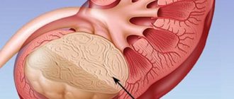
Kidney
The main sign of metastasis in the kidneys and adrenal structures is hematuria, which is characterized by the presence of blood in the patient’s urine.
An additional sign of metastasis in the kidneys is pain in the lumbar region, constant temperature and weakness, elevated blood pressure and progressive anemia.
Spleen
Metastases in the spleen are extremely rare, because the organ itself is capable of producing substances that destroy malignant cells.
Obvious signs of metastasis include fever, thrombopenia, increased organ size, heaviness and pain. As the secondary tumor grows, the condition worsens and the body becomes exhausted.
Pleura
The pleura lines the chest wall and lungs on the inside. It produces a special lubricant that facilitates pulmonary function during breathing. Metastasis to pleural tissue is accompanied by cough, low-grade fever and pain in the sternum.
Stomach
Metastasis in the stomach is quite rare, with tumors spreading here from the uterus, esophagus, breast or lung. Metastasis is accompanied by hyperthermia and lack of appetite, anemia and taste changes, pain in the stomach, etc.
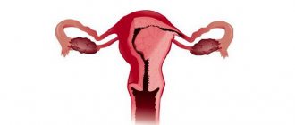
Ovarian
At the initial stages, ovarian metastases do not manifest themselves in any way. Some cancer patients experience a lack of appetite and general weakness, menstrual irregularities and hyperthermia. When the metastasis increases, painful sensations and a bursting feeling appear in the lower abdomen.
Adrenal glands
Many tumors metastasize to the adrenal glands, for example, from the lungs, kidneys, mammary glands, etc.
Such tumor spread causes adrenal insufficiency.
Large secondary formations are almost always accompanied by necrotic processes.
For uterine cancer
Metastasis in uterine cancer begins at stage 3 of the oncological process. The spread of malignant cells occurs through the lymphogenous route, and hematogenous spread is possible at the last stage of cancer.
Patients complain of spotting between periods, lumbar pain and pain in the lower abdomen, especially during exercise.
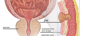
Bladder
Metastatic spread of malignant cells into the bladder structures occurs through the lymphogenous route, mainly from the pelvis or ureter.
At first, symptoms most characteristic of cystitis appear, including frequent urges, lumbar pain, and painful urination.
With the development of metastasis, the condition worsens, constant hyperthermia appears, blood in the urine, etc.
Pancreas
Pancreatic metastasis is characterized by such manifestations as sudden weight loss and lack of appetite, nausea and vomiting syndrome, epigastric pain and frequent diarrhea.
Sometimes metastases in the pancreas cause some yellowing of the skin and girdle pain in the abdomen.
Throat
Metastatic formations in the throat usually appear from tumors of the mouth, respiratory and digestive organs. Most often, such localization of metastases causes the following symptoms:
- Sores and sores in the throat;
- Swelling of oral tissues;
- Problems speaking, breathing, swallowing;
- Enlarged lymph nodes, etc.
Signs of metastases
The main organs of secondary damage are: spine, costal region, skin, muscle tissue, heart muscles, abdominal cavity. The respiratory system (lungs), chest (breast), uterus, ovaries, kidneys are affected. The adrenal glands, gastrointestinal tract, and bladder are affected. Each organ has its own symptoms; they must be distinguished in order to recognize them in time and seek medical help, prolonging your life.
Cancer is characterized by the following symptoms when the following are affected:
- Spine and ribs – pain appears with any movement, radiates into the bones, compression of the vertebrae occurs, coordination of movements is impaired, leading to paralysis. Pain and intensity increase, subsequently weakening of the bone tissue leads to systematic fractures.
- Muscle tissue - proceeds calmly, without pain and changes in the muscles, later they themselves become deformed, compactions and unpleasant sensations appear that turn into pain.
- Skin – increased formation of pigmentation develops. Multiple birthmarks appear on the skin, its color changes, appetite disappears, weight decreases, with increased fatigue.
- Cardiac muscle - the first signs similar to arrhythmia appear, disorders and inflammatory processes in the myocardium and pericardium are possible, leading to a stroke.
- Abdominal cavity - initially there may be no symptoms, then sudden changes in temperature begin, weight decreases, nausea appears, the skin on the body and the whites of the eyes turn yellow, at the 4th stage of cancer, general poisoning and death develop when the cancer disintegrates.
- Lungs - initially there are zero symptoms, then a cough occurs, shortness of breath with fever, they are mistaken for a common cold (they are treated for it). Possible pain in the neck (throat) coupled with bloody discharge and weight loss. This should alert the patient and the doctor.
Metastases in the lungs
- Mammary gland - diffuse changes in the tissues begin, compactions form, with increasing pain, with the addition of dizziness and neurological symptoms.
- The cervix and ovaries are practically asymptomatic; the appearance of the organ changes, invisible to the normal eye. A pulling sensation and additional bleeding appear.
- Kidneys and adrenal glands - changes begin in the structure and structure of the organ, unpleasant nagging pain, blood spots and discharge during urination may occur.
- The gastrointestinal tract proceeds calmly, there may be no symptoms, only minor disturbances (gas formation, a feeling of satiety). Taste preferences change.
How to detect in the body?
Detection of metastases requires a thorough diagnosis, including:
- ;
- Ultrasound;
- MRI;
- Survey radiography;
- PAT;
- Radioisotope diagnostics.
Such procedures make it possible to determine the degree of metastasis, the size of secondary tumors, germination into other tissues and the presence of purulent processes or decay, growth patterns, etc.
Are they visible on ultrasound?
Ultrasound diagnostics is one of the main methods for detecting the metastatic spread of malignant processes.
Such a study is considered quite informative and is widely used in modern diagnostic practice.
Diagnosis and treatment
Sometimes a secondary focus of a malignant tumor is initially diagnosed, and from it the main affected organ is calculated; for the patient this only means negative dynamics in the future, since it indicates the third or fourth stage of the development of pathogenesis. Initially, the spread of daughter cells and nodes is asymptomatic; their appearance can only be diagnosed using cumulative methods during complete medical examinations, which are carried out by medical institutions every four years. If cancer is suspected, doctors palpate the painful area, and in case of some alarming complaints, additionally prescribe:
- Ultrasound;
- MRI;
- blood and urine tests for tumor markers;
- CT;
- biopsy (puncture);
- laparoscopy;
- endoscopy;
- scintigraphy.
Taking a biopsy sample
In the first stages of cancer, treatment is divided into stages (identification of the main organ, checking for metastases, treatment of pathologies, rest, re-treatment, observation) and is aimed at stopping the process of formation of metastases. It is the appearance of additional foci of malignant tumors that leads to death. There are various ways to do this:
- Chemotherapy – medications are introduced into the body, they block the growth of not only pathogenic cells, but also healthy ones. At the end of the course, the functionality of healthy cells returns. It is difficult to tolerate by patients, many tissues are affected (hair loss), and is used in conjunction with removal and irradiation.
- Tumor removal – surgical, ultrasound, laser. The tumor is excised with a margin to protect nearby tissues. Laser intervention is preferable, since the laser removes all malignant cells in the resulting wound and in the blood.
- Radiotherapy is radiation that affects cellular DNA, destroying and stopping tumor growth. But it affects both healthy cells and diseased metastases. Most often used in combination with removal (when it is impossible to completely remove the entire tumor due to metastasis).
- Hormonal therapy is the administration of hormones. This is only an additional method used in conjunction with one of the main ones.
- PDT (photodynamic therapy) - drugs are administered that, when exposed to light waves of a certain length, stop the malignant process. The method is quite new and is used at the initial stages of pathogenesis. It is most effective for skin and cervical cancer (only the cancerous tumor is killed, the ability to conceive is preserved), and for tumors localized in the spine.
- Immunotherapy – medications with a high content of interferon are administered, which stimulates the immune system to more intensively fight pathology.
- Cryodestruction method - the word “cryo” refers to the effect of negative temperatures. The cancerous tumor is frozen (cold is applied with a special device), and then the dead cells come out naturally. The method can be used to influence only urological and gynecological types of cancer, sometimes for lung cancer without metastases, the development of a malignant process in the liver, and for the removal of tumors on the face.
Carrying out cryodestruction of skin formations
The use of one method is unjustified; a combined type is chosen (tumor removal followed by chemotherapy or radiation). The main thing at this time is to find a good oncology center and choose a worthy attending physician who is trustworthy. Otherwise, patients, trying to get rid of metastases, begin to try traditional methods of treatment. Eg:
- Vitamin B17 – it is believed that its deficiency causes a weakening of the immune system and leads to cancer.
- Increased iodine intake.
- Consuming calcium with magnesium.
- Refusal of dairy products.
- Treatment with propolis. It is believed that the bee product blocks the growth of the pathogenic environment, then kills it.
- They use a tincture of the poisonous hemlock herb. It supposedly works in a similar way to a chemical medicine.
- Tinctures of herbs and mushrooms, including fly agarics.
Not all of the listed remedies are useless! Taking vitamins brings relief. Tinctures of poisonous herbs and mushrooms are not able to stop cancer and have the opposite effect, poisoning the body even more. Or they directly send the person to the next world.
You shouldn’t waste your precious time into emptiness - it’s better to fight the disease medically. The earlier treatment is started, the greater the chance of defeating the disease and eradicating the tumor. Despite the fact that during the remaining life you will have to regularly go for screenings and additional studies (for example, MRI).
How to cure?
Treatment of cancer pathology with metastasis is determined by the location, size and number of secondary foci. Several different techniques are used: surgical removal, radiotherapy and drug therapy.
Surgical treatment
Initially, doctors try to remove the primary formation, which in the future may act as a source of metastases.
Then they proceed directly to the removal of the metastatic foci themselves. To do this, the lymph nodes and adjacent tissue are removed.
When removing secondary formations, the surgeon also cuts off part of the healthy tissue, which may also contain micrometastases.
Radiofrequency ablation
Radiofrequency ablation is now successfully used in the treatment of metastatic spread of tumor processes.
This method involves destroying the tumor through high temperatures created by special electrodes. Electromagnetic currents heat malignant tissues and destroy them. Then the dead cells dry out, and a scar forms in their place.
Medicines
Drug treatment of metastatic tumors involves the use of methods such as chemotherapy, immunotherapy, targeted and hormonal therapy.
Chemotherapy with antitumor drugs stops the growth and spread of metastases. This technique is often combined with radiation or radiofrequency ablation.
Treatment
There is no specific treatment for metastases. The main methods of treatment are divided into two large groups: therapeutic and surgical .
Therapeutic treatment can be carried out with chemotherapy or using radiation and laser technologies . These methods can be used independently in the treatment of cancer or performed before surgery.
Chemotherapy treatment and radiation therapy are designed to suppress the occurrence of new metastases and slow down the growth of existing ones. Therapy is carried out either with one drug or several, and in this case it will be polychemotherapy.
After surgery, chemotherapy and radiation therapy help suppress the growth of possible new metastatic lesions.
Surgical treatment is called radical if during the operation the primary lesion, lymph nodes and all metastases are removed. Such operations are carried out in compliance with the principles of ablastics (preventing tumor cells from entering nearby tissues) and antiblastics (removal of all tumor cells in the area of the operation). The extent of the operation depends on the size of the lesion, the number and location of metastases. In cases with single metastases, only surgical method can be used.
Sometimes methods are used that do not involve the use of a scalpel. Such methods include cryosurgery, laser surgery and photodynamic treatment .
If there are complications (compression of the spinal cord substance, pathological fractures), palliative treatment . One important aspect of palliative care is pain control. Also the objectives are to treat symptoms: nausea and vomiting, infectious complications and bleeding.
Surgical treatment of tumors with multiple distant metastases is often not performed, since it is impractical if it is not possible to remove all metastases and the lymph nodes adjacent to them.
How long do people live with metastases: prognosis
Typically, the presence of metastases in lymph nodes and other organic structures indicates an unfavorable prognosis for oncological pathology.
- Prognosis of metastases in the abdominal cavity. The mortality rate for such metastasis today is 5%. Timely detection of abdominal metastasis and mandatory chemotherapy with appropriate rehabilitation greatly increases the patient’s chances of a favorable outcome of oncology treatment.
- To the adrenal glands. Adrenal metastases are usually combined with damage to other organs, so the prognosis depends on the specific clinical situation.
- Mediastinum. Such metastasis, if detected early, can have a positive outcome, however, if detected late, the prognosis is unfavorable.
- Intestines. With timely access to an oncologist, there is a tendency to a successful outcome of the disease. Cure with timely surgical intervention in combination with radiotherapy and chemotherapy occurs on average in half of the patients. In the later stages, the prognosis is disappointing.
- Liver. Without treatment for metastatic liver lesions, survival is 4 months. If the patient receives the necessary help, the life of the patient is extended by one and a half years; additional chemotherapy can give the cancer patient about another year of life.
- Lungs. Unfavorable factors for pulmonary metastasis are its appearance earlier than 12 months after removal of the primary cancer focus, as well as the rapid increase in metastatic tumors. The 5-year survival rate for single metastases and after adequate treatment is about 40%.
Almost all patients suffering from cancer die within a decade, regardless of the presence of metastatic processes. And if they are present, then life expectancy is significantly reduced.
If the patient has terminal (fourth) stage of oncology and there are metastases, then life expectancy is several weeks, and sometimes days, depending on the type of tumor.
How do metastases appear?
The clinical picture of metastatic cancer is determined by the location of the metastases:
- Damage to the peritoneum (peritoneal carcinomatosis) leads to ascites, which requires laparocentesis;
- Damage to the pleura (pleural carcinomatosis) causes the development of exudative pleurisy, which requires thoracentesis;
- Damage to the bones and spine leads to pain in different parts of the body, fractures;
- Lung damage manifests itself as shortness of breath;
- Brain metastases lead to headaches, dizziness, convulsions, and impaired consciousness;
- Liver damage leads to obstructive jaundice.
In addition, toxins released during the constant death of tumor cells cause cancer intoxication of the body.
In my head
Metastases in the head, or more precisely in the brain, can develop at stage 4 of almost any cancer.
More often, metastases to the brain are caused by lung cancer, malignant tumors of the large intestine, breast, and kidney. Often the primary cause of brain metastases is skin melanoma. Secondary lesions in the head are one of the most serious complications of cancer.
The life expectancy of patients when cancer cells penetrate the brain rarely exceeds several months. The symptoms of brain metastases are quite varied. In addition to headaches and neurological disorders, signs such as speech impairment, memory impairment, gait changes, convulsions, and constant nausea may be observed. Treatment of metastases involves neurosurgery, radiosurgery, and radiation exposure.
Chemotherapy is practically not used in the treatment of intracranial metastases, since most drugs cannot cross the blood-brain barrier.
Photo: Metastases in the brain
What oncological diseases provoke the spread of metastases to the brain can be found in this section.
Do you know what liver metastases look like on ultrasound? You can see it clearly here.
Classification of metastases
There are several signs by which secondary foci of oncology can be divided into types. Modern medicine takes into account the path of transfer of diseased cells and the size of new formations. The last sign plays an important role in the patient’s life. The smaller the cancer metastases, the higher the chance of getting rid of them. Large ones have a great impact on the survival of patients. Another sign is the distance from the main tumor. According to it they are divided into:
- satellites - near the maternal tumor;
- regional – in areas of regional lymph flow (mostly lymph nodes are affected);
- distant - other organs and lymph nodes located far away; this group includes the 5 types of pathologies described below.
Virchow's pathology
Appears with carcinoma of the stomach and pancreas. A less common cause is cirrhosis of the liver. Metastases form on the neck, in the upper zone of the clavicle. Doctors explain this by the direction of lymph outflow. The cells reach the cervical ganglion. They cannot move further from it and form a new tumor here. The Virchow lesion has a round, regular shape with clear contours. It is dense and large. There is no pain when palpated.
Krukenberg metastasis
Diseased cells are transported in the lymph flow, often ending up in the ovaries. According to statistics, this pathology accounts for 40% of secondary neoplasms in them. It is caused by carcinoma of the stomach, bile ducts, breast, intestines, and pelvic organs. Both halves of the ovaries are often affected. The lesions are small and smooth. If you feel them, there is no unpleasant sensation. Mortality in the first year is 95%.
Schnitzler
This rare pathology also appears with a tumor in the stomach. Diseased cells create foci of cancer in the rectum. The affected areas are lymph nodes and tissue. The problem can be determined by palpating the rectum. New lesions are round or egg-shaped and have smooth borders. Cancer cells can move to this point with lymph through all 3 outflows from the stomach at once. Local symptoms of the Schnitzler process are similar to paraproctitis - inflammation of the tissue of the rectum.
Solitary metastasis
This name hides single lesions that are located far from the main tumor. They often occur in the lungs or brain. Other organs are rarely affected. The size of the solitary metastasis is at least 3 cm. According to statistics, this type of secondary cancer appears with a frequency of 90% in smokers. It is detected during an x-ray or MRI. The solitary focus in the lung is always surrounded by parenchyma.
Osteoblastic and osteolytic
Such foci of cancer appear in the bones. They are dangerous because they cause irreversible cellular mutations and change the composition of tissues. They got their name from the type of their activity. They stimulate the activity of osteoblasts or osteoclasts. These are the cells that produce the basic substance of bone tissue or destroy the old structure. Each of these types of cancer lesions has its own characteristics:
- Osteoblastic - against the background of increased production of the main substance, calcium deposits appear. Secondary tumors appear due to sarcoma, prostate, breast, and thyroid cancer. The osteoblastic type is dangerous due to poor patient survival.
- Osteolytic – provokes active tissue destruction. It often affects the bone tissue of the lower extremities, ribs, and pelvis. Less commonly, the pathology affects the brain.
Pathogenesis of tumor metastases
Metastasis is a complex process. There are five main stages in it.
In stage I, invasive growth occurs. This is a common feature of all malignant tumors - they can grow into surrounding tissues and spread to neighboring organs [1][10]
At stage II, intravasation occurs—the penetration of a tumor cell into a blood or lymphatic vessel. This is a complex process; various signaling molecules, proteins, receptors, and enzymes are involved. The number of blood vessels in the area of the malignant tumor and their diameter play a role. In addition, tumor cells skillfully manipulate healthy tissues, as a result of which they help them metastasize [1][11].
Further, at stage III, tumor cells migrate in the body with the flow of lymph (lymphogenous) or blood (hematogenously). Most cancer cells die in the process. They can spread individually, but most often travel in groups called clusters[1].
At stage IV, tumor cells that have successfully completed their journey through the blood or lymphatic vessels leave them and penetrate the organ tissue. This process is called extravasation. It is quite complex; blood flow features and various cellular and molecular mechanisms play a role. It has been proven that tissue structure is of great importance, so metastases are most likely to occur only in certain organs [1].
At the last stage V, tumor cells settle in a new location and form secondary foci. After this, the growth of metastases begins. The likelihood of this happening depends on the properties of the cancer cells themselves and the organ into which they spread [1].
Bone metastases
Bone metastasis occurs when cancer cells spread to the bones from their original sites. Almost all types of cancer can spread to bone tissue. But some cancers are more likely to spread to the bones: breast cancer and prostate cancer.
Bone metastasis can occur in any bone, but most commonly occurs in the spine, pelvis, and hips. Bone metastases may be a sign of primary cancer or may occur years after cancer treatment.
Once cancer invades bone tissue, it cannot be cured. Treatment only helps to reduce pain and stop metastases in bone tissue. Bone metastases can cause paralysis and hypercalcemia (high levels of calcium in the blood).
Some cancers may start directly in the bones (bone cancer). These cancers are significantly different from bone metastases. Bone metastases are much more common than original bone cancer, especially in adults.
Treatment of metastases
Therapy always begins with eliminating the main focus. So, in case of breast cancer with metastasis to the liver, it is necessary to remove the tumor from the mammary glands. At the same time, the doctor must prevent new diseased cells from forming or spreading. The main problem in the treatment of metastatic cancer is that most types are incurable. The doctor chooses treatment tactics based on the clinical picture of the disease:
- At almost all stages of cancer, drug therapy is indicated, especially with multiple metastatic nodes.
- Radiotherapy (radiation therapy) is carried out in the last stages with a large volume of lesions. This is effective in metastasizing cancer to the brain, stomach, and lymph nodes.
- Severe cancer at the last stage requires palliative treatment. This is psychological support, the use of symptomatic therapy. All methods prolong the patient’s life, but do not eliminate death. Often this tactic is used for secondary lesions in bone tissue.
Dormant metastatic nodes are rarely observed. They develop slowly and may not show signs of growth for many years. They do not cause immediate death, but also require treatment after removal of the main tumor. For this purpose, a course of chemotherapy is carried out. If this is not done, dormant lesions may become active under the influence of stress, sunlight, or decreased immunity.
Systemic drug therapy
The most well-known method of treating cancer is chemotherapy. Cytostatics are used for this. They disrupt the process of growth and division of diseased cells. They are prevented from spreading and the growth of secondary lesions is stopped. Often this treatment is supplemented with radiation. The effect of chemotherapy depends on the type of cancer. In case of kidney and brain carcinoma, it often does not make sense. Even in drug therapy for this pathology, the following options are distinguished:
- Targeted therapy (biological) – blocks the growth of cancer cells by interfering with the mechanism of action of target molecules responsible for tumor growth and carcinogenesis (development). Treatment includes thyroxinase inhibitors (Erlotinib, Vintafolide), serine kinase inhibitors (Afinitor), and monoclonal antibodies (MabThera).
- Hormonal – effective only against a tumor that is sensitive to estrogen and progesterone. This is oncology with metastasis to the lymph nodes and nervous system. During menopause, women are prescribed Tamoxifen; in premenopausal women, Goserin is prescribed. It is possible to use the new drug Anastrozole. Treatment is carried out for six months.
- Immunotherapy – vaccines from cancer cells, interferons, interleukins, immunostimulants, antitumor proteins are used. Such drugs can fight micrometastases and prolong the life of patients in the final stages of cancer. The group of these drugs also includes monoclonal antibodies, which are used in targeted therapy.
- Biosphosphonates (Zometa, Bonefos) are medications that are required for bone tissue damage. They stop the oncological process and improve the general condition of the patient. Used orally or intravenously.
- Stem cell transplantation is an injection into the bone marrow. Prescribed when chemotherapy or radiation treatment cannot be performed. Effective against cancer metastasis to lymph nodes.
Surgery
If multiple metastases from cancer threaten the patient’s life or greatly impair its quality, surgery is possible. It has many contraindications and low effectiveness. For this reason, it is carried out mainly for single, small secondary lesions. Indications are their appearance in the liver, peritoneum, pleura, and spine. The doctor chooses the tactics, taking into account the exact diagnosis and the patient’s condition. Much depends on the organ in which the tumors appeared:
- Removal of the main cancer and excision of the secondary one - in most cases this is a classic abdominal operation. So, the surgeon can cut out the lymph nodes, part of the nearby tissue. Be sure to remove a small amount of healthy tissue, since micrometastases may be present there.
- Organ transplantation is performed in patients with extensive metastasis to the liver.
- Stenting of the bile ducts is also for secondary formation in the liver. An endoscopic and low-traumatic technique in which a special tube is inserted into the duct cavity. It restores the movement of bile.
- Laparoscopic removal of the ovaries - when breast cancer has metastasized there.
Micro- and macrometastases in the brain, up to 4 pieces. and sizes less than 35 mm also require surgery. The method of radiosurgery is used here. High-intensity radio wave beams are directed at the tumor. This destroys diseased tissues, but does not affect healthy ones. For secondary lesions in the brain, it is advisable to combine surgery with radiation therapy and chemotherapy. The same method can be used for other organs in the last stage of the disease.
Prognosis for cancer metastasis
If no action is taken, the average remaining life for the patient will be 1–6 months. Metastases bring the final stage of cancer closer. They disrupt the functioning of vital organs and systems and destroy them. The outcome depends on the number of secondary lesions. When alone, the chances of survival are higher. Multiple more often lead to death. Forecasts for areas affected by metastases in cancer:
- Abdominal cavity – risk of death is 5%. After chemotherapy, the chances of a favorable outcome (cure) are high.
- Intestines - in the later stages the risk of death is high, in the early stages the prognosis is favorable. About 50% of patients survive after surgery, radiotherapy and chemotherapy.
- Liver – if the patient does not receive timely treatment, he will die in 3-5 months. With chemotherapy, the lifespan will be 2.5 years (less commonly, up to 5 years).
- Lungs - survival rate for single metastatic nodes (if treatment was carried out) will be 40%. The average lifespan is 10 years. In later stages it is reduced to 5 years.
- Kidneys – about 3–40% of patients live 5 years after metastasis is detected.
- Bone tissue – life expectancy ranges from 6–13 months. If metastasis goes from thyroid carcinoma to the spine, the period increases to 3 years.
- Brain, spinal cord - after irradiation the patient can live up to 2 years.
- Lymph nodes - with cervical and groin lesions, 50–60% of people live longer than 5 years. Frequent relapses in the armpits shorten life to 1–2 years. The most dangerous option is abdominal nodes - the prognosis is unfavorable.
Mechanism of spread of cancer cells
The process of metastasis formation is not fully understood. Many experts believe that this is due to the restructuring of the vessels that feed the tumor. The process itself has 4 stages. Initial – the cancer cell penetrates the vessel. It is then transported through the blood or lymph. This path is the most common. Less often, it comes into contact with the tissue of a healthy organ and grows there. Modern medicine identifies 5 ways of forming metastases:
- Lymphogenic - pathological cells are transported with the lymph flow to regional nodes. First they are partially destroyed. Gradually, the number of metastases grows, the lymph cannot cope and secondary foci of oncology appear. This situation often occurs during the transition from stage 1 of cancer to stage 2. The lymphogenic pathway is characteristic of tumors of epithelial origin. These are sarcomas and melanomas, oncology of the cervix, larynx, intestines, and stomach.
- Hematogenous - malignant cells spread throughout the body through the bloodstream through veins and vessels. This happens with connective tissue tumors. Possibly at a late stage of sarcoma, with oncology of the pelvic organs, abdominal cavity. Hematogenous formations appear in the lungs and liver. They can be secondary in nature: first they spread with lymph, then with blood.
- Implantation – malignant cells come into contact with the cavity of a healthy organ and settle on the walls inside it. The process is called seeding. Implantation metastases form in the peritoneum, pericardium, and pleura.
- Liquorogenic - some doctors connect it with implantation. Tumor cells spread throughout the central nervous system through the cerebrospinal fluid or cerebrospinal fluid (CSF).
- Intracanalicular – perineural (along the cavity near the nerve trunk) spread of pathological cells. “Transport” is tissue fluid. This route of metastasis is rare and not all specialists identify it.
Metastasis depending on the location of the lesions
According to statistics, doctors often detect metastases in the lungs, liver and lymph nodes. This is the picture in many patients with different types of cancer. The heart muscle is slightly less commonly affected, but this situation is more dangerous. Treatment here is difficult. In rare cases, secondary lesions form in the adrenal glands, brain, and spine. Where they appear depends on the location of the primary tumor:
| Type of cancer (by location) | Endpoint of metastasis |
| Ovarian | abdominal cavity, liver, lungs |
| Stomach | |
| Intestine (large intestine) | |
| Pancreas | |
| Uterus | |
| Lungs | liver, adrenal glands, healthy lung |
| Mammary gland | lungs, liver and bone tissue |
| Kidney | |
| Prostate | |
| Thyroid gland | central nervous system, bones, lungs, liver |
| Melanoma | liver, lungs, muscle tissue, skin |
Description
Metastasis processes are actively launched in more than 80% of those suffering from cancer of one or another department, diagnosed in the later stages of the disease.
Such single or multiple screening of cellular fragments of a malignant tumor is a life-threatening phenomenon. Let's look at what these malignant foci look like in various organs and systems of functioning of the human body.
In the lungs
Lung oncology is the leader among diagnoses with early and aggressive metastasis.
The formations are multiple, regular, round in shape. They have a denser structure than the organ itself and a more delicate, pinkish tint.
Similar to spherical moving shadows, they are able to capture almost the entire surface of the organ in a short period of time.
Their distinctive feature is a clear pulmonary pattern, which retains its natural shape even in those places where it encircles formations.
The largest malignant lesions are about 6 cm in size, the smallest are within 1 cm.
In the liver
Metastases of this organ differ from others in that they can have either a simple or complex structure, so outwardly they can look completely different.
In the first case, these are lesions that are homogeneous in structural content, characterized by smooth, regular outlines and having different echogenicity. Sometimes they are surrounded by a dark rim and resemble the eye of a fish.
With their complex structure, the composition of the tissues is different - the formation inside is denser, and the texture is heterogeneous, which is clearly visible in the photo.
Basically, they are characterized by a multiplicity of processes and are easily diagnosed by ultrasound.
In the bones
The affected, irreversible areas of hard tissue in the photo are darker in color. The contour of the organ in the zone of development of the anomaly is deformed, and the lesion itself has a red tint and a somewhat voluminous shape, protruding above the surface of the bone by 1 - 2 mm. The consistency is softer and looser than the place where they are located. They appear multiple times, the size varies from 0.5 mm to 4 – 5 cm.
Diagnosis of breast lipoma: treatment and prognosis. Is breastfeeding effective in preventing breast cancer? Here is the opinion of experts.
What should the diet of patients with bowel cancer be like? Follow the link https://stoprak.info/vidy/zhkt-kishechnik/tonkaya/racion-ozdorovitelnogo-pitaniya-pri.html menu examples.
On the skin
They are located on the surface of the skin and look like small spots or regular rounded nodules. Upon palpation, their denser structure is felt in comparison with the skin epithelium in the normal state.
The nature of the spread is multiple, aggressive, anomalies can quickly increase in size.
The color is predominantly beige, similar to the natural shade of the skin, which, as the cancer grows, changes to a darker color - red, with a bluish tint, purple, or even black.
Often the halo that envelops them looks inflamed, while the wounds can swell and secrete an unpleasant smelling, bleeding fluid.
It is characterized by rather hard, fibrous scars of various sizes.
In the lymph nodes
Metastasis processes are extensive and manifest themselves in the form of fairly large nodular compactions, which are not only palpable, but also clearly visible to the naked eye.
Multiple tuberous compactions protrude high above the surface of the neck and are mobile upon palpation. They are distinguished by a purple skin halo that encircles the places where nodular connections appear.
In the spine
They have a medium-density consistency, a delicate shade, with relative external fragility, quickly destroy bone tissue and reach the surface of the intervertebral sections of the organ. The photo shows that the affected bone areas somewhat change their shape and size. They are usually located in the central compartment of the spine.
In my head
In fact, metastases located in the head section are characterized by a lighter color, but to diagnose them, the patient is injected with a coloring pigment, as a result of which the anomalies in the photograph, on the contrary, are darker.
This makes it easier to visualize them. They can be either single or multiple, varying in size. The smallest are lesions less than 0.5 cm in diameter, large ones are more than 7–8 cm. The structure of the tumor is heterogeneous, lumpy, closer to the central part the formation is denser and darker.
In the intestines
In the intestinal sections, metastases are larger and have a rich dark, sometimes black, color. The external structure resembles a nut, but has a soft and somewhat elastic consistency. Forming in the inner part of the intestine, after a short time they break through the organ and come to the surface, squeezing the tissues and interfering with the digestive processes.
In the abdominal cavity
It spreads focally, fixing on the inner side of an organ that has a membrane structure. Once fixed, they then become motionless. Quite large lesions have a somewhat elongated shape and are concentrated mainly in the lower part of the peritoneum, where the amount of fluid lubricating the membrane is limited.
If you find an error, please select a piece of text and press Ctrl+Enter.
Metastasis in breast cancer
At the early stage of the disease, the immune system is good at suppressing single pathological cells in the bloodstream. Secondary tumors appear at stage 4 of the disease. It is considered incurable and affects 10% of women worldwide each year. Diseased cells in breast cancer are spread in 2 ways:
- lymphatic - this is how they reach the lymph nodes;
- hematogenous - they can travel through the blood to both bone tissue and the brain.
Metastases from breast cancer are dangerous because they can appear anywhere. Diagnostics must be carried out for the whole organism. According to statistics, the lymph nodes and skin are the first to be affected. Next – liver, kidneys, lungs. Bone tissue and brain are the last to be affected. If you do not take into account the symptoms of damage to individual organs, the problem will manifest itself as a “small symptom syndrome”. It includes:
- loss of appetite;
- sleep disturbance;
- weight loss;
- headache;
- rise in temperature.
Regional and distant metastases
Secondary foci of oncology are classified by distance from the main foci. With breast cancer, regional metastases often appear - in areas of close lymph outflow. These are nodes in the armpits, near the sternum. Next to them will be affected those located on the neck, above the collarbones. Distant foci occur in other organs and systems. Symptoms depend on their location:
| Localization | Clinical picture |
| Regional |
|
| Remote | Symptoms are determined by the organ where the metastasis process has completed:
|
Distant metastases - what can be done
Metastasis is not a death sentence at all. The presence of distant foci of cancer sharply reduces the prognosis for life, but this does not mean that one must surrender to the mercy of an arrogant and unceremonious parasite. Modern therapy makes it possible to suppress tumor foci and create conditions for recovery, but a lot depends on:
- Type of cancer;
- Size of the primary lesion (worse when the volume of the tumor exceeds 2 mm or 103)
- A specific body;
- Qualifications and experience of the doctor;
- Availability of special equipment for treatment;
- A person’s desire to defeat the doctor (psychological factor).
No folk or non-traditional methods: no matter what negative consequences chemotherapy and radiotherapy have, these are the methods that help suppress distant metastases and create conditions for recovery.
Causes of metastasis
Cancer can migrate from where it started and spread to other and distant parts of the body. Normal cells produce intercellular adhesion molecules that allow them to stick to each other.
What is MRI? How is magnetic resonance imaging performed?
These molecules can be compared to glue. However, cancer cells do not have these molecules, which makes it easier for them to spread. Almost all cancers have the ability to metastasize, but whether they spread to other parts of the body depends on many individual factors.
How and with what to treat metastases in oncology?
Any patient who has been diagnosed with active metastasis from cancer is concerned about the possibility of its cure. Most often, patients ask about this. Are there truly effective methods to get rid of a deadly disease? It is worth saying that treatment of stage 4 cancer with metastases is a very complex and lengthy process, which does not always end in victory for doctors. How high its effectiveness will be directly depends on the extent of the metastatic lesion and the correct choice of therapeutic tactics.
The main therapeutic measures for this terrible disease, as well as for eliminating primary foci, are:
- surgical intervention;
- chemotherapy;
- irradiation.
It is worth saying that the treatment of the metastatic process is complicated by the fact that secondary lesions have increased resistance to generally accepted therapeutic methods.
Important! We should not forget how important a role nutrition plays in cancer with metastases. When secondary malignant lesions appear, it is recommended to pay increased attention to the diet, since an appropriate diet, selected by a specialist, will allow you to quickly stabilize the condition and achieve remission.
Surgical treatment
The main therapeutic method to relieve the patient from the metastatic process is surgical removal of the metastases. Although it is worth saying that most often stage 4 cancer with metastases is classified as inoperable (multiple metastatic lesions cannot be resected completely), and patients undergo only palliative interventions aimed at alleviating their condition. But in some cases, curative surgery to remove cancer metastases is also acceptable. Planning for surgical resection of metastatic nodes and making a decision on its admissibility is carried out taking into account the possibilities of using chemotherapy and radiation, which allows us to select the most adequate treatment protocol for each individual patient.
Worth knowing! Surgical treatment of metastatic cancer is possible only in the presence of a single focus, since with numerous germinations of abnormal elements into lymph nodes, tissue structures, and spread to the bones, making their complete removal impossible.
Radiation and radiotherapy
Treatment of cancer metastases only through irradiation is practically not used in clinical practice. Radiation is mainly used for palliative purposes, to relieve painful symptoms. Achieving high results in relieving severe symptoms is achieved with regular irradiation according to the scheme of 1 procedure every 5 days.
Chemotherapy
Treatment of cancer metastases with antitumor drugs is considered the most common therapeutic method. With its help, it is possible to effectively reduce the size of the metastatic node and subsequently control its growth. But it is worth knowing that chemotherapy for cancer with metastases is always used in combination with surgery and radiotherapy. Only such a symbiosis of therapeutic measures can produce visible results.
Traditional treatment and use of soda
If a patient is diagnosed with stage 4 cancer with metastases, traditional medicine offers him mainly palliative treatment methods. That is why patients, seeking salvation, look for information about how cancer metastases are treated with folk remedies. There is a lot of such information on the Internet, but I would like to warn you right away that many of the recipes are simply useless, and some are even life-threatening. Before starting treatment with soda, kerosene, fly agaric or herbal infusions, you should consult a specialist and weigh the pros and cons. And we must not forget that traditional medicine can only be a complement to traditional treatment.
What it is?
Metastases are secondary foci that are malignant in nature and are considered the most severe manifestation of cancer relapse. They can spread throughout the patient’s body from the location of the tumor through the bloodstream, lymphatic system, as well as increasing in size and germinating into neighboring organs and functioning systems.
You can learn how metastasis occurs from this video:
The process is characterized by a multiplicity of options that fundamentally differ from each other, the nature of origin and the method of distribution throughout the human body. In addition, its various manifestations penetrate into certain parts of the body with varying frequencies, and often their development is not accompanied by severe symptoms.
Virchowsky
Virchow's pathology is formed in the upper region of the clavicle in the neck; the provoking factor is considered to be a malignant tumor of the stomach.
Fragments of structures affected by cancer move through the lymphatics, flowing into the cervical node. Due to the fact that their further path is blocked, diseased cells begin to form a new formation in this place with the same nature.
Doctors believe that the main cause of this type of metastasis is a tumor of the abdominal system, pancreas, or cirrhosis of the liver.
It looks like a large compaction, regular, round shape, defined contours, palpation of the source of inflammation does not cause pain.
Krukenbergsky
It is characterized by the lymphogenic nature of the formation and is located in the ovaries. It makes up about 40% of pathological neoplasms of the secondary type from the total amount of metastasis in a given organ.
Occurs with cancer diagnoses:
- stomach;
- breasts;
- intestinal sections;
- bile ducts;
- bladder and cervical anomalies.
Often isolated, and this is not an indicator of the neglect of its staging. Basically, both parts of the organ are affected. They are distinguished by their small size, smooth surface and complete absence of discomfort when pressing on the formation.
Schnitzler
Schnitzler's processes are the development of focal tumors in cellular tissues in the area of the rectum and pareractal lymph nodes.
They can be felt by pressing with your fingers, there is no visible pain syndrome. This type of metastasis is provoked by gastric cancer. From a scientific point of view, it is interesting because it is localized in remote segments of the body.
It is a compaction resembling the shape of an egg and is characterized by even, smooth outlines. The movement of atypical cells occurs through the lymphatic system through the following gastric tracts:
- from the right side of the organ;
- from an outflow extending beyond the cavity;
- from pathways ending in the peritoneal lymph.
This article contains information about rectal carcinoma.
Osteoblastic
This type develops in bone tissue and activates the activity of osteoblasts, the excessive activity of which provokes increased calcium deposits in hard tissues, thereby causing their rapid growth.
Causes of tumor lesions:
- breast cancer;
- prostate oncology;
- lesions of the thyroid gland;
- sarcoma.
Osteoblastic metastasis differs from other types of secondary tumor formations in its extremely unfavorable prognosis for the effectiveness of treatment and the low percentage of patient survival.
Solitary
Solitary type - single formations of a malignant nature located in the lungs or brain tissue. Extremely rarely - in other organs. The size is more than 3 cm, diagnosed by x-ray examination. If located in the lung, it is enveloped in the parenchyma of the organ and is an active manifestation of a non-small cell form of cancer.
Its appearance has a strong connection with smoking - 90% of patients with solitary metastasis have a long-term nicotine addiction. The penetration ability of lesions into organs is rapid and practically impossible to correct.
Osteolytic
Secondary pathology, just like the one described above, is concentrated in bone tissue, however, the specificity of its negative effect on tissue has its own peculiarity. Systematically destroying the structural component of bones and provoking increased activity of osteoclasts, it causes mutation and a qualitative change in the composition of tissues at the molecular level.
The ribs, pelvic bones and lower extremities are most often affected . Sometimes the anomaly also affects the brain, partially limiting its activity. A distinctive feature from analogue pathologies is not the proliferation of tissues due to the appearance of bone tumors, but, on the contrary, their persistent destruction.
Lymphogenic metastases
The main “target” of metastasis is the neck area , or rather, the lymph nodes of the neck , through which lymph flow passes both from the upper part of the body (head, chest organs, upper limbs) and from the structures and organs of the lower half of the human body (abdominal organs, torso , lower extremities).
Typical pathways of metastasis
Due to the peculiarities of the topography of the lymphatic system, the most common “routes” of lymphatic metastasis are the following:
- cancer of the lower lip, anterior parts of the tongue and oral cavity, and upper jaw metastasizes primarily to the mental and submandibular lymph nodes;
- tumors of the posterior parts of the tongue, floor of the mouth, pharynx, larynx, thyroid gland - into the lymph nodes along the neurovascular bundle of the neck;
- Lung cancer and breast cancer metastasize to the supraclavicular region, to the lymph nodes located outside the sternocleidomastoid muscle.
Fungal metastasis of lung cancer to the skin of the shoulder
- Abdominal cancer metastasizes to the supraclavicular region, to the lymph nodes located inward of the sternocleidomastoid muscle, between and behind its legs
- metastases of stomach cancer spread so exponentially that metastases to each of the targets have their own classification depending on the “target”: metastases to the lymph nodes in the left supraclavicular region (Virchow’s metastases, Virchow’s nodes), to the lymph nodes of the pelvic region (Schnitzler’s metastases, Schnitzler’s nodes), lymph nodes axillary region (Irish metastasis), ovarian metastasis (Krukenberg metastasis), navel (metastasis from Sister Mary Joseph)
Cancer of the right ovary (1) with metastases in the abdominal cavity: metastases in the intestine (2), metastases of the diaphragm (3) and omentum (4)
The second place in the frequency of concentration of lymph nodes to which metastases are sent is the axillary region. Their examination is required if the patient has breast cancer, skin cancer of the trunk and upper extremities (including melanoma).
Metastases to the lymph nodes of the groin area give rise to malignant tumors of the external genitalia, lower extremities, and sacro-gluteal region.
Why are metastases dangerous?
The spread of metastases of the primary tumor throughout the body and their uncontrolled growth is the most dangerous process from the point of view of the destruction of malignant structures. Most often, premature death occurs precisely because of the active germination of secondary foci of neoplasia from the maternal neoplasm.
Metastases in stage 4 cancer have an increased degree of danger, which experts associate with a number of unfavorable factors:
- With the advent of daughter degenerated structures, the human body almost completely loses its ability to fight a dangerous disease.
- Tissues that have degenerated from healthy to deviant provoke further progression of the oncological process.
- Secondary neoplasia affects any vital organs, leading to disruption of their functioning.
Worth knowing! The danger of metastatic lesions lies in the fact that it is not possible to determine the location of all metastases. For this reason, complex therapy, consisting of antitumor drug treatment and radiation, is not able to completely relieve the patient of a life-threatening illness.
Is it possible to cure metastases, and what does it give?
The main goal of active chemotherapy and radiation therapy is to prevent the occurrence of cancer metastases at the earliest possible stage. Treatment is based on the general principles of tumor treatment, including chemotherapy, radiation therapy, and surgical treatment (for single metastases).
Treatment of cancer metastases has certain difficulties. Therefore, in some cases, we treat metastases in order to relieve symptoms and prolong life. We use systemic therapy (chemotherapy, hormone therapy, targeted therapy), and local treatments (surgery, radiotherapy). Treatment inhibits tumor growth, reduces its size, and slows down the process of metastasis. This helps give the patient extra months, and sometimes years.
At the same time, maintenance therapy is carried out to help cope with the symptoms and side effects of chemotherapy. For pain, strong painkillers are prescribed.
Abroad, percutaneous transhepatic radiofrequency ablation is often used for metastatic foci in the liver. This technique is available to our patients at the European Clinic.
As mentioned above, treatment in such cases is a means of alleviating the patient’s condition. The likelihood of achieving remission is very low.
At the European Clinic we also use a technique such as embolization of arteries that feed large metastases in various organs.
Find out about modern methods of treating cancer metastases in the liver, which are used by doctors at the European Clinic, as well as about the results of treatment - the stories of patients at the European Clinic.
Features of tumor recurrence and metastasis
Various clinical observations and results of statistical studies have proven that the frequency and characteristics of relapses and metastases, which determine the prognosis of the disease, are influenced by the following factors:
- Tumor stage at the start of specialized treatment
Theoretically, in patients who received radical treatment (surgery or radiosurgery) at the FIRST stage of the disease, tumor cells did not penetrate beyond the tumor into the vessels of the lymphatic or circulatory system. This means there is no reason to expect metastases or tumor recurrence.
Metastases to the skin, the primary tumor is gastric adenocarcinoma
However, there is no accurate information about whether single cells penetrated into the bloodstream/lymph flow, whether the tumor was completely excised, or whether a radiosurgical dose of ionizing radiation from CyberKnife or Gamma Knife was delivered to the full volume of the tumor lesion.
Therefore, patients who received treatment at the first stage of cancer are subject to mandatory examinations.
- Tumor localization
Modern treatment methods make it possible to achieve treatment effectiveness, for example (except for melanoma), of 70-80%. The same figure in patients at the first stage of non-melanoma skin cancer reaches 100%. In this case, the location (localization) of the primary tumor affects only the frequency of metastasis, but also the “targets” to which it “sends” metastases.
Examples of metastasis:
- cancer of the anal rectum - in the inguinal lymph nodes;
- tumors of the middle and upper ampullary parts of the intestine - up the mesentery and into the lymph nodes of the pelvic tissue;
- prostate cancer - in the skeletal system (pelvis, sacrum, spine).
Due to the peculiarities of the anatomical structure of each organ, even the location of the tumor in a certain part is a factor influencing the prognosis of spread. For example, if a breast tumor develops in the inner quadrant, the prognosis may be worse than if it is localized in the outer quadrant, etc.
- Form of tumor growth and histological structure of the tumor
Superficial forms of skin cancer grow slowly, without metastasizing for many years. Tumors of the infiltrative type grow quickly and metastasize early. Unfavorable results of treatment of patients with lung cancer were observed in poorly differentiated forms of cancer. Melanoma metastasizes extremely actively. Exophytic tumors of the gastrointestinal tract (polyp-shaped, mushroom-shaped) are less malignant than infiltrative forms of cancer of the same organ.
- Nature and scope of radical treatment
The way the patient received treatment for the primary tumor has a direct impact on the likelihood and nature of metastasis. Modern oncology has repeatedly proven that the greatest effect (including reducing the frequency of relapses and metastases) can be achieved through combined treatment, which uses a combination of methods: surgery, radiosurgery (CyberKnife, Gamma Knife), chemotherapy, targeted treatment, etc.
- Patient age
Tumor growth and metastasis in younger people, compared to older patients, proceeds similarly to other biological processes - faster and more intense.
Metastases in the liver
One of the organs to which cancer cells most often spread through the circulatory system is the liver. There are different treatment options depending on the number and size of liver metastases and the type and size of the original cancer. In many cases, liver metastases are treated in the same way as the original cancer (with the same drugs). Surgical removal of the tumor increases the effectiveness of treatment.
The liver is one of the most susceptible organs to radiation, so even a small dose of radiation can seriously impair its function and increase the patient's risk of death from liver failure. The functions of the organ are also impaired if metastases spread to the entire liver.
Complications of tumor metastases
It is complications that cause the death of most cancer patients.
One of the common complications of advanced cancer is ascites, an abnormal accumulation of fluid in the abdominal cavity. Most often, this condition occurs in malignant tumors of the ovaries, breast, colon, pancreas, and stomach. At first, there are no symptoms of ascites. When a lot of fluid accumulates in the abdomen, it increases in volume, the patient gains weight, quickly gets full, is bothered by nausea, and swelling appears in the legs. Half of cancer patients diagnosed with ascites die within 1–4 months [13].
Obstructive jaundice is a complication that develops as a result of a violation of the outflow of bile. In this case, the skin and whites of the eyes turn yellow, the urine becomes dark, the stool becomes discolored, the patient is bothered by itching and general malaise. Obstructive jaundice worsens the patient’s condition, contributes to liver dysfunction, and makes it difficult to carry out active antitumor treatment [14].
Hypercalcemia develops with bone metastases. The level of calcium in the blood increases due to the destruction of bone tissue and the production of biologically active substances by tumor tissue. The patient is concerned about thirst, dry mouth, nausea and vomiting, constipation, and the amount of urine increases. In severe cases, severe renal dysfunction and confusion develop. There is a threat to the patient's life.
Spinal cord compression due to spinal metastases occurs in 5–10% of cancer patients. The first symptom of this condition is pain in the spine, arms, and legs. Movements are impaired, muscles become weaker, and a feeling of numbness and tingling occurs. If nothing is done, paralysis of the legs or arms develops (depending on the level at which the spinal cord is compressed), and the patient loses control of the bladder and rectum [8].
Edema, which leads to increased intracranial pressure, occurs with metastases in the brain. In mild cases, symptoms such as headache, worsening with changes in body position, dizziness, nausea, and vomiting occur. More severe manifestations: double vision, drowsiness, periodic visual disturbances, persistent hiccups, impaired consciousness. Dangerous signs are a decrease in heart rate, breathing, and an increase in systolic blood pressure. With a strong increase in intracranial pressure, part of the brain is literally squeezed into the hole through which the spinal cord exits. The functioning of the nerve center responsible for breathing is disrupted, and this leads to the death of the patient [6].
Symptoms of metastatic cancer
Currently, in Russia, most patients are given a primary diagnosis of stage III-IV cancer.
This means that the patient has distant metastases. They need treatment that will improve their condition and increase their life expectancy. The Yusupov Hospital treats patients with metastases, thereby increasing life expectancy. In the presence of metastases, we are talking about an advanced stage of the disease. It manifests itself with the following general symptoms:
- sleep disturbance;
- weight loss;
- loss of appetite;
- headache;
- severe or general weakness;
- anemia;
- increase in body temperature.
- Fever, chills
- diarrhea, constipation
- nausea, vomiting
What do metastases look like? Local symptoms depend on the location of the metastases. In the presence of metastatic foci in the lungs, patients are bothered by cough, chest pain and shortness of breath. Metastases in soft tissues are detected in melanoma. When liver metastases are diagnosed, the symptoms will be as follows:
- pain in the right hypochondrium;
- increase in abdominal volume;
- yellowness of the skin and mucous membranes
- bitterness in the mouth
- itchy skin
Cancer of the intestines, stomach, breast and others can metastasize to the liver. Krukenberg metastases are malignant cells that “settle” in the ovaries and form a secondary tumor. With metastases in the bones, a person feels constant pain that is not relieved by analgesics, and pathological fractures appear.
Particularly insidious metastases in the spine. At first they have symptoms similar to those of osteochondrosis, without causing concern in the patient. People do not know how to distinguish osteochondrosis from metastases. And only after weakness of the muscles of the limbs appears, paresis and paralysis occur, doctors discover significant changes in the vertebrae and signs of compression of the spinal cord.