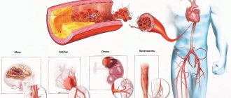
When metabolic processes in the heart muscle are disrupted, the production of energy compounds decreases and toxic substances accumulate. This leads to the development of myocardial dystrophy. It is accompanied by pain in the heart, tachycardia and arrhythmia, fatigue, and shortness of breath. For treatment, drugs are used that improve myocardial nutrition, normalize rhythm and blood circulation.
Reasons for the development of myocardial dystrophy
Myocardial dystrophy is always a secondary process. It occurs in the presence of heart disease and non-cardiac pathologies. The most common reasons include:
- chronic alcoholism;
- long-term treatment with medications;
- exposure to radiation, chemical poisoning, hazardous conditions at work;
- hormonal imbalance (thyroid disease, diabetes, metabolic syndrome, obesity, menopause, pheochromocytoma);
- vitamin deficiency, deficiency of protein, microelements in the diet, strict diets;
- autoimmune diseases;
- foci of chronic infection, especially tonsillitis;
- damage to the liver, pancreas and intestines;
- intense physical activity that exceeds the body's reserve capabilities;
- in infants – intrauterine hypoxia, encephalopathy, infections;
- hypertonic disease;
- tachycardia;
- anemia.
Disease prevention
To avoid such a disease, you must first of all not neglect and promptly treat those pathologies that can lead to its occurrence. It is also very important to maintain a healthy lifestyle, which consists of proper nutrition and daily routine, giving up all bad habits, and the absence or at least minimizing various worries and stress.
Moderate physical activity also helps reduce the chances of dystrophic changes in the myocardium. It must be remembered that it is much easier to prevent any disease by taking very simple measures than to treat it later.
Classification of metabolic-dystrophic changes in the myocardium
Depending on the damaging factor, cardiac muscle dystrophy occurs:
- nutritional (eating disorders, beriberi disease, scurvy);
- dishormonal;
- diabetic;
- anemic;
- catecholamine (adrenal hyperfunction);
- thyrotoxic (excess of thyroid hormones);
- tonsillogenic;
- alcoholic;
- toxic;
- mixed.
We recommend reading the article about dysmetabolic myocardial dystrophy. From it you will learn about the causes and mechanism of development of pathology, forms, methods of diagnosis and treatment, prognosis for the identified disease. And here is more information about fatty degeneration of the myocardium.
How does pathology affect the functioning of the body?
Hypertrophic changes in the myocardium do not occur in one day; this process can last for years. When the pathology is still at the initial stage of formation, a person may not experience discomfort and live a normal life. But gradually the disorders are gaining momentum, having a noticeable impact on overall well-being and negatively affecting the performance of individual organs. Here is a list of the most characteristic symptoms of cardiac pathology:
- Periodically there are difficulties with breathing, shortness of breath with any physical exertion.
- Pain in the chest or head.
- When a person is in a horizontal position, he is overcome by an unexpected cough, as well as a feeling of lack of air.
- The person feels dizzy and may faint.
- Blood pressure fluctuates from high to low.
- Constant unreasonable fatigue.
- I always want to sleep during the day and find it difficult to fall asleep at night.
- Attacks of hypertension are difficult to control with medication.
- Hands, feet and face swell at the end of the day.
- Cyanosis of the fingertips, lips, and area around the mouth may be observed.
- Heart rhythm is disturbed.
Symptoms of dystrophic processes in the ventricular myocardium
The initial manifestations are usually minor; if the process continues for a long time, then the functioning tissue in the myocardium is replaced by coarse connective fibers (cardiosclerosis). This reduces the contractile properties of the heart muscle, leading to circulatory failure, significant disturbances in the rhythm and conduction of cardiac impulses.
Clinical forms of various diseases have common symptoms - shortness of breath, tachycardia, general weakness; depending on the background pathology, they also have distinctive features.
Thyroid diseases
With thyrotoxicosis, the formation of energy compounds (ATP, creatine phosphate) decreases, and protein metabolism changes. In this case, an excess of hormones activates the work of the sympathetic nervous system. In patients, the pulse and blood circulation accelerate, and the volume of blood released per minute increases. The heart works under high load conditions, and not enough energy is produced for this.
Symptoms are associated with various forms of arrhythmia: tachycardia, extrasystoles, atrial flutter. Long-term hyperfunction of the thyroid gland leads to weakness of myocardial contractility, heart pain, shortness of breath, edema and congestion in the liver.
Hormone deficiency slows down the rate of metabolic processes, leads to fluid retention in myocardial cells, increased sodium and decreased potassium levels. Patients experience a rare heart rhythm, impulse blockade, constant dull and aching pain in the heart, and dizziness.
Alcoholism
It has been proven that daily consumption of 60 - 80 ml of ethyl (3 bottles of beer or 200 ml of vodka, 5 glasses of wine) is guaranteed to lead to myocardial dystrophy after 7 - 8 years.
In case of liver dysfunction, stress overload, infections, this period is reduced to 2 years or occurs when taking smaller quantities of alcoholic beverages. In this case, changes in the heart muscle are reversible only until circulatory failure develops (shortness of breath with slight exertion, palpitations, general weakness, edema).
Toxic form
A toxic form of myocardial dystrophy
Appears with prolonged treatment with hormones (Hydrocortisone, Prednisolone, Betamethasone, Dexamethasone) or non-steroidal anti-inflammatory drugs (Ibuprofen, Indomethacin), cytostatics (Methotrexate, Cyclophosphamide), antibiotics (Doxorubicin, Tetracycline), antidepressants, tranquilizers.
Acute poisoning is caused by carbon monoxide, salts of phosphoric or arsenic acid, and chloroform.
Manifestations of toxic myocardial dystrophy are:
- pain in the heart - aching, stabbing;
- hot flashes, sweating or chills with cold extremities;
- low exercise tolerance, severe weakness;
- headache;
- arrhythmia - tachycardia, extrasystoles, attacks of atrial fibrillation, atrial fibrillation, conduction block;
- swelling in the legs, accumulation of fluid in the pericardium, abdominal cavity;
- cough, attacks of shortness of breath or suffocation;
- heaviness and pain in the liver.
Tonsillitis
Myocardial dystrophy develops with frequent sore throats. Accompanied by severe pain in the heart area, constant weakness, a feeling of irregular rhythm, and sweating. Patients often have elevated body temperature to subfebrile levels (37.1 - 37.4 degrees), difficulty breathing, pain in muscles and joints.
In athletes
The reason may be loads for which the body is not ready, or the presence of inflammatory processes (sometimes asymptomatic) in the ENT organs, lungs, digestive or genitourinary systems.
Such myocardial dystrophy occurs when there is insufficient blood supply to the hypertrophied myocardium, disturbances in electrolyte and hormonal balance. It is manifested by the rapid development of fatigue, general weakness, apathy or depression, palpitations and stabbing pain in the heart, arrhythmia.
Watch the video about the first symptoms of heart disease:
Anemia
If the hemoglobin content in the blood drops to 85 g/l and below, then all tissues of the body experience oxygen starvation. The reason for this may be iron deficiency, destruction of red blood cells, trauma, tumors, chronic bleeding (peptic ulcer, hemorrhoids, uterine), intravascular coagulation syndrome (DIC). Symptoms of anemic dystrophy of the heart muscle:
- pale skin,
- frequent dizziness,
- increased heart rate,
- pulsation of the arteries of the neck,
- dyspnea,
- tachycardia.
During the examination, expansion of the borders of the heart, vascular and cardiac murmur can be detected. Without treatment and progression of anemia, circulatory failure appears.
Climax
Low estrogen levels and fluctuations in hormone levels lead to the development of dystrophic changes in the heart muscle in the form of pressing pain, often radiating under the shoulder blade or to the left arm, tingling or discomfort in the heart area. Increased symptoms are observed during periods of hot flashes, sweating and rapid heartbeat. With concomitant arterial hypertension, the likelihood of developing heart failure cannot be excluded.
Definition of pathology
The left ventricle is the lower part of the heart. It has an elongated oval shape and a more muscular inner layer. In this chamber, two sections can be distinguished: the posterior cavity (it connects the ventricle with the atrium through the venous opening) and the anterior one in the form of a canal (precedes the entrance to the aorta).
From the aorta of the left ventricle of the heart, blood begins its movement in a large circle, carrying nutrients and oxygen through the main arteries, veins, capillaries to all organs and parts of the body. This camera is larger than all the others. The left ventricle can be thought of as a container that receives blood supplied from the arteries through the left atrium. Backflow is prevented by the mitral valve. The further function of the most important part of the heart is to push blood through the aortic valve into the largest vessel - the aorta. The force of contraction of the left ventricular myocardium depends on its filling during diastole.
Sometimes blood enters in excessive quantities, or when it is released, the walls of the chamber have to strain more than usual due to existing obstacles. This helps to overstrain the left ventricle and build muscle mass. This is how the heart adapts to the excess load. This is myocardial hypertrophy.
The increased volume of this layer requires more oxygen. But the coronary arteries are not designed for this, so myocardiocyte hypoxia develops. The next stage may be atrophy of the cardiac muscle tissue and heart failure.
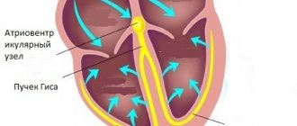
Diagnostic signs of diffuse and moderate changes on the ECG
The main method for detecting dystrophy in the heart muscle is to conduct a standard ECG and functional tests. In the presence of diffuse (widespread) changes, the following can be identified:
- decrease in ST;
- sinus tachycardia;
- extrasystoles;
- deformed or smoothed, sometimes negative T (with pheochromocytoma);
- low-amplitude waves, especially of the ventricular complex.
With obesity and hypothyroidism, the rhythm slows down, conduction blocks occur in the atria or ventricles, and QT prolongation occurs. With focal changes, such disturbances are detected only in 2-3 leads; they are usually weakly expressed. In order to discover the cause of myocardial dystrophy and distinguish it from diseases with similar symptoms, tests are performed. In typical cases, their results are:
- bicycle ergometry - no signs of ischemia during exercise;
- intake of potassium salts is normalized;
- Anaprilin – T positive or tends to normal;
- hyperventilation (fast breathing), sudden change in body position - T and ST decrease.
In addition to the ECG, when making a diagnosis, the appearance of systolic murmur over the base and apex, dull tones, and the absence of valvular defects on ultrasound are taken into account. If it is difficult to determine the disease, a radioisotope scan of the heart (scintigraphy) is performed.
In the resulting image, foci or diffuse changes are visible in myocardial dystrophy. They manifest themselves in the form of weak accumulation of drugs, which reflects a decrease in the number of functioning cells.
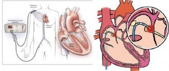
If all these methods turn out to be uninformative, then they resort to myocardial biopsy.
Diagnostics
The difficulty in making the correct diagnosis for myocardial dystrophy lies in the absence of changes during examination at the first stage of the process. At this stage, only the underlying disease can be identified, for which secondary myocardial dystrophy is typical.
| Examination method | Characteristic changes |
| Inspection by system | Enlarged heart size with a shift to the left Irregular pulse (sinus arrhythmias) |
| Auscultation (assessment of heart murmurs by ear) | Moderate muting of tones at all points Weakness of the first tone at the cardiac apex Weak systolic murmur |
| Electrocardiography (ECG) | Sinus arrhythmia (extrasystole, increased or decreased heart rate) Low voltage of the ventricular complex Partial bundle branch blocks Impaired repolarization (recovery after contraction) of the heart |
| ECG with drug tests (potassium, beta blockers) | Improvement of pathological changes |
| Velergometry (ECG under physical activity) | There is no increase in the volume of blood ejected during contraction Decreased exercise tolerance Inability to fully perform the required load |
| Ultrasonographic examination (ultrasound) | Heart enlargement Expansion of the cavities of the heart chambers at the third stage of the disease Decrease in the volume of blood released during myocardial contraction in the phase of organ dysfunction |
| Scintigraphy with Thallium 201 | Impaired passage of potassium and sodium ions through the cell wall Pathology of myocardial metabolic processes |
| Magnetic resonance imaging (MRI) with radioactive phosphorus | Decrease in the amount of energy reserves of cardiac muscle cells Change in pH (acidity) of the cellular fluid |
| Biopsy (tissue sampling) of the myocardium with histochemical analysis | Pathology of enzymatic metabolism in heart tissue Destruction of myocardial fibers Changes in organ cells |
The most accurate diagnostic method is the sampling of myocardial tissue, but given that the procedure requires cardiac puncture, the indications for it are extremely limited. A biopsy is performed only in difficult diagnostic situations when myocardial dystrophy cannot be established by other methods.
Treatment of dystrophic changes in the myocardium
To restore metabolic processes in the heart muscle, it is necessary to normalize hormonal levels and blood composition, treat infections, intoxication and anemia, completely stop drinking alcohol and smoking, and avoid physical and emotional stress.
Drug therapy includes:
- vitamin complexes (Neurorubin, Milgamma);
- lipoic acid (Tiogamma turbo, Espa-lipon);
- antioxidants (Vitamin E, Nicotinamide, Selenium);
- antihypoxants (Cytochrome, Mexidol, Corvitin);
- potassium and magnesium salts (Panangin, Magne B6, Kalipoz prolongatum);
- antiarrhythmic (Atenol, Breviblok, Cordarone);
- cardiotrophic agents (Riboxin, Potassium orotate, Preductal, Carnitine, ATP, Cardonate);
- adaptogens (ginseng tincture, Pantocrine);
- diuretics (Trifas, Veroshpiron), cardiac glycosides (Digoxin), ACE inhibitors (Prenesa) for heart failure.