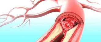
- What is intrauterine infection
- What is dangerous in pregnant women: consequences
- Causes and risk group
- Symptoms
- Diagnostics
- Treatment of intrauterine infection during pregnancy
- Prevention
What is this pathology?
In medicine, the term IUI (intrauterine infections) refers to a large group of diseases in which infectious damage to the fetus occurs. The provoking factor, as a rule, is the penetration of pathogenic microflora into the womb.
Such a process, if adequate therapy is not started in a timely manner, can lead to very serious consequences, including the death of the baby.
Basically, the infection reaches the developing fetus directly from the sick mother. The fact is that during pregnancy, the mother and the unborn child share a common blood supply system. For this reason, physiological fluids are freely exchanged between two organisms.
If we add to this factor the fact that with the onset of pregnancy, a woman’s body begins to produce less antibodies, one can easily understand why even a harmless bacterium can cause IUI.
Mechanisms
The degree of pathological impact is determined by the characteristics of the morphological development of the fetus at a particular stage of pregnancy, its reaction to the infectious process (maturity of the immune system), and the duration of microbial aggression.
The severity and nature of the lesion are not always strictly proportional to the virulence of the pathogen (the degree of its pathogenicity). Often, latent infection caused by chlamydial, viral or fungal agents leads to intrauterine death or the birth of a child with serious abnormalities. This is due to the biological tropism of microbes, i.e., the tendency to reproduce in embryonic tissues.
Infectious agents have different effects on the fetus. They can provoke an inflammatory process in various organs with the further development of a morphofunctional defect or have a direct teratogenic effect with the appearance of structural abnormalities and malformations. Of no small importance are intoxication of the fetus by products of microbial metabolism, metabolic disorders and hemocirculation with hypoxia. As a result, fetal development suffers and differentiation of internal organs is disrupted.
Classification
Depending on the type of pathogenic microorganisms that triggered the infectious process, the following forms of intrauterine infections are distinguished:
- Bacterial. Usually their development is provoked by listeria, tuberculosis bacillus, and treponema pallidum (the causative agent of syphilis).
- Fungal-parasitic infections are caused by candida, chlamydia, mycoplasma, toxoplasma and other members of the fungal family.
- Viral processes are the result of infection with herpes, enteroviruses, rubella, hepatitis and other pathogens of this class.
- Mixed forms are diagnosed in the majority of cases of IUI (approximately 50% of the total number of diseases).
This fact is explained by the fact that the expectant mother’s body becomes susceptible not to a single strain of pathogens or a separate group, but to all pathogenic microorganisms. That is, the protective barrier of the expectant mother weakens and cannot fully resist attacks from infectious agents.
But at the same time, any woman suffers all kinds of infections throughout her life, after which a strong immunity is formed. For this reason, in medical practice, most often there are cases when a group of 3-4 pathogens becomes the culprits of the invasion.
Clinical forms
Neonatal herpes
| IUI: neonatal herpes | |
| ICD-10 | 35.235.2 |
| ICD-9 | 771.2771.2, 054.xx054.xx |
| MeSH | D018445 |
Main article: Herpes
Of the herpes family viruses, all the main types can cause herpes infection in a newborn: herpes simplex virus types 1 and 2, herpes simplex virus type 3 (varicella zoster), type 4 - Epstein-Barr virus, hairy leukoplakia of the tongue, immune depression syndrome, 5- 1st type - cytomegalovirus infection, 6th type - roseola, 7th type - chronic fatigue syndrome, 8th type - Kaposi's sarcoma. However, the term “neonatal herpes” is used only in relation to diseases caused by herpes simplex virus types 1 and 2. The most dangerous for a child is HSV-2.
The likelihood of a child becoming infected depends on how long the mother has been infected. The “fresh” the infection, the more likely the child is to become infected. If at the time of birth the mother has a rash, this is an indication for a cesarean section[12].
Clinical manifestations
- 1. Local form (mucocutaneous) - damage to the skin, mucous membranes, and, less commonly, encephalitis.
- 2. Cerebral form - the clinical picture corresponds to the general signs of IUI, but there are also specific signs: damage to the eyes and mucous membranes; herpetic encephalitis, which is necrotic in nature, resulting in destruction of the brain down to the hemispheres; severe thrombocytopenia with hemorrhagic syndrome.
- 3. Disseminated neonatal herpes
Diagnosis
In the diagnosis of neonatal herpes, assessment of the mother's specific medical history is important. During a clinical examination of children born to mothers with acute or recurrent genital herpes, examination of the skin and mucous membranes must be done with special care. If a newborn has seizures of unknown etiology, a lumbar puncture is indicated (with herpetic encephalitis, lymphocytosis, monocytosis and a high protein concentration are noted). If a newborn develops sepsis and there is no effect of antibiotics, an examination for herpes is necessary. Among laboratory diagnostic methods, the gold standard is the isolation of the virus from blood, cerebrospinal fluid, and vesicles by the cultural method. In the cutaneous form, the contents of the vesicles or scrapings from the skin can be examined using the immunofluorescent method to detect the virus antigen. And in case of generalized infection and meningoencephalitis, blood and cerebrospinal fluid are examined using the PCR method. The level of IgG antibodies is not informative, since these are maternal antibodies. IgM levels indicate acute infection in the newborn.
Treatment
For all forms of neonatal herpes, systemic antiviral therapy is indicated, since the localized form may precede the generalized one. With early administration of antiviral drugs, the outcome is favorable. Regardless of the form of infection, acyclovir is used. Acyclovir (Zovirax, Virolex) intravenously for 2-3 weeks plus antiherpetic immunoglobulin for 2 weeks[12]. It makes no sense to stop breastfeeding, since HSV is unlikely to pass into mother's milk, except for rashes on the mother's breasts. Local remedies for ophthalmic herpes include vidarabine, florenal, and bonaftone ointment.
Congenital cytomegalovirus infection
Main article: Cytomegalovirus
The frequency of occurrence is 0.2-2.5%. The virus is transmitted by all secretions (saliva, urine, blood, tears). Clinical manifestations during primary infection in pregnant women are nonspecific and may resemble the clinical manifestations of acute respiratory viral infection. There are a number of factors contributing to the high incidence of intrauterine infection with cytomegaly virus. These include epidemiological features, such as significant genetic variability of CMV strains, the widespread distribution of CMV infection in the human population (in the vast majority - in the form of a latent-persistent course), the predominance of subclinical forms, both in primary and secondary infection, diversity mechanisms and routes of transmission of infection. The next factor is the immaturity of the immune system of the fetus and newborn. And finally, adaptive immune changes in a woman’s body during pregnancy (decreased functional activity of cellular immune mechanisms), during which reactivation of a latent-persistent CMV infection is possible.
Infection most often occurs during childbirth, or through mother's milk. During pregnancy, infection occurs only if the mother becomes infected for the first time during pregnancy.
Clinical manifestations
Congenital hepatitis with severe jaundice, severe thrombocytopenia with hemorrhagic syndrome, meningoencephalitis. Specific signs are calcifications in the subependymal parts of the brain and chorioretinitis. The long-term prognosis is determined by the degree of brain damage. If meningoencephalitis is suffered early, children are usually disabled; if hepatitis, cirrhosis develops early; if carditis, chronic heart failure develops.
Diagnostics
Children with symptoms of congenital infection, as well as without clinical manifestations of TORCH syndrome, if they are born to women at risk, are subject to examination for CMV infection. In newborns in the early neonatal period, if CMV is suspected, the pathogen is first identified by any available method. Most often, PCR or detection of virus antigens is used; the virological method is less often used. Any biological fluid (urine, saliva, blood, tears) can serve as material for PCR; however, active CMV infection is indicated only when the CMV genome is detected by PCR in the blood and cerebrospinal fluid. When viral DNA is found in other environments, an unambiguous assessment of the period of the disease cannot be given. To clarify the severity of the process, serological methods are used - anti-cytomegalovirus antibodies of classes M and G are determined. Moreover, the study of “paired sera” is mandatory, that is, monitoring the study of antibody titers after 3-4 weeks. Detection of IgM class antibodies in umbilical cord blood and in the blood of a child in the first weeks of life is an important diagnostic sign. And the detection of IgG in the child’s blood without comparison with maternal titers is not diagnostically significant, since transplacental transfer of antibodies from the mother’s body is possible.
Treatment
Therapy for congenital CMV infection consists of etiotropic and syndromic therapy. The indication for etiotropic therapy is the active period of congenital CMV infection. The drug of choice for etiotropic treatment is Cytotect. Children are administered anticytomegalovirus immunoglobulin (cytotect) intravenously 2 ml/kg 2 times a day every 2 days for 3 weeks [13].
If there is a danger to life, then ganciclovir is added intravenously for 14-21 days, although virostatics (antiviral drugs) such as ganciclovir and foscarnet are used extremely rarely due to their high toxicity.
Congenital toxoplasmosis
Main article: Toxoplasmosis
Frequency 1:1000 newborns Toxoplasma oocysts are usually found in the feces of cats and goats, from where they are released into the external environment. In pregnant women, the clinical course of the disease is similar to mononucleosis or influenza, with high fever or very long-term low-grade fever, and enlarged lymph nodes. Arthralgia or arthritis is often associated.
The likelihood of infection of the fetus: infection usually occurs if the infection is fresh and depends on the duration of infection. If the 1st trimester - the probability is 15%, in the second 30%, in the third - 60%.
Clinical manifestations
In the fetus and newborn, the infection can take two forms: damage to the eyes and brain or generalized toxoplasmosis. In addition to the general signs of infectious toxicosis, hepatitis, meningoencephalitis, eye damage (congenital cataract, possibly glaucoma, optic nerve atrophy) are added.
Diagnostics
Scheme of examination for toxoplasmosis of newborns: in the presence of clinical signs of toxoplasmosis, antibodies are examined. If no antibodies are detected, the test is repeated after 2 weeks; if there are no antibodies during the second test, then further monitoring is not needed. If detected, specific therapy is indicated. If IgM class antibodies are detected during the initial study, then etiotropic therapy is immediately indicated. If only IgG is detected, the test is repeated after 4 weeks. Therapy is indicated when the antibody titer increases. If the titer drops, the child does not need treatment, but further monitoring is necessary.
Treatment
Treatment of toxoplasmosis can be carried out antenatally - that is, treatment of a pregnant woman. If the infection is in the 1st half of pregnancy, spiramycin, claforan, rovamycin are used. If in the 2nd half of pregnancy - chloridine + sulfasalazine + folic acid. Treatment of children is effective during periods of circulation in the blood of non-cystic forms of the parasite; drugs do not act on cystic forms. There is no need for complete sanitation, since cystic forms (carriage) provide normal non-sterile immunity. The most effective drugs are pyrimethamine in combination with sulfonamides. There are combination drugs: Fansidar, Metakelfin. Co-trimoxazole is also used in age-specific dosages[14]. Treatment of newborns involves the following regimen: chloridine + sulfadimezine + folic acid. Course 4-6 weeks. During the first year, 4 courses with a break of 1.5 months, and during the break, spiramycin for 1.5 months [12][15][16].
Chlamydia
Main article: Chlamydia
WHO data indicate that 35-50% of newborns whose mothers are infected with C. trachomatis
, chlamydial ophthalmia develops (5 times more often than gonococcal ophthalmia), and in 11-20% pneumonia develops[17].
Infection usually occurs during childbirth, the probability of transmission is 40-70%. The disease does not appear immediately, but after 7-14 days. Clinical manifestations
There are three forms of infection in newborns:
- persistent
- latent
- acute (generalized infection - meningoencephalitis, intrauterine pneumonia, gastroenteritis)[17][18].
The main manifestations of the disease in a newborn are:
- nasopharyngitis 25%
- conjunctivitis resistant to the use of conventional remedies, responds only to treatment with tetracycline ointment,
- pneumonia 10-15% - mild toxicosis, but pronounced obstructive syndrome, paroxysmal painful cough.
- high eosinophilia
- proctitis, gastroenteritis - 5%
- vulvitis, urethritis 15%
Treatment
In addition to the child, it is necessary to treat both the father and mother. A newborn child is prescribed erythromycin in suppositories for 24 days or erigran orally. Azithromycin can also be used[12][19].
Mycoplasmosis
Main article: Mycoplasmosis
Mycoplasma infection usually occurs during childbirth. The frequency of detection of the pathogen in pregnant women is 20-50%, the risk of infection of the fetus is unknown. Pregnant women with seropositive mycoplasmosis are treated after the 16th week of pregnancy, which reduces the incidence of morbidity in newborns.
Clinical manifestations
In newborns, it manifests itself in the form of pneumonia, which begins imperceptibly with toxicosis, pallor appears, shortness of breath increases, and only then physical findings appear. On the radiograph, a specific sign is the “blizzard symptom” - bilateral fine-focal, sometimes confluent pneumonia. Mortality rate is 15%.
Treatment
Newborns are prescribed erythromycin or azithromycin, and in severe forms, chloramphenicol [20][12].
Congenital rubella syndrome
| Intrauterine infections | |
| Cataract in congenital rubella syndrome | |
| ICD-10 | 35.035.0 |
| ICD-9 | 771.0771.0 |
| DiseasesDB | 11729 |
| MedlinePlus | 001658 |
| eMedicine | emerg/388 |
| MeSH | D012410 |
Main article: Rubella
If the mother becomes infected in the first 12 weeks, it is better to terminate the pregnancy. Before pregnancy, it is necessary to be examined, and if the mother is seronegative, then vaccinated[12]. If a mother becomes infected with rubella in the 1st trimester, the child has a 25% chance, after the 5th month – 1-2%.
Clinical manifestations
A characteristic clinical manifestation is Greg's triad
:
- congenital heart disease such as patent ductus arteriosus, ventricular septal defect, atrial septal defect, pulmonary stenosis, myocardial necrosis
- eye defects (cataracts, microphthalmia, glaucoma)
- deafness.
In 2/3 children, congenital rubella manifests itself at the end of the perinatal period.
Treatment
There is no specific therapy; treatment is symptomatic.
Candidiasis of newborns
Main article: Candidiasis
The frequency of candidiasis in the structure of infectious and inflammatory diseases of newborns is about 15-30% of cases, and in half of them it remains unrecognized or diagnosed late[21]. Candidiasis can be caused by any of the species, but most often by Candida albicans. Risk factors for the development of candidiasis in newborns include: prematurity, diabetes mellitus in the mother during pregnancy, urogenital candidiasis in the mother during pregnancy, repeated courses of antibiotics, especially in combination with immunosuppressive therapy, immune disorders, especially neutropenia, the presence of mechanical ventilation in the early neonatal period, resuscitation measures, abdominal operations.
Clinical manifestations
Based on the time of infection, congenital
candidiasis that developed during antenatal or intrapartum infection and
postnatal
candidiasis. Depending on the localization of the process, candidiasis is divided into:
- cutaneous candidiasis is a lesion of the skin and its appendages. The lesion may be localized or widespread.
- candidiasis of the mucous membranes of the oral cavity, conjunctiva, external genitalia
- generalized candidiasis - damage to internal organs that do not communicate with the external environment with the addition of candidemia.
- visceral candidiasis - damage to internal organs and other systems that do not communicate with the external environment, for example carditis, hepatitis, nephritis.
- systemic candidiasis - affects one or more organs that communicate with the external environment - for example, candidiasis of the gastrointestinal tract.
- candidiasis - the presence of Candida in natural habitats in greater than the prescribed concentration.
Candidiasis is also classified according to the severity of the process as mild.
and
severe
forms, depending on the location and volume of the lesion, the presence of infectious toxicosis.
In addition, there are acute
(7-14 days) and
protracted
(more than 6 weeks) course of the disease.
Diagnosis
Diagnosis of neonatal candidiasis is based on the clinical picture. In the cutaneous and mucocutaneous form there is no need for laboratory confirmation. Laboratory diagnosis becomes crucial for generalized, visceral and systemic candidiasis. Laboratory criteria can be considered the identification of fungi in an active state by microscopy of the substrate, the isolation of antigens and DNA in sterile substrates, the isolation in quantities greater than those allowed for sowing of substrates that are the site of saprotification of fungi.
Treatment
For localized skin candidiasis, local therapy with antifungal ointments (clotrimazole, isoconazole, ketoconazole, natamycin) is used. In case of prolonged course, systemic antimycotics are prescribed - fluconazole orally. The daily dose is 5-8 mg/kg once a day. For mucosal candidiasis, the affected areas are treated with a 2% soda solution or a 0.1% hexoral solution. In case of relapse, fluconazole is used. For systemic candidiasis for the treatment of the gastrointestinal tract, respiratory, genitourinary system, as well as for visceral and generalized candidiasis, treatment begins with fluconazole, and if ineffective, amphotericin B or ambisome is prescribed intravenously for 5-7 days[22].
Early congenital syphilis
Main article: Syphilis
Against the backdrop of an epidemic increase in the incidence of syphilis in Russia in the 90s, the incidence of congenital syphilis increased sharply: in 1997, the overall incidence of syphilis exceeded the level of 1990 by 51 times, and that of congenital syphilis by 47 times. The dynamics of the increase in the incidence of congenital syphilis is similar to the dynamics of the proportion of pregnant women among women with syphilis. According to L.I. Tikhonova (1999), in 1995-97. in Russia this figure was constantly growing: 4.9%, 5.5%, 6.5%[23].
Congenital syphilis can be prevented by identifying and treating infected mothers during pregnancy. Therefore, pregnant women are examined serologically three times, including immediately before birth.
Early congenital syphilis is an IUI that manifests itself in a child under 2 years of age (according to ICD-10). Early congenital syphilis may be manifest
(with clinical manifestations) and
hidden
.
Clinical manifestations
In newborns with early congenital syphilis, the following symptoms are observed: syphilitic rhinitis, diffuse Hochsinger infiltration, chorioretinitis, hepatosplenomegaly, syphilitic pemphigus, roseolous and pustular rash, osteochondritis, periostitis, osteoporosis.
Diagnostics
In newborns born to mothers with syphilis, umbilical cord blood is taken for analysis at birth to carry out a set of serological tests. In addition, weighing and pathological examination of the placenta are carried out. With syphilis, the placenta is enlarged and there are signs of inflammation. A spinal tap must be performed. In the analysis of cerebrospinal fluid there are specific changes: lymphocytic cytosis above 20 cells in 1 ml, protein above 1.5-1.7 g/l, positive results of RIF and a complex of serological reactions. On the 7-8th day of the child’s life, serological blood tests are repeated - microprecipitation reaction, immunofluorescence reaction, immobilization reaction of Treponema pallidum, enzyme immunoassay to detect IgM.
Treatment
Treatment is carried out with one of the penicillin drugs for 2-3 weeks. The choice of drug depends on the analysis of the cerebrospinal fluid. After completion of treatment, the child is discharged under the supervision of a dermatovenerologist, and the diagnosis is reported to the district clinic only with the consent of the mother. In the KVD, clinical and serological control is carried out once every 3 months until the child reaches the age of 3 years[24].
Notes
- ↑ 1 2 V.V. Vlasyuk
Morphological diagnosis of intrauterine infections. Tutorial. St. Petersburg, 2010 - 47 p. ISBN - 5-00-001976-8/ - Okhotnikova I.M., Ageikin V.A., Lozovskaya L.S. The significance of intrauterine viral infection in organ pathology of infants // Med. scientific and educational-methodological. magazine. - 2001. - No. 5. - P. 81-87.
- Congenital malformations. Prenatal diagnosis and tactics / Ed. B. M. Petrikovsky, M. V. Medvedev, E. V. Yudina. - M.: RAVUZDPG: Realnoe Vremya, 1999. - 325 p.
- https://spravka.komarovskiy.net/vnutriutrobnye-infekcii.html Komarovsky. Intrauterine infections
- https://www.ic.omskreg.ru/~medstat/BIBLIO/opport/index.htm Dolgikh T. I., Noskova F. V. Opportunistic infections in children (issues of diagnosis, clinic and treatment). Omsk: Publishing house OGMA, 1999. - 99 p.
- ↑ 12
A. L. Zaplatnikov, N. A. Korovina, M. Yu. Korneva, A. V. Cheburkin A. L. Zaplatnikov, N. A. Korovina, M. Yu. Korneva, A. V. Cheburkin Attending physician 08- 2005 https://www.lvrach.ru/doctor/2005/08/4532901/ - A. Ya. Senchuk, Z. M. Dubossarskaya.
Perinatal infections: practical. allowance. - M.: MIA, 2004. - 448 p. - Diagnosis of intrauterine infections in newborns using the polymerase chain reaction method
- Intrauterine infections. Symptoms. Diagnostics. Prevention. | EUROLAB | Pediatrics
- Volodina N. N., Degtyareva D. N.
Diagnosis and treatment of intrauterine infections. - M.: Method. rec. for neonatologists, 1999. - https://www.rae.ru/ru/publishing/mono07_811.html MODERN PRINCIPLES FOR DIAGNOSIS OF INTRAuterine INFECTION OF THE FETAL
- ↑ 123456
Perinatal infections (issues of pathogenesis, morphological diagnosis and clinical and morphological comparisons) Tsinzerling V. A., Melnikova V. F. - Clinical and laboratory characteristics, pathomorphological features, diagnosis and treatment of cytomegalovirus pneumonia “Infectious Diseases”, 2004. - T. 2, No. 1. - P. 73-80. V. I. Shakhgildyan, O. A. Tishkevich, O. Yu. Shipulina. https://www.hivrussia.ru/pub/2006/16.shtml
- Cheburkin A.V. Clinic and differential diagnosis of congenital toxoplasmosis “Medical parasitology and parasitic diseases” No. 5, 1984, p. 53-57.
- On the detection and prevention of toxoplasmosis in Moscow. Methodological recommendations (No. 25). M., 2007
- Clinic, diagnosis and treatment of toxoplasmosis G. Yu. Nikitina, F. K. Dzutseva, Yu. V. Borisenko, L. P. Ivanova Attending physician 10-2008 https://www.lvrach.ru/doctore/2008/10/ 5828652/
- ↑ 12
Urogenital chlamydia Chlamydial Genitourinary Infections https://www.venuro.info/venera/chlamidioz.php - Chlamydia Uskov Alexander Nikolaevich https://queerlvov.narod.ru/hlamidioz.html
- https://www.hlamidioz.info/6.html Chlamydia in children
- Perinatal infections: practical. manual / ed. A. Ya. Senchuk, Z. M. Dubossarskaya. M.: MIA, 2005. 318 p.
- Protocols for diagnosis, treatment and prevention of intrauterine infections in newborns, Moscow, State Educational Establishment VUNMC Ministry of Health of the Russian Federation, 2001 p. 53
- Protocols for diagnosis, treatment and prevention of intrauterine infections in newborns, Moscow, State Educational Establishment VUNMC Ministry of Health of the Russian Federation, 2001 pp. 55-57
- https://www.ill.ru/news.art.shtml?c_article=142 Syphilis and pregnancy
- Protocols for diagnosis, treatment and prevention of intrauterine infections in newborns, Moscow, State Educational Establishment VUNMC Ministry of Health of the Russian Federation, 2001 pp. 59-64
Main routes of infection of the fetus
Of course, any expectant mother is most concerned about the question of how an infection can get to a newly born living lump.
There are four ways for pathogenic microflora to penetrate:
- the ascending path consists of the spread of infection through the genitals (chlamydia, enterococci);
- infectious pathogens from the fallopian tubes reach the fetus in a descending way if the woman had inflammatory processes in the appendages;
- hematogenous, that is, through the blood, is considered the most common way of spreading infection (viruses, toxoplasma);
- intrapartum transmission occurs when the fetus comes into contact with contaminated amniotic fluid or during childbirth.
Causes
The source of infection for the fetus is most often the mother. We should never forget about iatrogenic infection against the background of medical procedures.
The following routes of infection from the carrier to the fetus are distinguished:
- through the placenta;
- ascending;
- descending;
- contact.
With contact transmission, infection occurs during the passage of the child through the birth canal. Viral diseases are usually transmitted transplacentally because, due to their small size, they easily penetrate the barrier. Bacterial infections, as well as fungi, most often infect the fetus in an ascending manner, penetrating into the fetus through the uterine cavity, into which they enter from the genital tract.
Main markers of IUI during pregnancy
It is impossible to determine the type of pathogen that provoked the development of intrauterine infection based on clinical signs alone. Therefore, in medical practice, laboratory tests are used for this purpose. Moreover, for an accurate diagnosis it is necessary to take tests several times.
You should know that in medicine intrauterine infections are referred to as TORCH syndrome. This Latin abbreviation reflects all the most common infections.
Symptoms of intrauterine infection in a newborn and during pregnancy
When the fetus is affected by VUI, miscarriages and frozen pregnancies often occur, and the child may be stillborn or die during childbirth. A fetus that survives may have the following abnormalities:
- intrauterine growth retardation,
- hydrocephalus;
- microcephaly
- eye damage (a disease called cataract);
- anemia;
- fever;
- skin rash.
In a pregnant state, it is not so easy to detect infection of the fetus, so doctors do everything possible to do this. It’s not for nothing that a pregnant woman has to undergo so many different tests several times a month.
The presence of intrauterine infection can be determined by tests . Even a smear taken in the chair can show some picture of the presence of infections, however, they do not always lead to intrauterine infection of the fetus.
When an intrauterine infection affects a child shortly before birth, it can manifest itself in diseases such as pneumonia, meningitis, enterocolitis or another disease.
The signs described above may not appear immediately after birth, but only on the third day after birth, and only if the infection strikes the child while moving through the birth canal, doctors can notice its manifestation almost immediately.
HIV
The immunodeficiency virus (HIV), which poses a huge threat to the fetus developing in the mother’s womb, cannot be ignored. Thanks to the capabilities of modern medicine, today all expectant mothers are required to undergo screening for the presence of this dangerous infection. Therefore, doctors are able to identify it in a timely manner, which allows them to take all necessary measures to prevent infection of the fetus.
We also need to pay attention to this important point: today, doctors recommend that all parents planning to have offspring undergo a series of laboratory tests. This event helps to timely identify the presence of pathogens of dangerous diseases.
The danger of intrauterine infections
The main threat of this group of diseases is that invisible agents interfere with the development of the fetus, thereby causing enormous damage to the defenseless body.
Of course, such an intervention does not go unnoticed, because babies are born weak, with low body weight and all sorts of developmental defects.
IUI is especially dangerous in the first trimester of pregnancy, when all the organs and systems of the unborn child are formed. If infection occurs during this period, the baby may be born with obvious defects. Very often in such cases, babies are born who are completely unadapted to independent life.
Also, one of the most common complications of IUI is spontaneous miscarriage in the early stages of pregnancy or premature birth in later stages. Intrauterine infection can occur in both acute and chronic forms.
For an expectant mother, IUIs during pregnancy in the 2nd trimester are dangerous because they increase the likelihood of developing a septic process. There are many more risks for the baby: these are, first of all, various developmental anomalies, deformities, damage to the organs of hearing and vision, heart defects and other complications. Many of these consequences have already been discussed above.
Of course, the outcome of the infectious process and the prognosis for the future depend on a combination of the following factors:
- duration of the disease;
- gestational age (pregnancy);
- types and number of strains;
- degree of persistence of maternal immunity.
If IUI can be identified at the initial stages and appropriate therapy is initiated in a timely manner, then there is a chance to save the child’s life and minimize the consequences of the pathology.
Prevention
Avoiding IUI is quite simple; to do this, pregnant women just need to follow a few rules. Every woman is recommended:
- avoid planning pregnancy if active phases of any infectious diseases are detected;
- be vaccinated with complex vaccines;
- limit contact with children;
- reduce visits to public places;
- be more careful about personal hygiene and the hygiene of family members;
- thoroughly clean food (vegetables and fruits);
- avoid eating foods with insufficient heat treatment;
- exclude soft cheeses and semi-finished products from the diet;
- periodically undergo testing by doctors, including the level of immunoglobulins for TORCH infections;
- Be sure to contact a doctor after contact with carriers of infectious diseases.
Intrauterine infections are quite serious pathologies of pregnancy.
They become the main cause of various defects in the fetus, and in advanced cases can lead to its death. There are several reasons for the occurrence of the disease, but in order to not give the infection a chance, every pregnant woman needs to be attentive to her own health. In particular, do not forget about timely prevention of IUI, including comprehensive vaccination during pregnancy planning. Pregnancy Diseases TORCH infections during pregnancy
Causes of IUI
Science has not yet precisely established all the factors that provoke the development of intrauterine infections. But it is known for sure that they can contribute:
- pathologies of the female genitourinary system;
- suppressed immunity, including HIV infection;
- respiratory diseases, especially in the first trimester of pregnancy;
- exacerbation of chronic pathologies;
- any surgical procedures at any stage of gestation.
It must be remembered that in any case, infection of the fetus occurs only from the mother.
Epidemiology[ | ]
The true frequency of congenital infections has not yet been established, but, according to a number of authors, the prevalence of this pathology in the human population can reach 10%.
Intrauterine infections have the same patterns as infectious diseases in general.
They have a leading place in the structure of infant mortality.
The share of IUI in the structure of perinatal mortality in our country is almost 25%, however, transplacental infection of the fetus is considered one of the most likely causes of 80% of congenital malformations, which, in turn, account for about 30% of all deaths of children under 1 year of age [2 ][3]
TORCH[ | ]
In 1971, WHO identified the concept of TORCH syndrome
.
This is an abbreviation for the most common intrauterine infections : T - toxoplasmosis, O - others, which includes mycoplasma, syphilis, hepatitis, streptococci, candida and other viral and bacterial infections, R - rubella (rubella), C - cytomegalovirus, H - herpes
If not clear etiological diagnosis, they talk about TORCH syndrome.
At-risk groups
When asking about the risk of IUI during pregnancy, what it is and who is most susceptible, then, as a rule, patients are carriers of pathogenic agents:
- who have already given birth to children with signs of IUI:
- having children attending child care institutions;
- working in the medical field and in education;
- suffering from chronic inflammatory pathologies of any localization;
- women who have previously given birth to premature babies.
From all of the above, we can conclude that IUIs are most often found in women who are more at risk of contracting infections. The risk group for IUI during pregnancy (what it is is discussed above) should also include patients who have frequently had abortions.
Clinical symptoms
The difficulty in diagnosing IUI lies in the fact that the pathology predominantly occurs in a latent form. Often the disease is simply disguised as an elementary deterioration in general condition, which is usually mistaken for the course of toxicosis. Therefore, doctors are able to identify the pathological process when it takes a generalized form.
Among the symptoms that should alert the expectant mother are the following clinical manifestations:
- increase in background temperature;
- inflammation of the lymph nodes;
- joint pain;
- skin rash;
- conjunctivitis;
- various manifestations of a cold (cough, runny nose).
If the listed symptoms persist for a long time, the woman should definitely tell the doctor about it.
You can suspect fetal infection due to the following signs:
- slow growth and development;
- change in the volume of amniotic fluid;
- the appearance of signs of hydrocephalus in the fetus;
- placental defects;
- polycystic disease;
- The size of the fetus is less than the norm for the term.
Ultrasound diagnostics helps to identify the deviations listed here.
Symptoms
The clinical manifestations and severity of the infection are determined by many factors: the type and characteristics of the pathogen, the mechanism of its transmission, the strength of the immune system and the stage of the pathological process in the pregnant woman, the gestational age at which the infection occurred. In general, this can be represented as follows (table):
Symptoms of intrauterine infection are noticeable immediately after birth or in the first 3 days. But it should be remembered that some diseases may have a longer incubation (hidden) period or, conversely, appear earlier (for example, in premature babies). Most often, the pathology is manifested by the newborn infection syndrome, manifested by the following symptoms:
- Weakening of reflexes.
- Muscle hypotension.
- Refusal to feed.
- Frequent regurgitation.
- Pale skin with periods of cyanosis.
- Changes in the rhythm and frequency of breathing.
- Muffled heart sounds.
Specific manifestations of pathology include a wide range of disorders. Based on the tissue tropism of the pathogen, intrauterine infection during pregnancy can manifest itself:
- Vesiculopustulosis: a rash on the skin in the form of blisters and pustules.
- Conjunctivitis, otitis and rhinitis.
- Pneumonia: shortness of breath, bluish skin, wheezing in the lungs.
- Enterocolitis: diarrhea, bloating, sluggish sucking, regurgitation.
- Meningitis and encephalitis: weak reflexes, vomiting, hydrocephalus.
Along with the local pathological process, the disease can be widespread - in the form of sepsis. However, its diagnosis in newborns is difficult, which is associated with the low immune reactivity of the child’s body. At first, the clinic is quite sparse, since only symptoms of general intoxication are present, including those already listed above. In addition, the baby is underweight, the umbilical wound does not heal well, jaundice appears, and the liver and spleen are enlarged (hepatosplenomegaly).
Diagnostic methods
To accurately determine the signs of IUI during pregnancy, doctors use a whole range of measures. These include such mandatory procedures as:
- Taking swabs for culture from the vagina.
- Test for IUI during pregnancy (blood is checked for antibodies).
- Cardiotocography.
- If necessary, the pregnant woman's physiological fluid is taken for analysis to do a DNA test.
It has already become clear what IUI is during pregnancy. By the way, signs of the disease are also determined by ultrasound. In addition, the doctor carefully collects information about previous diseases and the presence of chronic processes in the woman. Subsequently, based on the results of all of the above types of examination, a diagnostic conclusion is made. In this case, the severity of the damage to the fetus is necessarily assessed, and the method of further treatment is determined.
After the baby is born, blood is immediately drawn from the umbilical cord, and amniotic fluid is also examined. In special cases, spinal secretions, urine and saliva are taken from newborns for examination. Such tests allow us to see a broader picture of the ongoing process.
Diagnostics
Diagnosis of IUI includes two mandatory components: 1) clarification of the nature (etiology) of the infection and 2) proof of the intrauterine genesis of the disease. Diagnosing IUI is extremely difficult. Anamnesis data and features of the course of pregnancy can only suggest the possibility of intrauterine infection. Accurate diagnosis involves examining 1) the mother, 2) the placenta, and 3) the fetus (newborn, child). The study of the placenta (placenta, membranes and umbilical cord) must be of high quality, which involves studying at least 2 pieces of the umbilical cord, 2 rollers of the membranes (twisted from the site of rupture to the place of attachment to the placenta) and 10 pieces of the placenta. It is necessary to conduct bacteriological and immunohistochemical (IHC) studies of the placenta and membranes. The introduction of IHC studies into the practice of a pathologist is absolutely necessary. This is the only way to overcome the existing overdiagnosis of chlamydia, mycoplasmosis, toxoplasmosis, “deenkova” and other infections. The immunofluorescence method when examining the placenta gives a large number of false positive results. Methods for laboratory diagnosis of IUI can be divided into direct and indirect.
Direct ones include:
- microscopy
- culture method, virus replication on tissues
- Detection of antigens by RIF, ELISA and IHC.
- PCR[8]
Indirect diagnostic methods are serological studies using enzyme-linked immunosorbent assay (ELISA), qualitative and quantitative analysis of IgM, IgG, IgA. The newborn's blood is examined. The presence of IgG may indicate transplacental transfer of maternal antibodies, so the newborn’s blood is tested again after 3-4 weeks. An increase in IgG titer of 4 times or more is diagnostically significant[9]. The detection of IgM in the blood of a newborn indicates the presence of an active infection in the child. From additional studies, a general blood test can detect leukocytosis with a shift to the left, leukocytosis with neutropenia, toxic granularity of neutrophils, anemia. In addition, children with suspected IUI need to undergo abdominal ultrasound to detect hepatosplenomegaly and neurosonography [10] [11].
Principles of treatment
If the diagnosis of IUI during pregnancy is confirmed, doctors develop an individual treatment program. Typically, such treatment includes:
- Use of antibiotics to minimize damage to the fetus and prevent complications.
- If a fungal infection is detected, then a course of treatment with penicillin drugs is carried out.
- For viral infections, medications such as Acyclovir are used.
- General strengthening agents, as well as immunomodulators, must be included in the treatment of IUI during pregnancy. They will help restore the normal potential of protective forces.
- To eliminate negative symptoms, antipyretic and pain-relieving medications are used in a dosage that is acceptable for the expectant mother.
Children born with signs of IUI are subject to long-term observation (up to 6 years).