During the perinatal period, a woman undergoes three mandatory screenings. This is necessary to assess the health of the expectant mother and baby. Ultrasound examination makes it possible to monitor the development of a child and promptly detect possible pathologies. One of the required screening parameters is the infant's heart rate (HR). If a more in-depth assessment of cardiac activity is necessary, echocardiography or ultrasound of the fetal heart is performed separately during pregnancy.
Purpose of examination and diagnostic modes
Ultrasound of a child’s heart is an innovative diagnostic method, the main goal of which is to obtain the most objective data on the state of the cardiovascular system. The results of the study allow us to predict the further course of pregnancy:
- if pathologies incompatible with life are identified, have an abortion in a timely manner;
- if operable diseases are detected, perform intrauterine surgery (by the time the child is born, the child’s heart will function in the correct mode);
- in case of cardiac dysfunction, prepare the woman for a caesarean section (natural childbirth can become an unbearable burden for the baby).
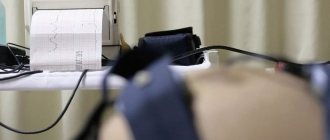
In the latter case, the expectant mother is prescribed additional ultrasound monitoring and mandatory CTG (cardiotocography) in the third trimester.
Cardiotocography is performed in the third trimester, starting at 32 weeks
Ultrasound diagnostics of the fetal heart is carried out in one of three modes:
- One-dimensional. Black and white image with a clear graph of fetal cardiac activity.
- Two-dimensional. In addition to functionality, it reflects the structure of the organ and its formation.
- Ultrasound with Doppler. The performance of the heart, its anatomical characteristics, and the condition of the blood vessels are assessed.
The choice of regimen is determined by the gynecologist leading the pregnancy.
Procedure Details
Of course, all mothers and children are different, so if the number of beats per minute differs from the normal values by several units in one direction or another, then you should not be scared or panic. In addition, the age of the embryo may be incorrectly determined, and what is abnormal for the 10th week of pregnancy is quite common at an earlier stage. Also, the heart rate is affected by the time of day at which the study is carried out, the health of the mother and the activity of the child, who can be actively moving or sleeping peacefully at this moment.
Ultrasound and examination of the fetal heart help identify the following developmental pathologies:
- Tachycardia, that is, an accelerated fetal heart rate. This pathology occurs in approximately 1% of all children in the womb and indicates that the child does not have enough oxygen. Women with cardiovascular disease are more at risk;
- Brachycardia, that is, a slowing of the rhythm and a general decrease in fetal activity. A low heart rate indicates a compressed umbilical cord and low blood pressure, which is very dangerous during pregnancy. Brachycardia in the fetus is common during childbirth, when the woman is given hormones and analgesics, which cannot but affect the child;
- Noises and irregular rhythm. This most often indicates a heart defect. Sometimes, with a multiple pregnancy or an incorrect position of the child, it is difficult for the doctor to isolate the right one from the many sounds, and the diagnosis may be made incorrectly. The final verdict is made only after the baby is born.
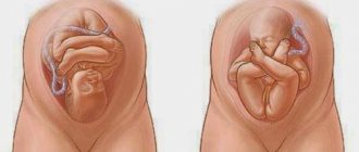
Many mothers want to find out the sex of the child already during pregnancy. They say that if the fetal heart rate is slowed down to 140-150 beats per minute, but the rhythm is even, then there will be a boy. And vice versa, if the heart rate approaches the upper limit of normal and the heart beats irregularly, then a girl will be born.
There is a sign for determining gender by the location of the child on the right or left in the uterus. It says that girls are on the left side and boys are on the right. You can trust these signs or not, but you can accurately determine the sex of a child only after birth.
Paid clinics often offer not only to print out an ultrasound image, but also to record a video of the sound of the child’s heartbeat. This is a relatively expensive service, but many are willing to pay for the unique opportunity to add such a video to the family archive and get to know the child before he is born.
Indications for the procedure
Fetal echocardiography is performed if certain risk factors are present. This may be testimony from the mother or from the child. The first include:
- genetic predisposition (heredity) to diseases of the cardiovascular system;
- unfavorable medical history (spontaneous abortion, presence of older children with cardiac pathologies);
- chronic diseases of the endocrine system;
- infections suffered at the beginning of the perinatal period;
- woman age 35+;
- taking medications during the period of conception and in early pregnancy (antibiotics, non-steroids, antidepressants, etc.);
- pregnancy after IVF (in vitro fertilization);
- bad habits (alcohol and drug abuse).
The reasons for the appointment on the part of the future baby are: deviations from the norm in terms of TVP (size of the nuchal translucency in thickness), non-compliance of heart rate with standards, suspected genomic anomalies: extra chromosome syndrome (Downism), abnormal chromosome set (Edwards syndrome), hereditary Turner and Patau syndromes, severe complication of multiple embryonic pregnancy (feto-fetal transfusion syndrome). You can undergo additional research on your own initiative in order to calm the excessive anxiety of the expectant mother.
Indications
Indications for the echocardiotocography procedure in a pregnant woman:
- severe shortness of breath that occurs with the slightest physical exertion;
- a history of heart and blood vessel diseases;
- feeling of changes in heart rate;
- a woman’s hereditary predisposition to cardiovascular pathologies;
- the woman experiences surges in blood pressure;
- a history of a heart attack;
- inflammatory processes in the kidneys;
- brain diseases.
Indications for fetal echocardiotocography:
- hereditary predisposition of the child to cardiovascular pathologies;
- previous miscarriages in a woman;
- presence of diabetes mellitus in the mother;
- diagnosis of lupus erythematosus in the mother;
- suffered toxoplasmosis during pregnancy;
- woman's age over 30 years;
- maternal rubella suffered during pregnancy;
- if the expectant mother took antibiotics in the first weeks after conception;
- the child has other chromosomal pathologies;
- the presence of fluid in the child’s cavity organs.
Timing of the examination
The doctor decides on an individual basis at what time to perform an ultrasound. For some patients, this procedure is prescribed repeatedly, as the child’s heart forms and develops. The device detects the first heartbeat at 4–5 weeks. This sign also determines the number of embryos in the uterus (multiple pregnancy), in this case the beating of two or more hearts will be heard. Heart rate is first determined around the seventh week.
Ultrasound will show the cardiac chambers (atria, ventricles) no earlier than at 14 weeks. Smaller vascular structures are visualized in the middle of the second trimester (starting at 18 weeks). The choice of how many weeks to undergo the examination depends on the information the doctor needs. The optimal period from which week to monitor the baby’s cardiac activity is considered to be 12–22.
How long does it take to do it?
Ultrasound of the fetal heart can be done in the second trimester of pregnancy. By the 12th week, the size of the fetal heart is still very small, but thanks to the presence of specially adapted parameters of cardiac programs in ultrasound machines, an ultrasound doctor can make a conclusion about the presence of the following cardiac malformations:
- defect in the left heart chambers;
- tricuspid valve atresia;
- underdevelopment of the pulmonary artery trunk, as well as its branches;
- valve pathology;
- defects of the open AV canal.
As a rule, these are severe and uncorrectable congenital heart defects. Unfortunately, in the early stages it is not possible to detect malformations of large vessels in the fetus.
If a heart defect is detected or suspected, ultrasound diagnostics will be required at 16 and 19 weeks. The most optimal time to do echocardiography is the period from 18 to 22 weeks. This is due to the fact that before this period the fetal heart is still extremely small, and then the volume of amniotic fluid decreases, which makes visualization difficult.
Using echocardiography, it is possible to identify most fetal heart defects, but it is necessary to understand that there are defects that are difficult to identify at the prenatal stage of development. These include open Bottal duct, defects in the septum of the heart, coarctation of the aorta, and minor disorders in the valvular apparatus of the heart.
Preparation and execution
There is no special preparation for echocardiography. The day before the study, the patient is recommended to drink soothing herbal remedies or other medicine that can affect the functioning of the cardiovascular system. There are no contraindications to ultrasound diagnostics. The procedure is absolutely harmless to the mother and fetus. Ultrasound waves are not foreign to the human body. Reflecting from the internal organs with a return echo signal, they are transformed by an ultrasound machine, and an image is displayed on the monitor.
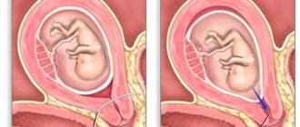
Depending on the duration of pregnancy and the individual characteristics of its course, an ultrasound of the heart can be done in two ways. Transabdominal (externally through the anterior wall of the peritoneum). In the first half of pregnancy, the procedure is performed with a full bladder. After the 20th week, amniotic fluid is sufficient, so there is no need to drink water before the examination. Transvaginally (by inserting an ultrasound probe into the vagina). In this case, the bladder should be empty.
The second option will show a more detailed picture, therefore it is preferable. You should not be afraid of this type of diagnosis. This does not pose a danger to the unborn baby. Diagnostics using the transabdominal method is performed with the patient in a horizontal position on her back. In the case of a transvaginal examination, the woman is positioned as during a regular gynecological examination, only on a couch rather than in a chair.
Why should an ultrasound of the fetal heart be performed?
Echocardiography of the fetal heart allows doctors to detect many different heart conditions early. Based on this, treatment tactics and appropriate measures that can be taken during and after childbirth are selected. However, sometimes it is necessary to resort to termination of pregnancy. This is performed in cases where the heart defect is absolutely incompatible with life, in addition, it may threaten the woman’s life.
However, if fetal echocardiography reveals abnormal heart function, you should not immediately panic. This could be a device error, an artifact during testing, etc. Therefore, after 1-2 weeks it is prescribed again. The obstetrician-gynecologist will indicate a more precise date. When the pathology has already been accurately diagnosed, a mandatory consultation with other specialists will follow. For example, if surgical intervention is necessary for a defect, then it is worth discussing everything with a cardiac surgeon. Another related specialist whose consultation is necessary is a neonatologist.
Not all defects detected on fetal echocardiography require mandatory surgical intervention. Some of them go away on their own, for example, a patent septum between the ventricles or atria may no longer be visible on a subsequent ultrasound examination.
Evaluation parameters and norms
During the examination, the doctor determines the following indicators and their compliance with accepted standards:
- anatomical location of the heart (correct/incorrect);
- organ size (must be equal to 1/3 of the cross section);
- the angle of the heart axis in relation to the midline of the chest (ideally 45 degrees);
- chambers of the heart (relatively equal in size);
- purity of the sound of the fetal heartbeat (without pronounced acoustic phenomena in the form of creaking, whistling, etc.).
- interventricular and atrial septa (without deformations);
- organ tissue (without pathological changes);
- rhythmicity of cardiac work (clear, without interruptions).
Heart rate is determined according to the table, according to the perinatal period.
The permissible deviations of the fetal heart rate are shown in parentheses.
Who should undergo fetal cardiac echocardiography?
There are certain risk groups for pregnant women who may give birth to a child with congenital heart disease:
- alcoholism, drug addiction, smoking while pregnant;
- the presence of congenital heart defects in the mother, father or other close blood relatives;
- diabetes mellitus, whether gestational or not;
- systemic lupus erythematosus or other autoimmune diseases;
- taking medications that are prohibited for pregnant women (antibiotics, antidepressants, antiepileptic drugs, nonsteroidal anti-inflammatory drugs, etc.);
- the mother has so-called torch and other infections (rubella, cytomegalovirus, parvovirus, toxoplasmosis, Lyme disease, etc.);
- hyperfunction of the thyroid gland;
- maternal hypertension;
- diseases affecting connective tissue;
- age over 35 years;
- polyhydramnios.
Common pathologies
The most commonly diagnosed abnormalities and pathologies shown by ultrasound of the unborn baby’s heart include:
- Rapid heartbeat (tachycardia). There are two main types of this deviation: reciprocal tachycardia of the supraventricular type and ectopic tachycardia. In the first case, excess contractions appear in the atria, in the second, they can occur in various parts of the organ.
- Decreased heart rate (bradycardia). Two types are also defined: basal and decelerant. The first type does not pose a danger; failure of the second type indicates oxygen starvation of the baby.
- Benign tumor of striated muscle tissue (rhabdomyoma). It is detected in the left or right ventricle or on the interventricular septum. It is a dangerous pathology because the tumor can block the blood flow and cause the death of the child.
- Functional noise, that is, between beats and during myocardial contraction, an extraneous sound is heard. This phenomenon is not always a pathology and requires special treatment. On the other hand, the presence of acoustic phenomena may be a sign of heart disease. Therefore, in case of a heart murmur, CTG is most often prescribed additionally.
- Not abnormal, but a special structure of the heart (hyperechoic focus or HEF). As a monosymptom it is not dangerous, but in combination with other abnormalities it requires regular monitoring.
- Congenital defect of the EVC (single ventricle of the heart);
- A narrowing of the opening of the aorta in the valve area, which makes it difficult for blood to flow out of the left ventricle (aortic stenosis).
- Congenital pathology of the formation of walls separating the chambers of the heart from each other or their absence (atrioventricular canal, otherwise endocardial cushion defect).
A stable heartbeat with a frequency of 140–160 beats/min is observed from the 14th week
The treatment tactics for diagnosed diseases are determined by the doctor. The woman may be referred for consultation to other medical specialists. Ultrasound of the fetal heart is one of the most important examinations during the perinatal period. Timely and competently performed diagnosis helps to avoid complications in the baby in the future, and possibly save his life. A woman can decide for herself where to have the procedure done in a antenatal clinic at her place of residence or a paid diagnostic center. The examination cannot be ignored.
Ultrasound for pathology
If a deviation of the cardiovascular system from normal development is suspected, specialists prescribe an ultrasound of the heart, as this is the fastest and most informative examination. Diagnosis is carried out after three preliminary checks:
- auscultation of the heart,
- Ultrasound,
- echocardiography,
If any indicators and results of preliminary diagnostics indicate pathology, it is necessary to immediately conduct an ultrasound examination of the fetal heart.
Echocardiography of the fetus consists of an ultrasound examination using additional sensors that record the activity of the heart. This procedure does not pose any danger to either the fetus or the mother. This research method allows us to identify the presence of defects in a child even before his birth, during the prenatal period of development.
Execution technique and interpretation of results
Performing an ultrasound is technically very simple, but interpreting and evaluating the results requires experience and skill. The procedure uses various sensors that allow examination through the abdominal wall (transabdominal) or inside (transvaginal). For better passage of ultrasonic rays, a special gel is used.
To obtain reliable ultrasound results, the doctor takes measurements of the fetus and compares the data with the norms for each week. The coccygeal-parietal (CTP) and biparietal size (BPR) must be specified. In the first trimester, these values will help to accurately determine the gestational age. The following studies additionally measure the circumference of the head (CG), abdomen (AB), the length of the bones of the limbs and their correspondence to the weeks of pregnancy.
It is necessary to measure the fetal heartbeat: in the 1st trimester the parameter can be 160-190 beats, from the 11th week - 140-160 beats/min. The conclusion should indicate the amount of amniotic fluid, the location of the placenta, and the length of the cervix.
Modern diagnostic methods are capable of identifying genetic abnormalities at an early stage. With the help of echocardiography, you can find out about heart defects and quickly operate on the child. An ultrasound will help accurately determine the sex of the baby, and techniques such as 3D and 4D will even show the baby’s facial features in a photo or video.
Although there are various debates surrounding the safety of the method, the study provides a lot of useful information about the health of the expectant mother and fetus.
Varieties of echo kg fetus
Many expectant mothers are interested in the question of how to make an echo of a kg fetus. The method includes several different types of techniques, the choice of which is made based on the purpose of the study.
These can be the following types of echocardiography method:
- A two-dimensional echocardiogram (or 2-D) allows you to study the anatomy of the main structures of the heart (valves, chambers, vascular system).
- M-mode (one-dimensional) allows you to observe the state of the leaflets, valves and walls of the heart during their operation.
- Pulsed wave Doppler echocardiography makes it possible to study and evaluate hemodynamics and heart rhythm.
This study also makes it possible to study the direction and speed of blood flow in the heart vessels. It also helps to assess the degree of vasoconstriction and the level of blood volume leaving and returning to the heart, which is very important when diagnosing malformations of this organ.
To echo the fetal heart, new generation devices are used, which are completely safe for both the woman and her unborn baby.