Basophils are large, sedentary cells belonging to the smallest variety of leukocytes. They are formed, like all other formed blood cells, in the red bone marrow, from a common stem protocell - the hemocytoblast. Exposure to certain inducers stimulates these primary cells to divide, which takes 4 days. After which, within 5 days, morphological maturation occurs, during which basophils receive a unique functional specialization and a special structure that distinguishes them both from erythrocytes and platelets, and from other leukocytes - neutrophils, eosinophils, monocytes and lymphocytes.
Basophil cells are unique cells of the human body. They are represented by 3 separate types: basophilic segmented blood granulocytes or basophilic leukocytes, tissue basophils or mast cells, and pituitary basophils.
Unlike pituitary basophils and mast cells, basophilic leukocytes circulating in the blood enter it from the bone marrow already in a mature state, while pituitary and mast basophils are formed from granulocytes of the parietal pool and mature directly in the blood.
All three types of basophils, although they are direct relatives, are different from each other, and each performs its own specific job.
Functions
Basophils are capable of producing active substances that affect the processes of inflammation, blood circulation and thrombus formation, which contributes to the following functions:
- phagocytosis – capture and destruction of pathogenic microorganisms;
- release of histamine to improve vascular permeability and migration of other types of leukocytes from the bloodstream into the tissue when pathogenic microorganisms and antibodies are detected;
- release of heparin to maintain normal blood circulation in areas of allergic inflammation;
- improvement of tissue trophism and formation of new capillaries;
- participation in antiparasitic immunity;
- increased inflammatory processes in the epithelium of the intestines and internal organs in the presence of helminths, as well as local inflammation in the presence of ticks;
- synthesis of biologically active substances necessary for the formation of allergic reactions and changes in blood composition during inflammation;
- cleansing the body of the effects of inflammation.
The structure of basophilic leukocytes
Basophilic leukocytes are the largest granulocyte cells. They are significantly larger in size than their “classmates” – neutrophils and eosinophils. The diameter in a drop of blood is 9 microns, and in dry smears from 7 to 12 microns. The cell shape is round.
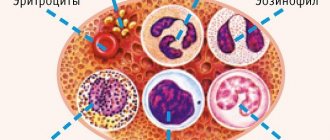
The photo shows all types of leukocytes circulating in the blood
All basophils received their name because of their ability to be stained with basic dyes during laboratory testing. After such manipulation, in the cytoplasm of the cells one can distinguish blue-violet granules of different sizes, sometimes with a purple tint, reminiscent of black caviar (basophilic granulation).
The nucleus of basophils is located in the center and consists of 2 segments, which, as a rule, resemble the letter S. The nucleus contains little heterochromatin, and therefore is poorly stained, and, due to the large number of colored granules, it is practically invisible. In immature cells, it looks like a rod, which is why such cells are called rod-nucleated granulocytes.
Structure of a basophil cell
Basophilic leukocyte granules contain:
- free salt-like compounds of heparin and histamine;
- serotonin, factors of anaphylaxis, chemotaxis and platelet activation;
- leukotriene C4, prostaglandins, acid glycosaminoglycans.
Some of these substances are constantly present in the cell, while others are synthesized and released only during the interaction of basophilic leukocytes with allergenic antigens.
On the surface of the plasma membrane there are highly qualified immunoglobulin IgE molecules, as well as Fc-epsilon-RI receptors and tetrametric isoforms (αβγ2). It is the reaction of releasing the content of basophilic grains - degranulation that occurs under the influence of these membrane receptors that determines the main tasks that basophilic leukocytes perform.
Causes of elevated basophils in adults
An increase in the level of basophils can be both the body’s reaction to physiological processes and one of the symptoms of the development of a number of systemic pathologies and diseases of internal organs.
Natural reasons that result in an increase in the number of basophils in peripheral blood are as follows:
- menstrual cycle in women;
- use of contraceptives with a large amount of estrogen in the composition;
- small doses of radiation (frequent x-ray procedures);
- recovery period after infections;
- pregnancy.
Pathological causes of increased basophils in the blood can be classified as follows:
- allergy;
- blood diseases;
- pathology of the thyroid gland.
Allergic reactions
After an allergen enters the body, basophils form a hypersensitivity reaction, which differs in type:
- anaphylactic - general or local reactions of the body to an allergen, accompanied by the release of histamine from basophils.
- cytotoxic - the appearance of antibodies to the allergen causes an autoimmune response at the intracellular level and hemolytic pathologies.
- immunocomplex - the formation of immune complexes in tissues that cause damage to blood vessels, joints, kidneys, etc.
- Delayed-type hypersensitivity is the slow development of allergies to molecular compounds formed as a result of prolonged exposure to internal and external allergens (for example, the use of medications or skin contact with chromium).
Diseases that are accompanied by allergic reactions and increased levels of basophils:
- Allergies to wool, metal, food, pollen and flowering plants, etc.
- Goodpasture syndrome is an autoimmune disease with diffuse alveolar hemorrhage and glomerulonephritis caused by the accumulation of specific antibodies in the basement membranes of the renal and pulmonary capillaries.
- Serum sickness is an allergic reaction that occurs after the administration of injections, solutions and other medications. An immune response to foreign antibodies and an increased level of basophils in the blood can be observed when using anti-diphtheria, anti-botulinum, anti-tetanus and other serums.
- Exogenous allergic alveolitis is an inflammatory process in the alveoli and bronchioles of the lungs, caused by external factors such as fungal spores, plant and house dust, bacterial spores, and some medications.
- Rheumatoid arthritis is a systemic disorder of a genetic or acquired nature and is accompanied by the development of chronic inflammation of the connective tissue of small joints.
- Systemic lupus erythematosus is an autoimmune pathology with inflammatory reactions of connective tissue in the vessels of internal organs, skin, etc.
- Glomerulonephritis is an inflammation of the kidneys that occurs as a result of a number of systemic diseases (systemic lupus erythematosus, systemic endocarditis). Glomerulonephritis causes edema, hypertension, and urinary retention.
- Vasculitis is an autoimmune inflammation of all types of blood vessels, which leads to disruption of the structure and functions of all internal organs.
- Contact dermatitis is skin irritation as a result of the negative influence of the external environment. With contact dermatitis, basophils accumulate in the affected tissues and cause swelling, blisters, erosion, and peeling.
- Bronchial asthma is an allergic disease of the respiratory tract caused by functional changes in the functioning of immune cells. Asthma is accompanied by mucus secretion, coughing, difficulty breathing, and suffocation.
- Type 1 diabetes mellitus is a disease of the endocrine system with damage to pancreatic cells and insufficient secretion of insulin. The main symptoms of type 1 diabetes include hypoglycemia, decreased digestibility of food, and increased thirst.
Blood diseases
Blood diseases (myeloproliferative) are disorders in the functioning of the bone marrow, which result in an increase in the production of stem cells, as well as improper maturation of granular leukocytes. With myeloproliferative pathologies, basophils and other forms of leukocytes increase significantly, and the number of red blood cells and platelets also changes:
- Chronic myeloid leukemia is a pathological proliferation of tissues that form all types of granular leukocytes. As a result of the development of myeloid leukemia, the level of monocytes, basophils, and eosinophils in the plasma increases. At an advanced stage, the disease is manifested by dystrophic changes in the heart, kidneys, liver, spleen, as well as intoxication, frequent bleeding, and purulent-inflammatory processes.
- Erythremia (polycythemia) is a chronic disorder of hematopoiesis with a pathological increase in the number of red blood cells and some forms of leukocytes, hypertrophy of the bloodstream and decreased functionality of internal organs. Erythremia manifests itself mainly in adults aged 40 to 60 years.
- Myelofibrosis is a disorder of hematopoietic processes resulting from the replacement of bone marrow with connective tissue. With myelofibrosis, a large number of immature forms of basophils, monocytes, lymphocytes, eosinophils and erythrocytes are observed in the plasma, and anemia, malfunctions of the spleen, liver, etc. are diagnosed.
- Lymphogranulomatosis (Hodgkin's cancer) is the appearance of malignant tumors in the lymph nodes. In some forms of lymphogranumatulosis, a significant increase in all forms of leukocytes is manifested.
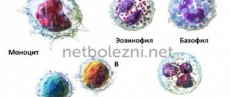
Eosinophils, neutrophils, lymphocytes and monocytes
The basophil content of all leukocytes is only 0.5%. In addition to this type of corpuscle, the following types can be found in the blood:
- Neutrophils. The most numerous group of leukocytes that fight severe infectious conditions.
- Lymphocytes. They are part of the immune system, preventing invading viruses and bacteria (pathogens).
- Eosinophils. They help fight parasitic infections. The functions of basophils and eosinophils play an important role in the body.
- Monocytes fight infections in the bloodstream, fight antigens, promote the restoration of damaged tissue, and also destroy cancer cells.
Let us take a closer look at the functions of basophils in immunology.
conclusions
Basophils are protective blood cells, indicators of allergies, inflammatory and oncological processes. In a healthy person, no more than 1% of white blood cells should be detected among all leukocytes and no less than 0.1 * 10⁹/l. If the number of cells is less, they speak of basopenia. The condition rarely indicates a serious illness, but this option should not be ruled out.
p, blockquote 36,0,0,0,0 —>
The level of basophils increases in allergic, inflammatory reactions, as well as in oncohematological diseases. Despite the insignificant content of cells in the blood, it is necessary to pay attention to their values so as not to miss serious pathologies. Read more about basophilia in the article: “Elevated basophils - what it is and its causes in adults and children. Is the condition dangerous?
p, blockquote 37,0,0,0,0 —> p, blockquote 38,0,0,0,1 —>
If according to the results of the OAC there are other abnormalities, you need to consult a doctor for help.
Content
Basophils 0 in an adult - what does this mean? Basophil 0 in the human body is considered by many to be “blood scouts”. They are cells that are a type of white blood cell. Basophils 0 in the blood perform a sufficient number of functions if they are in normal quantities. However, when it comes to raising or lowering them, it is worth contacting a specialist for advice or further examination.
Norms
The level of basophils is determined through a clinical blood test. The concentration of such bodies is established as a percentage of the total number of lymphocytes in the form of absolute and relative values. The absolute amount, regardless of age, is from 0.01 to 0.065 * 109 g/l, the relative amount directly depends on the age of the person and has the following indicators: 0.75% - newborns; 0.5% - baby from one month; 0.6% - one-year-old children; 0.7% - two-year; 0.5-1% - adult category.
Mast cells
Major populations of human mast cells
Mature mast cells are most often found in the skin, lymphoid organs, thymus, lungs, uterus, gastrointestinal tract, bladder, conjunctiva, nasal mucosa, synovial membranes, mesentery. Based on the presence of tryptase (trypsin-like protease) and chymase (chymotrypsin-like protease) in cells, two subpopulations (see table above):
- mast cells containing both enzymes (TKTX; present in the skin, intestinal submucosa and connective tissue);
- mast cells containing only tryptase (TKT; predominantly localized in the lungs and gastrointestinal tract).
These groups also have functional differences, although data on this are still rather scarce.
Enzyme double-positive skin cells are capable, unlike lung and intestinal cells containing only tryptase, to easily release histamine (read the link for what it is) in response to a wide range of different stimuli. The noted subpopulations also differ in the content of cytokines. For example, IL-4 is found primarily in TKTX, and IL-5 and IL-6 are expressed primarily in TKT. At the same time, both subpopulations of cells carry the FcεRI receptor on their surface and participate in IgE-dependent allergic and antiparasitic reactions.