home
→
Gynecology and obstetrics
→
Pregnant women
→
Fetal size according to ultrasound and weight
Urgent appointment with a gynecologist
Gynecologist consultation and examination
A complete diagnosis of pregnancy these days is impossible without an ultrasound examination. This study is the main diagnostic method during pregnancy. It is quite informative, allows you to reach a large number of women and, most importantly, is safe. In addition, by recording onto a USB drive and archiving ultrasound sessions, today you can monitor the development of the fetus week by week over time.
The frequency of fetal ultrasound is determined by the gynecologist who monitors the woman. The purposes of performing an ultrasound examination of the fetus differ depending on at what stage of pregnancy it is performed.
Main types of fetal ultrasound
- transabdominal – the sensor of the device is located on the woman’s stomach;
- transvaginal – the device’s sensor is inserted into the vagina.
When performing a transvaginal ultrasound, a more accurate determination of fetal parameters is possible. Recently, three-dimensional and four-dimensional ultrasound of the fetus have become popular, which allows one to obtain additional information about the process of bearing a baby.
4D is a real color film about a baby that you can watch, save and keep as a keepsake.
Encyclopedia of Ultrasound and MRI
Ultrasound examination has been used in world obstetric practice since 1966, when ultrasound was first approved for use in diagnosing the course of pregnancy. Since that time, ultrasound diagnostics has not lost its relevance, and on the contrary, it is still one of the most harmless methods of examining pregnant women.
The method combines the ability to study the state of internal organs and the fetus, without penetrating into the internal environment, with safety and high performance. Interpreting ultrasound during pregnancy is a matter for a professional.
Fetal ultrasound norms by week
First ultrasound
carried out at approximately 10-14 weeks. During the examination, the doctor determines whether there is an ectopic pregnancy, measures the thickness of the collar zone and examines the parameters and condition of the internal organs of the unborn baby, identifying congenital malformations, if any. The fetal heart rate is also measured to determine possible heart defects. In some cases, at this stage it is already possible to determine the sex of the fetus by ultrasound.
Second ultrasound
carried out at 20-24 weeks. Based on the results of this study, it is already possible to accurately determine the sex of the child and ensure that there are no obvious signs of pathology. The doctor measures the fetal brain structure and studies the development of internal organs - the liver, heart and intestines.
Third ultrasound
The gynecologist does it in the third trimester, at about 30-32 weeks. During this study, you can find out the weight of the fetus by ultrasound, assess the position of the fetus in the uterus, and check the maturation of the placenta. The condition of the lungs and heart rate are also checked. Interpretation of fetal ultrasound also always includes data on the condition of the uterus, umbilical cord and amniotic fluid.
The volume of amniotic fluid is calculated through the amniotic index. If the index is increased, this indicates polyhydramnios, but if it is decreased compared to the norm, this means that the woman has oligohydramnios. Turbidity in the amniotic fluid may indicate that there is an infection in the body. All this requires the help of a doctor and adequate treatment.
All of the above studies are included in the so-called “screening” program, which is carried out on average three times per pregnancy in order to identify fetal malformations on ultrasound and chromosomal pathologies. If necessary, and to confirm previously obtained data, the doctor may additionally prescribe a consultation with a geneticist, biochemical screening, as well as chorionic villus biopsy, amniocentesis and cordocentesis.
Causes of congenital fetal pathology
What factors can adversely affect the fetus? The causes of various pathologies are:
- burdened heredity;
- Mom's age is over 35 years, dad's age is over 40;
- unfavorable environmental conditions;
- working with toxic substances;
- bad habits - alcohol abuse, smoking, drug use;
- viral and bacterial diseases, especially in the first trimester - influenza, rubella, toxoplasmosis, hepatitis B, sexually transmitted diseases;
- taking certain medications;
- excessive psycho-emotional stress.
The more factors are present, the greater the likelihood of fetal pathologies. That is why you need to start planning for the birth of a child in advance, at least six months before conception. This time is enough to undergo a comprehensive examination, identify and cure diseases, and prepare for the conception of a healthy baby.
To highlight the impact of bad habits on the fetus, we present some numbers. With maternal alcoholism, toxicosis is observed in 26% of cases, antenatal death and asphyxia in 12%, miscarriages in 22%, premature birth in 34%, birth injuries in 8%, and intrauterine growth retardation in 19%.
Fetal size chart by ultrasound
BRGP (BPR) – biparietal head size. DB – thigh length. DGrK – chest diameter. Weight - in grams, height - in centimeters, BRGP, DB and DGrK - in millimeters.
| A week | Height, cm | Weight, g | BRGP | Dlb | DGrK |
| 11 | 6,8 | 11 | 18 | 7 | 20 |
| 12 | 8,2 | 19 | 21 | 9 | 24 |
| 13 | 10,0 | 31 | 24 | 12 | 24 |
| 14 | 12,3 | 52 | 28 | 16 | 26 |
| 15 | 14,2 | 77 | 32 | 19 | 28 |
| 16 | 16,4 | 118 | 35 | 22 | 34 |
| 17 | 18,0 | 160 | 39 | 24 | 38 |
| 18 | 20,3 | 217 | 42 | 28 | 41 |
| 19 | 22,1 | 270 | 44 | 31 | 44 |
| 20 | 24,1 | 345 | 47 | 34 | 48 |
| 21 | 25,9 | 416 | 50 | 37 | 50 |
| 22 | 27,8 | 506 | 53 | 40 | 53 |
| 23 | 29,7 | 607 | 56 | 43 | 56 |
| 24 | 31,2 | 733 | 60 | 46 | 59 |
| 25 | 32,4 | 844 | 63 | 48 | 62 |
| 26 | 33,9 | 969 | 66 | 51 | 64 |
| 27 | 35,5 | 1135 | 69 | 53 | 69 |
| 28 | 37,2 | 1319 | 73 | 55 | 73 |
| 29 | 38,6 | 1482 | 76 | 57 | 76 |
| 30 | 39,9 | 1636 | 78 | 59 | 79 |
| 31 | 41,1 | 1779 | 80 | 61 | 81 |
| 32 | 42,3 | 1930 | 82 | 63 | 83 |
| 33 | 43,6 | 2088 | 84 | 65 | 85 |
| 34 | 44,5 | 2248 | 86 | 66 | 88 |
| 35 | 45,4 | 2414 | 88 | 67 | 91 |
| 36 | 46,6 | 2612 | 89,5 | 69 | 94 |
| 37 | 47,9 | 2820 | 91 | 71 | 97 |
| 38 | 49,0 | 2992 | 92 | 73 | 99 |
| 39 | 50,2 | 3170 | 93 | 75 | 101 |
| 40 | 51,3 | 3373 | 94,5 | 77 | 103 |
During the study, it is necessary to take into account that each device can be configured for different tables with fetal sizes by week, so ultrasound protocols may have significant differences. It is best to clarify all the obtained indicators with your doctor at your appointment. Only a doctor will tell you in detail about the normal size of the fetus according to ultrasound and, if problems arise, will tell you how to deal with them.
How is ultrasound interpreted?
Interpreting the results of an ultrasound scan of pregnant women is an individual matter for each expectant mother.
How to interpret an ultrasound during pregnancy? This is the task of both a doctor specializing in ultrasound diagnostics and the obstetrician-gynecologist himself.
Medical data obtained from ultrasound examination are correlated with medical history data and the individual characteristics of the pregnant woman.
Only after this the doctor decides how physiological the data obtained by the measurement is, how this or that ultrasound result during pregnancy is interpreted and whether adjustments need to be made to its management.
A specialist in the field of ultrasound diagnostics conducts the examination itself and the necessary measurements, the results of which are transmitted to the obstetrician-gynecologist. Therefore, the description of the results is carried out by two specialists. The result sounds like “pregnancy, term.” If there is a pathology, it is also indicated in the conclusion.
This protocol is a standard measure for monitoring fetal health in the first trimester
Come to us
The work of gynecologists at the international medical center ON CLINIC is aimed at caring for a woman during her pregnancy and her unborn child. When you come to see us, you can be sure that the doctors will answer all your questions, conduct the necessary research, help calculate the weight of the fetus using ultrasound, and tell you what the normal size of the unborn baby and all its organs should be week by week.
All examinations at the center are carried out according to international standards. Our center is equipped with the most modern diagnostic equipment and is ready to receive you any day, seven days a week.
Features of diagnostics of the fetus and uterus
Ultrasound scanning is considered a universal, non-invasive, convenient and safe technique for examining patients. Its essence lies in the analysis of the transformation of mechanical vibrations of structures of various densities above the audible frequency. Ultrasound equipment uses the acoustic impedance of sound waves with a frequency of 2–10 MHz. During pregnancy, the examination does not cause discomfort or pain to either the mother or the baby - it is performed using a special sensor.
- anatomical features of the developing baby;
- placenta – “child’s place”;
- umbilical cord - umbilical cord;
- amniotic fluid surrounding the fetus;
- the uterine cavity, its ligamentous apparatus and appendages.
The purpose of ultrasound is to assess the condition of the pregnant woman and the unborn baby, and to diagnose possible congenital and genetic syndromes. This is especially important in cases where there is a special indication - a hereditary predisposition to such anomalies.
Interpretation of ultrasound during pregnancy allows you to determine the size of the fetus, the amount of amniotic fluid, the degree of premature aging of the placenta, its integrity and the place of attachment to the uterine wall. Practitioners use the indicators of this examination to choose tactics for managing the gestation period and preparing for the process of expulsion of the fetus.
Key points of fetometric research
The key data of a fetometric study are the following indicators:
- DB - thigh length;
- BPR - biparietal size;
- DP - shoulder length;
- KTP - coccygeal-parietal size;
- DN - nasal bone length;
- LZR - fronto-occipital size;
- OG - head circumference;
- DG—tibia length;
- AB—abdominal circumference;
- TVP is the thickness of the collar space.
A decoding of the designations of the studied parameters is provided, since the fetometric data is written in the table in Latin.
A video about the stages of ultrasound was presented by the 1st Medical Quarter of Crede Experto on Taganka.
Child's weight
By the 12th week, the baby’s body weight is normally only 19 g; by the middle of pregnancy, the baby will weigh about 345 g, and by the 32nd week - almost 2 kg.
If you promptly pay attention to the problem with the discrepancy between the fetal body weight and the standard, and take preventive measures, then it will be possible to correct the situation relatively easily. The rate of weight gain is greatly influenced by genetic factors. The gynecologist makes sure that the dynamics are positive.
KTR (Latin analogue of CRL) means coccygeal-parietal size, that is, the height of the child. It is calculated from the crown to the end of the coccyx.
If this indicator differs slightly from the norm, then the fetus is not in danger. An increase in CTE over several weeks by the same value indicates that the fetus is relatively large in size.
The letters BPD indicate the width of the fetal head. This is the maximum distance between the parietal bones. The size can be determined by taking measurements along the smallest axis of the circle between the child's temples. BDP allows you to determine the exact duration of pregnancy.
The parameter of biparietal fetal head size (BSD) helps to identify developmental abnormalities already during the first trimester. The data obtained characterize the state of the fetal nervous system.
The LZR or fronto-occipital size is calculated between the most distant points of the forehead and the back of the head.
The volume of a child's chest is determined by calculating the diameter of its circumference. A size that does not correspond to the norm should not cause any particular concern; most likely, this is a genetic feature. Perhaps the baby will simply be born large. It is necessary to take into account the physical characteristics of the mother and father.
Abdominal circumference is measured at 20 and 32 weeks of pregnancy. The parameter is calculated along the line of the liver, stomach and umbilical vein. When the difference in indicators exceeds the permissible norm, the doctor will diagnose intrauterine growth retardation. However, to confirm it, the size of the coolant is calculated in relation to other parameters - the size of the head, femur, and BPR. If most indicators are normal, then this indicates a delay in the development of the asymmetric form.
If a discrepancy in the thigh length is detected, this also does not indicate the presence of pathology. Much depends on individual characteristics. For example, when the length of the femur and shin bones is longer than normal, it means that the parents of the unborn baby or other relatives have a long leg.
PMP (PVP)
PVP is the estimated body weight of the fetus. During an ultrasound examination, there is a possibility of an error in weight. To eliminate errors, this figure is calculated using various medical formulas.
Calculation methods:
- Zhordania (Lebedeva) - PMP = height of the uterine fundus × abdominal circumference.
- Bublichenko - PMP = 1/20 of a woman’s weight.
- Lankowitz - PMP = (height, weight of mother, abdominal circumference, height of the uterine fundus) × 10.
- Jones - PMP = (height of the uterine fundus - 11) × 155. The value 11 is a conditional coefficient for a pregnant woman weighing up to 90 kg.
- Yakubova - PMP = (abdominal circumference, height of the uterus) × 100/4.
Calculations are made after 38 weeks of pregnancy.
The Family TV channel presented a video about performing an ultrasound in the third trimester.
What is PMP, why is this indicator calculated during pregnancy?
This abbreviation stands for “estimated fetal weight.” The second term doctors use, EFT (or estimated fetal weight), has a similar meaning. Why did doctors need to measure PMP at different stages of pregnancy? Estimated fetal weight is a very important evaluation criterion. A harmoniously developing child has normal body weight. Fetal weight that does not correspond to the established gestational age, its slow or too rapid weight gain indicate possible disorders and are a reason for a more thorough examination of the expectant mother.
A fetus with abnormal body weight is exposed to great risks during intrauterine development, during childbirth and after birth. By determining the approximate weight of the fetus, doctors can diagnose certain disorders and begin treatment at an early stage. The goals of establishing the PMP indicator in obstetric practice:
- assessment of fetal development in accordance with gestational age;
- anomaly detection;
- diagnosis of frozen pregnancy;
- identification of certain diseases in the mother (for example, excessively high body weight of the child during gestation may be a symptom of diabetes mellitus);
- predicting the course of labor and the likelihood of using a cesarean section.
Third planned study, basic standards
A third ultrasound at 32-34 weeks is necessary to identify malformations that appear only in late pregnancy (for example, an aneurysm of the vein of Galen - a disorder of the structure of the vascular wall of a large cerebral vessel). It allows you to assess the functional state of the fetus, make a diagnosis of developmental delay syndrome (FGR), which makes it possible to carry out a set of necessary therapeutic measures, and identify indications for timely and careful delivery. The presence of FGR requires mandatory monitoring after 7-10 days during active therapy.
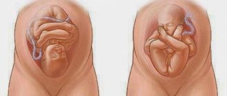
An important point is the presentation of the fetus (cephalic or pelvic), which significantly affects the method of delivery. It is also mandatory to determine the expected weight of the fetus, which should be taken into account in the tactics of further management of pregnancy and especially childbirth.
To assess the condition of the fetus in the third trimester, determination of the biophysical profile of the fetus during ultrasound can be used (Table 10).
When assessing the tabular parameters, the sum of points is determined, on the basis of which a conclusion is made about the condition of the fetus:
- 12-8 is normal;
- 7-6 – questionable condition of the fetus, possible development of complications;
- less than 5 – severe intrauterine hypoxia (lack of oxygen supply to the fetus, leading to varying degrees of impairment of its vital functions) with a high risk of perinatal losses (fetal loss during the period from 22 weeks of pregnancy to 168 hours after birth).
Ultrasound examination during screening periods makes it possible to identify a large number of pathologies and take preventive measures to eliminate them as much as possible in the prenatal period, and if elimination is impossible, to reduce the consequences.
The baby's insides are carefully examined in the 3rd screening study. The heart, its valves, kidneys, liver, and genitals are examined.
| Main ultrasound indicators, values for 38 weeks | Norm |
| BPR | 100 mm |
| PAP | 128 mm |
| Heart rate | 120-160 beats per minute |
| Height | 49-50 centimeters |
| Weight | 2800-3000 grams |
| BRGP | 92 |
| coolant | 336 mm |
| OG | 309-357 mm |
At the 3rd study, parents receive confirmation of the child’s gender. Cases when it is not possible to determine the sex of a child are very rare. By this period, the child should turn correctly in the womb; if this does not happen, there is an indication for a caesarean section.
The condition of the placenta occupies an important place in the study: the distance from the internal os of the uterus is determined (normally 7 cm). Placenta previa occurs at a shorter distance. The maturity of the placenta is different at each stage of pregnancy. By the 36th week, maturity reaches III degree, the thickness reaches 35-45 millimeters. Aging of the membrane, which occurs prematurely, leads to discomfort for the child. The occurrence of this fact requires intervention from specialists.
At 3 ultrasound, the fact of entanglement with the umbilical cord, if any, is determined.
Amniotic fluid serves as an indicator of the normal course of pregnancy. The AI index is an assistant in this matter. A high indicator indicates polyhydramnios, a low indicator indicates oligohydramnios. These pathologies are often associated with fetoplacental insufficiency. An ultrasound can also reveal signs of intrauterine infection of the fetus.
The size of the uterus, its tone, length, diameter, and cervical canal must correspond to the norms at each stage of pregnancy. Screening studies focus on these parameters.
What determines the weight of the fetus during pregnancy?
The weight of the child depends on the functioning of the placenta and the incoming nutrients with oxygen. Starting from the second trimester, the fetus increases to 80 grams. In the later stages, the seven-day gain reaches 200 grams, but before labor the pace slows down significantly due to the death of the placenta.
The indicator also depends on the following factors:
- unbalanced maternal diet;
- stressful situations during pregnancy;
- chronic diseases;
- toxicosis;
- smoking and alcohol abuse;
- genetic predisposition.
The weight of the baby during intrauterine development also depends on gender. Boys are born larger than girls.
What affects a child's weight?
Many factors can affect fetal weight. Among them:
- Heredity. A child's weight gain largely depends on the build of the parents. If dad and mom have a fairly large build, then the child’s weight will be significantly greater than that of parents with an average body size. But if the parents are thin and short, then the baby will be born with low weight.
An important indicator is the weight of the parents at birth.
- Nutrition. During pregnancy, a woman should consume the entire range of useful vitamins and elements that her baby will receive with her. This has a beneficial effect on the development of its mass.
Abuse of fried, fatty and floury foods can lead to a lack of important substances for the child and developmental delays.
Excessive intake of vitamins can negatively affect growth.
- Bad habits. When the fetus consumes alcohol, nicotine and narcotic substances during pregnancy, not only is there insufficient weight, but also various developmental pathologies.
- Stress. If the expectant mother often worries and is nervous, this cannot but affect the baby’s weight gain.
- Chronic diseases. For example, women with diabetes are more likely to give birth to children with excess body weight, and with heart pathologies, on the contrary, there is a lack of weight.
- Extreme conditions. If the pregnancy is difficult and there is a constant threat to the baby’s life, then it is most likely that he will lose weight.
- Multiple pregnancy. If a woman is carrying 2 or more children, their weight will deviate from the norm. But in this case it is considered natural.
- Condition of the placenta. If the uteroplacental blood flow is disrupted, the baby does not receive enough oxygen and its growth slows down.
- Age of the pregnant woman. Particular attention should be paid to women in labor under 18 and after 35 years. They are more likely to have insufficient fetal weight.
- Taking medications. Some medications, especially antibiotics, can have a serious effect on the growth and development of the fetus. The most dangerous period is the 1st trimester, since at this time the baby is developing important structures.
- Pregnancy order. It is known that the size of the first child may be slightly smaller than in subsequent pregnancies.