Indications for diagnostics
Colonization of the gastric mucosa by Helicobacter pylori bacteria is accompanied by the development of ulcerative lesions, carcinogenic tumors, chronic gastritis, manifestations of dyspepsia, gastroesophageal reflux and irritable bowel syndrome.
The bacterium Helicobacter pylori (H. Pylori) under an electron microscope
Enzyme immunoassay allows you to quickly identify bacterial colonization of the mucous membrane and correctly develop the optimal treatment regimen or further examinations.
Anti-Helicobacter pylori analysis is carried out:
- for diagnostic purposes:
- Peptic ulcer of the stomach and duodenum.
- Esophageal ulcers.
- Non-ulcer dyspepsia.
- Esophagitis.
- Atrophic gastritis.
- Stomach cancer in close relatives.
- Helicobacteriosis infection of close relatives.
- during preventive examination to identify patients at risk;
- to assess the dynamics of treatment;
- for symptoms that raise suspicion of infection:
- Failure to accept protein foods.
- Heaviness in the stomach.
- Frequent vomiting of unknown origin, nausea, persistent heartburn, belching.
- Pain in the abdominal region (lower and upper abdomen), decreasing after eating.
- Flatulence.
- Weight loss of unknown origin.
- Constipation and diarrhea.
- Blood in vomit or stool.
Localization of the bacterium Helicobacter pylori
How it appears in the body
Helicobacter pylori is a gram-negative bacterium that inhabits the gastrointestinal tract and can exist there without air.
Once in the body, the bacterium settles in the stomach. It belongs to the only species that is not exposed to gastric juice. Transmitted in the following ways:
- through saliva;
- through mucous secretions;
- with dirt;
- through unwashed foods.
The main cause of Helicobacter pylori infection is considered to be weak immunity throughout the body and locally in the digestive organs.
Helicobacter does not always lead to devastating consequences. In its dormant state it is harmless. Here the degree of expression of the body’s own defenses is of great importance.
About the bacteria
Among the features of Helicobacter pylori are:
- it is resistant to gastric juice and hydrochloric acid, which is due to its rapid movement and production of ammonia;
- upon penetration into the mucous tissues of the stomach, it begins its destructive effect, leading to the formation of ulcers or foci of inflammation;
- during reproduction, intoxication of the body occurs, which leads to inflammation of the gastric mucosa.
What is a blood test to determine antibody levels?
ELISA consists of examining blood serum and determining antibody titers (concentrations), the presence of which is an indicator of a person’s infection with Helicobacter pylori. They are formed in response to the introduction of genetically alien proteins, in this case, to the microorganism Helicobacter pylori.
The formation of antibodies is part of natural defense mechanisms aimed at eliminating pathogens. If antibodies are detected in the blood during the study, this means that the immune system has reacted to the presence of a harmful microorganism in the body.
More accurate data is obtained by studying the titers of three immunoglobulins A, M, G:
- IgG antibodies to Helicobacter pylori work as a marker confirming the presence of bacteria in the body. Immunoglobulins of this type are detected from the third to fourth week after infection. But high levels of IgG titers are maintained for months after the pathogen has been eliminated;
- IgM antibodies are an indicator of early infection. They, like IgA, are found quite rarely. Their presence signals the onset of an early infection and a pronounced inflammatory process on the mucous membrane.
What is Helicobacter pylori
The spiral-shaped organism produces a special film that protects it from the effects of the immune response and antibacterial agents. It survives in unfavorable external conditions due to the ability to transform into a spherical coccal form. With the help of flagella, Helicobacter pylori moves through thick mucus on the surface of the stomach and quickly colonizes the antrum and pylorus.
Lipopolysaccharide complexes and protein components increase the adhesion of microbial cells to the gastric mucosa, and enzymes break down mucus, causing an inflammatory reaction. An additional damaging factor is the production of urease by the bacterium. As a result of a chemical reaction, ammonia is formed, which increases the pH level and destroys the epithelial layer of the organ.
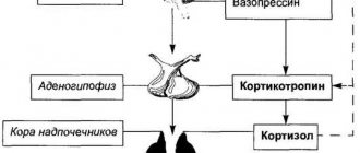
How does the immune system respond to the presence of Helicobacter pylori?
The presence of a foreign agent in the body causes a reaction of specific and humoral immunity. The adhesion of pathogenic microorganisms leads to increased division of protective cells, which are localized in the area of inflammation. The degree of accumulation of neutrophils, macrophages and other elements determines the severity of the pathological process with the further formation of various complications. Another link determines the production of antibodies, which indicate the duration of the disease and the degree of damage.
Advantages and disadvantages
Advantages of ELISA for the presence of Helicobacter pylori
The advantages include the following:
- high efficiency (more than 92%) of the study; IgG is detected in 95–100% of cases of Helicobacter pylori infection, IgA – in 67–82%, IgM – in 18–20%;
- detection of the pathogen in the early stages of infection;
- the ability to monitor deviations from the norm and the dynamics of the disease by comparing immunoglobulin titers in different periods;
- availability of analysis.
An analysis of antibody concentration is relevant if endoscopy is not planned.
An IgG test is used to identify primary infection in a patient with initial symptoms of digestive dysfunction. In this case, a high IgG content gives reason to suspect the development of an active infection.
A positive test result in a patient (with or without symptoms) who has not previously received any treatment will also indicate infection with Helicobacter pylori.
Disadvantages of the method
ELISA can show the immune response to infection, but does not diagnose the presence of the bacterium itself.
IgG antibodies are detected only 20–30 days after the introduction of Helicobacter pylori into the body, since the immune response is triggered with some delay. This leads to the following disadvantages of enzyme immunoassay:
- The likelihood of a false negative test result in infected patients. This occurs if the microbe first entered the body, but the defense system has not yet responded to the expansion of pathogens by producing antibodies.
- False positive result in cured patients. IgG antibodies remain present in the blood after complete destruction of the microorganism and in the absence of gastrological manifestations. This is especially true in older people. This means that the Helicobacter ELISA result may be a reaction to a long-treated infection.
- The likelihood of a false positive result if antibiotic treatment was already carried out before the analysis. Or antibacterial drugs that act against infection were used for other purposes. The IgG concentration remains elevated for up to one and a half years in 50% of patients cured of helicobacteriosis. Therefore, if the test result is positive in a patient who has previously taken antibiotics, it is difficult to distinguish between the state of the infectious process in action and the infection being suppressed and weak, which requires additional research.
- Low titers are detected when using certain cytostatic agents.
- It is difficult to make an accurate differential diagnosis between passive colonization of the gastric cavity by pathogens H. pylori and the disease in its acute form. This is impossible without taking other data into account.
The shortcomings of the study are compensated by the total analysis of IgG, IgM and IgA antibodies.
With the development of helicobacteriosis, the concentration of immunoglobulin IgG in the blood serum depends on the level of disease activity and decreases after the elimination of the pathogenic bacterial environment. Unlike immunoglobulin type G, antibodies A and M are detected in the blood much earlier after infection. In addition, IgA can be detected in the gastric juice and saliva of a person infected with Helicobacter pylori, which is a symptom of infection with the highest degree of activity.
Tactics for determining Helicobacter bacteria using stool analysis
The doctor prescribes a specific test for helicobacteriosis. The specialist takes into account the individual clinical picture of the patient’s health status. You can get tested for Helictobacter pylori yourself if you suspect a functional deviation of the digestive tract.
Molecular diagnostics
Molecular analysis reveals the specific DNA of the bacterium. The code is duplicated on a special device. When the bacteria reaches the desired size, it is compared with ethanol. If parts of the DNA match the original, then the test result is positive (Helictobacter pylori infection has been detected). The technique is characterized by a high degree of information content.
Cultural analysis
Bacterial inoculation (or cultural analysis) belongs to the category of microbiological research methods. During the procedure, biological material is placed in an environment that is most favorable for the proliferation of pathogenic microorganisms. After a week, the stool is examined using a microscope.
To identify Helicobacter pylori, the color of the bacteria and their ability to enter into certain reactions are taken into account.
Features of the technique:
- the procedure allows you to identify a pathological antigen;
- in the process of studying biological material, a sensitivity test to antibacterial drugs is carried out;
- The results of the study allow us to select the most effective treatment regimen.
Determination of Helicobacter pylori antigen in feces by immunological method
The main purpose of immunological tests when identifying Helicobacter pylori is to determine antigen + antibody complexes. Labeled antibodies are used for the study. When they react with feces, such substances undergo certain changes. Antibodies are able to detect the presence of not only pathogenic bacteria in the stool, but also their parts, as well as waste products.
Advantages and disadvantages of PCR diagnostics
PCR diagnostics has a number of advantages and disadvantages. Some types of pathogenic bacteria can only be detected using this technique (for example, cytotoxic types of microorganisms). Collecting material for analysis does not imply causing pain or discomfort to the patient. The procedure shows the most reliable results.
Advantages of the technique:
- the procedure allows you to identify various strains of pathogenic bacteria;
- the method has a high degree of sensitivity;
- the analysis is safe for the patient (no medical instruments are used to collect material);
- non-invasiveness and simplicity of the process of collecting biological material;
- high degree of specificity of the test;
- The method allows you to determine not only spiral-shaped, but also coccal types of bacteria.
The high degree of sensitivity of the test is its simultaneous advantage and disadvantage. The analysis accurately determines the presence of bacteria, but if the biological material or container is contaminated, the results of the procedure may be false positive.
To exclude false indicators, you should follow all the rules for collecting stool for testing.
Other disadvantages:
- relatively high price of the procedure;
- inability to determine the sensitivity of bacteria to antibacterial drugs;
- lower sensitivity compared to the procedure for examining a biopsy of the gastric mucosa.
Preparing for the test
Preparation includes the following:
- on the eve of the ELISA study, it is prohibited to drink alcohol or fatty foods;
- exclude physical activity during the day;
- You must donate blood before breakfast; you are allowed to drink water in the morning;
- the interval between the last meal and testing is at least 8–10 hours.
- The test should be taken before starting medication (if possible) or no earlier than 8–14 days after completion of therapy. If treatment is carried out, the referral for analysis includes a list of medications taken and doses.
Conducting the study, cost
The material for analysis is blood serum, which is taken by venipuncture. The collected biomaterial is poured into a test tube containing a special coagulant gel, which makes it possible to isolate plasma (blood serum) for research.
Complications during the blood collection procedure for testing are minimized. In cases of bruising at the site of a vein puncture, dry heat is used to quickly resolve the hematoma.
In different laboratories across the country, the cost of the study ranges from 340 for one type of immunoglobulin to 900 rubles for a total analysis.
You can get a laboratory answer for immunoglobulin G at most 24 hours after taking blood. The study of IgA continues longer. The result is obtained after 8 days.
Blood collection for ELISA (enzyme-linked immunosorbent assay)
Interpretation of results, normal indicator
There is a quantitative and qualitative determination of immunoglobulins G, A and M to Helicobacter pylori in blood plasma.
- A qualitative indicator indicates the presence and absence of antibodies without quantitative assessment. Normally, if the patient is not sick, there are no antibodies. In this case, the laboratory statement indicates that the test for antibodies to H. Pylori is negative.
- Indicators of the amount of IgG, IgA and IgM are based on reference (threshold) values indicating the norm, with which the obtained data are compared.
Reference standards in laboratories differ in numerical indicators and are assessed in different units. However, on the analysis results form, numbers are entered for the “norm” and deviations from the reference values. When deciphering, you need to take into account what immunoglobulin titers: below the threshold value means a negative test result, above - a positive one.
Table No. 1: Reference values of immunoglobulins in units of measurement U/ml
| Immunoglobulin type | Normal concentration, units/ml |
| A | < 0.9 |
| G | < 0.9 |
| M | < 30 |
Many laboratories note indicators at which the result of the ELISA analysis is regarded as “doubtful”. This is a reason to repeat the test after 14–20 days to clarify the diagnosis.
Table No. 2: Interpretation of the test for immunoglobulin G titers to Helicobacter pylori
| Result | Indicators in S/CO units (signal/cutoff) | Indicators in units/ml |
| Negative | < 0.9 | < 12.5 |
| Doubtful | 0.9 – 1.1 | 12.5 – 20.0 |
| Positive | > 1.1 | > 20.0 |
A refined diagnosis is established after a cumulative assessment of the results of an ELISA blood test for Helicobacter pylori with the study of indicators of the presence of three classes of antibodies to this bacterium.
Structure of antibodies - immunoglobulins A, G, M against Chyloribacter pylori
Table No. 3: Decoding of antibody titers of ELISA analysis in IFE units of measurement
| Type | Positive ≥ 30 IFE (for IgG and IgA) | Norm | Negative – less than 30 IFE |
| IgG |
| 30 |
|
| IgA |
| 30 |
|
| IgM | Early stage of acute infection (antibodies appear 7–8 days after infection). | Availability |
|
If immunoglobulin IgA is not detected, but the pain does not subside even with a negative test result for Helicobacter pylori, the test is repeated.
Elevated titers of three classes of immunoglobulins G, A and M indicate the aggressiveness of the infectious process.
A decrease in IgG concentration to 2% within six months indicates the destruction of H. pylori. But if titers do not decrease, this does not mean that the treatment is bad. The absence of IgG antibodies during a repeat test indicates the effectiveness of therapy and suppression of the bacterium. It is advisable to carry out the analysis after completion of therapy, at 10–12 weeks. In this case, the titer of immunoglobulin G to Helicobacter pylori when the infection is suppressed is reduced by 50% or more.
When pathogenic bacteria are suppressed, there is a tendency to a clear decrease in the severity of the inflammatory process in the gastric cavity.
If there are no gastrointestinal symptoms when H. pylori is detected, this is an indication that the stomach is colonized by pathogenic microbes, but the development of helicobacteriosis does not occur.
How to determine the presence of Helicobacter in the stomach
It is not possible to see such a tiny organism with a length of only 2-3 microns with the eye, nor is it possible to carry out diagnostics at home.
The patient can only assume the presence of gastritis based on the corresponding symptoms: epigastric pain after eating, heaviness and discomfort in the stomach, heartburn, belching of air or sour, metallic taste in the mouth. These signs of increased acidity very often accompany gastritis associated with a pathogenic microbe.
But it is possible to reliably determine whether the Helicobacter pylori bacterium has settled in the body or not only in the diagnostic department of an outpatient clinic, hospital or laboratory.
Detection of a pathogen in smears from a section of the inner wall of the stomach or cultivation of the microorganism on nutrient media.
Detection of antibodies in the blood, microbial antigens in the stool.
Detection of N. rulori under a microscope by coating a research sample with special dyes.
Polymerase chain reaction methods.
Urease test, breath test.
All of the above methods can be classified into two large groups:
- Invasive. Diagnostic methods based on endoscopic examination - FGDS, with taking a biopsy sample. A section of the inner wall of the stomach can then be subjected to cytological, cultural examination, and a urease test.
- Non-invasive. Other methods of detecting infection in which FGDS is not performed.